Comparative anatomy review for Exam 1 and 2
1/93
Earn XP
Description and Tags
Based on the review we did in class. All questions are from said reviews
Name | Mastery | Learn | Test | Matching | Spaced | Call with Kai |
|---|
No analytics yet
Send a link to your students to track their progress
94 Terms
Provide a reason why the skeleton is important to comparative anatomy
Bone is readily fossilized
- minimally susceptible to taphonomic varianceSkeleton is generally conservative (evolutionarily)
Skeleton is evolutionarily "plastic" enough to respond to major environmental and habitat changes
What is the function of a secondary palate? Provide one group which possesses a secondary palate
To separate the nasal cavity from the oral cavity so you can eat and breath
Crocodilians, Mammals
Which types of vertebrae are found in amphibians?
Cervical, Trunk, Sacral and Caudle
Provide three modifications to the skeleton found in birds and the region of the skeleton these are found in.
The sternum is changed to the keel by becoming longer outward wise to allow for more flight muscle attachment. Axial skeleton
Caudle vertebrae are merged to form the Pygostyle . Axial skeleton.
small fibula and elongated tibia called a tibiotarsus. Appendicular skeleton
What is cranial kinesis and what does it help with? Provide a group which does not have cranial kinesis.
Extra point of articulation in the skull to help with feeding. Crocodilian and mammals do not have it.
Provide 2 functions of the skeleton in vertebrates
Support the body
Mineral storage
Which group contains vertebrae in which the intercentra are dominant and the pleurocentra are ost?
Amphibians
What is another term for muscular limbs with well- defined joints and digits? In which group did this first appear?
Chiridium
early tetrapods
Provide 3 modifications to the skeleton of snakes and the region of the skeleton these are found in
Loss of limbs. Appendicular skeleton
Extra zygoatocity. Axial skeleton
Loss of jugal. Cranium
What are the two main types of ossification
Intramembranous Ossification and Endochondral Ossification
The skull of vertebrates can be divided into 3 major regions/components. What are those regions/components?
Chondrocranium, Splanchnocranium, Dermatocranium
Define aspidospondyly
Noting is connected in vertebrae
Provide a characteristic of the appendicular skeleton in Chondrichthyes.
Chordina bar or claspers
Briefly define endochondral ossification
Precursor of cartilage is replaced by invading bone
What important structures do the neural and hemal arches surround?
Neural arches: dorsal hollow nerve cord
Hemal arches: dorsal aorta
Provide a trend we observe in the evolution of the cranial skeleton of vertebrates
Chondrocranium and Splanchnocranium decrease while Dermatocranium stays the same
Provide 3 modifications to the skeleton in mammals and the region of the skeleton these are found in.
Limbs shift under the body. Appendicular
Secondary Palate
No cranial kinesis
Provide 2 major changes to the skull that occur in the evolution of synapsid.
loss of Post orbital
Chondrocranium and Splanchnocranium decrease while Dermatocranium stays the same
Loss of cranial kinesis
Briefly define intramembranous ossification?
No precursor of cartilage. Begins in the dermis
Which types of vertebrae are found in Mammals?
Cervical lumbar, thoracic, sacral, caudle
Which regions of the skull are found in all vertebrates?
Chondrocranium and Splanchnocranium
What are the two hypotheses for the origin of paired
fins?
Gill Arch theory
Ventral lateral Fin-Fold Theory
Provide the support and the anti support for the Gill arch theory
Support
Comparative anatomy and embryology:
• 1. Mandibular, hyoid and gill arches all develop from visceral arches
• 2. Spiracle, associated with mandibular arch, could represent “vestigial” gill-slit
between first and second arch (mandibular and hyoid arch)
• 3. Pseudobranch (vestigial gill filaments) could represent gill filaments of the
mandibular arch
• Points 2 and 3 suggest that the original function of the mandibular arch (V1) was
respiration (vs. current role as Jaws)
anti-support
Fossil record: Lack of “intermediate” fossil forms
• Developmental genetics: V1 in living agnathans does not contribute
to the gill arches but instead to the cartilages that surround the mouth and support the tongue
• Comparative anatomy:
• Cranial nerve V (trigeminal) innervates jaws in gnathostomes but innervates an area anterior to the pharynx in modern agnathans and not associated with gill function
Provide the support and the anti support for the Ventral lateral Fin-Fold Theory
This theory proposes that paired fins (pectoral and pelvic) evolved from continuous lateral folds of skin and muscle running along the sides of early vertebrates. Over time, parts of these folds became specialized and were retained as paired fins.
✅ Support for the Fin-Fold Theory:
Embryological Evidence:
In many vertebrate embryos, pectoral and pelvic fins develop from continuous lateral thickenings — similar to what the theory suggests.
These ridges appear simultaneously on both sides and at both fin positions (pectoral and pelvic), hinting at a common origin.
Genetic Evidence:
Shared expression of Hox genes and T-box genes (like Tbx4 and Tbx5) in both pectoral and pelvic regions suggests a shared genetic regulatory pathway, supporting a common evolutionary origin from a continuous fin fold.
Fossil Support:
Fossils like Osteostracans and early jawless fishes show long fin-like lateral flaps, possibly remnants of fin folds.
Some early jawed fishes (e.g., placoderms) have both pectoral and pelvic fins, showing an early and coordinated appearance of paired fins.
❌ Anti-Support / Criticisms of the Theory:
Lack of Direct Fossil Evidence for a Continuous Fold:
While lateral flaps exist in some fossils, there's no definitive fossil showing a true continuous lateral fin fold from which paired fins evolved.
Some argue the folds seen are too inconsistent or irregular to support the theory.
Segmented Development:
Pectoral and pelvic fins do not always develop in continuity with each other in embryos; instead, they arise as distinct buds at specific positions, suggesting separate origins rather than from a continuous structure.
Alternative Theories (e.g., Gill-Arch Theory):
Competing hypotheses propose different origins, like the gill-arch theory, which argues paired fins evolved from modified gill supports, casting doubt on the fin-fold idea.
🧠 Summary:
Support 🟢 | Anti-Support 🔴 |
|---|---|
Embryonic lateral ridges | No clear fossil evidence of a full fin-fold |
Shared genetic control (e.g., Tbx genes) | Independent development of fins in some embryos |
Fossils show fin-like flaps | Competing theories (e.g., gill-arch theory) |
In which group did endochondral bone first appear?
Osterithyians
Provide 1 major trend in the evolution of the axial skeleton of vertebrates
Specialization of the vertebrae
What 3 groups of vertebrates are capable of flight?
Briefly explain the differences in the appendicular
skeleton of each.
Aves: reduce digits, Arm is specialized for flight, carpels and metacarpals are fused
Pterosaurs: Thumb is specialized for flight leaving the other digits free for use
Bats: Digits are specialized for flight by elongating to form the wing
Provide 3 modifications of the skeleton that occurred during the transition from aquatic to terrestrial environments.
Limbs with digits
zygapophyses to prevent twisting
pelvic girdle attached to vertebrae
In which group does the humerus and femur first appear?
Rhipthian fishes
Which types of vertebrae are found in teleosts
Thoracic and Caudle
Provide the 3 types of cartilage found in vertebrates.
Hyaline cartilage, Fibrocartilage, Elastic cartilage
Provide 3 modifications to the skeleton of Testudines and the region of the skeleton which is modified.
Ribs expanded in shell Axial
No teeth Cranial
Vertebrae in shell Axial
Where can dermal bone be found in the postcranial
skeleton?
Pectoral Girdle
Which group contains vertebrae in which the pleurocentra are dominant and the intercentra are lost?
amniotes
What are the first two visceral arches referred to as?
What is their function?
Mandibular Arch-Jaws
Hyoid Arch- support
In which group did the exoskeleton first appear?
Ostractoderms, conodonts
In which group did pelvic fins first appear?
Placoderms
Provide the 3 major types of cells found in bone and
what they do.
Osteoblast-synthesize bone
Osteocytes-maintain bone
Osteoclast-breakdown bone
Provide an extant group of vertebrates which possess a notochord throughout their lives
Cyclostones and Petromyzontiformes (hagfish)
Explain the difference between the jaws of Chondrichthyes and Osteichthyes?
Chondrichthyes: meckel's cartilage
Osteichthyes: Novel Jaw including Premaxilla, Maxilla and Dentary
Provide 3 modifications to the skeleton observed in either Amphibians, Actinopterygians or Chondrichthyes (mix and match) along with the group and the region of the skeleton the modification occurs in
🐸 1. Amphibians – Axial Skeleton
Modification: Reduction in the number of trunk vertebrae and fusion of caudal vertebrae into a urostyle
Details: In frogs, many tail vertebrae are reduced and fused into a rod-like structure called the urostyle, aiding in jumping and shock absorption.
Region: Axial skeleton (vertebral column)
Group: Amphibians
🦈 2. Chondrichthyes (Cartilaginous Fish) – Cranial Skeleton
Modification: Cartilaginous skull with no dermal bones and highly developed rostrum
Details: Sharks and rays lack bony skulls; instead, their chondrocranium is made entirely of cartilage. Many have an elongated rostrum (snout) used for sensory detection.
Region: Cranial skeleton
Group: Chondrichthyes
🐟 3. Actinopterygians (Ray-finned Fish) – Appendicular Skeleton
Modification: Highly mobile fin rays (lepidotrichia) supported by modified pectoral girdles
Details: The pectoral fins of many ray-finned fish (like teleosts) are adapted for fine maneuverability, thanks to bony fin rays and specialized girdle articulation.
Region: Appendicular skeleton (fins and girdles)
Group: Actinopterygians
Provide 3 of the 5 types of caudal fins found in vertebrates and a group which possess each.
🦈 1. Heterocercal Tail
Description: The vertebral column extends into the upper lobe of the tail, making it asymmetrical.
Function: Provides lift during swimming, useful for animals without a swim bladder.
Example Group: Chondrichthyes (e.g., sharks)
🐟 2. Homocercal Tail
Description: The external appearance is symmetrical, although the vertebral column may angle slightly into the upper lobe.
Function: Efficient for fast, sustained swimming in open water.
Example Group: Actinopterygians (e.g., most modern bony fishes like tuna and bass)
🐠 3. Diphycercal Tail
Description: The vertebral column runs straight to the end of the tail, and the fin is symmetrical above and below.
Function: Common in species with slow, precise movements or those that dwell near the bottom.
Example Group: Lobe-finned fishes (e.g., coelacanths and lungfish)
What trend is observed in the evolution of the appendicular skeleton of both archosaurs and mammals. Explain the importance of this.
🔄 Trend: Shift from sprawling to upright limb posture
📘 Summary:
Group | Limb Posture Evolution | Importance |
|---|---|---|
Archosaurs | Sprawling ➡ Upright (dinosaurs, birds) | Efficient walking/running, flight adaptations |
Mammals | Sprawling ➡ Upright | Better support, endurance, diverse locomotion |
What are heterotopic bones? Provide one example
bones that develop in unusual or abnormal locations — that is, outside the typical skeleton. They form in soft tissues like muscles, tendons, or skin, often due to genetic programming, injury, or mechanical stress.
Patella (kneecap)
Skulls of amniotes are cool! What are the two different temporal fenestration types in amniotes, and which bones contribute towards the temporal bars in each condition?
Synapsids
Temporal bar (bordering the fenestra):
Formed mainly by the postorbital and squamosal
diapsids
Upper (supratemporal) fenestra
Lower (infratemporal) fenestra
🦴 Temporal bars (bones forming the fenestra borders):
Upper bar: Postorbital + Squamosal
Lower bar: Jugal + Quadratojugal
The evolutionary origin of the bones of the middle ear is cool! What is the evolutionary origin of the bones of the middle ear? (i.e., what bones in fishes are homologous to the bones of the inner ear). When in the evolution of vertebrates did each element become incorporated into the middle ear?
Jawed fishes had multiple bones in their lower jaw. The articular and quadrate formed the jaw joint, and the hyomandibula braced the jaw against the skull.
In early tetrapods, the hyomandibula lost its jaw-support role and became the columella, acting as a sound-conducting bone from the outer skull to the inner ear.
In synapsids (mammal ancestors), the dentary bone expanded, taking over the jaw joint role. The articular and quadrate became smaller and gradually shifted function.
In mammals, the articular and quadrate completed their migration into the middle ear, evolving into the malleus and incus respectively.
📜 Homologous Structures & When They Changed
Middle Ear Bone
Fish Homolog
Original Function in Fish
When It Became Part of Middle Ear
Stapes / Columella
Hyomandibula
Supported jaw/skull articulation and helped with gill breathing
🐟 Early tetrapods (~360 MYA) – First used to transmit sound
Incus
Quadrate
Upper part of jaw joint (connected to articular)
🦎 In early mammal-like reptiles (therapsids) – Began shifting toward the middle ear (~250–200 MYA)
Malleus
Articular
Lower jaw joint bone
🐀 Fully incorporated in mammals (~200 MYA), as the dentary took over the jaw joint
Flying is cool! What are the three hypothesis of the origin of flight and briefly discuss them?
Arboreal Hypothesis
Early birds hopped from tree to tree to escape predators
Feathers aided in longer jumps and eventually
glidingOver time, gliding gave way to active flight
Insect-net or Ground Up Hypothesis
Early birds were cursorial dinosaurs that ran and jumped to capture quick moving prey (insects)
Feathers helped with higher jumping and batting down prey
Wing Assisted Incline Running (WAIR) Hypothesis
Feathered forelimb aided bird ancestors in climbing steep inclines or vertically up trees
Based on evidence from newly hatched modern birds which use their developing wings for this purpose
Provide all of the possible bones that can be found in the pectoral
and pelvic girdle of vertebrates.
🟦 Pectoral Girdle (Shoulder Girdle)
Connects the forelimbs to the axial skeleton (vertebral column or skull)
🦴 Dermal Bones (from the dermis)
Clavicle – collarbone; often braces the scapula
Cleithrum – prominent in fish; reduced or lost in most tetrapods
Supracleithrum – found in bony fish
Postcleithrum – found in some fish
Interclavicle – midline bone, especially in reptiles
Posttemporal – connects girdle to the skull in some fish
🦴 Endochondral Bones (from cartilage)
Scapula – the shoulder blade
Coracoid – supports the glenoid fossa; reduced or fused in mammals
Procoracoid – a separate coracoid element in some reptiles and amphibians
Suprascapula – dorsal extension of scapula in amphibians/reptiles
Glenoid Fossa (joint socket, not a separate bone but an important feature)
🟩 Pelvic Girdle (Hip Girdle)
Connects the hindlimbs to the axial skeleton (via the sacrum)
🦴 Endochondral Bones (entire girdle is endochondral)
Ilium – articulates with the vertebral column
Ischium – forms the lower rear part of the pelvis
Pubis – forms the lower front part of the pelvis
Acetabulum – socket for the femur (again, not a separate bone but a critical joint feature)
📝 Summary Table:
Girdle | Bone Type | Possible Bones |
|---|---|---|
Pectoral | Dermal | Clavicle, Cleithrum, Supracleithrum, Postcleithrum, Interclavicle, Posttemporal |
Pectoral | Endochondral | Scapula, Coracoid, Procoracoid, Suprascapula |
Pelvic | Endochondral | Ilium, Ischium, Pubis |
Provide 2 synapomorphies of Osteichthyes
Presence of endochondral bone.
Presence of a gas bladder or lung (used for buoyancy or respiration)
What is the plesiomorphic condition of the
integument in vertebrates
The plesiomorphic (ancestral) condition is a multilayered epidermis with a non-keratinized outer layer and dermal bone elements (like dermal scales or plates).
What are the 3 subphyla of chordates and what is
our current understanding of their evolutionary
relationships
Cephalochordata (e.g., lancelets)
Urochordata (e.g., tunicates)
Vertebrata (e.g., fishes, amphibians, mammals)
Current understanding: Urochordates are more closely related to vertebrates than cephalochordates are, making Cephalochordata the basal chordate lineage
Briefly define von Baer’s law regarding embryonic
development
Von Baer's law states that general features of a group of animals appear earlier in development than the more specialized features; embryos of different species resemble each other more in early stages than in later stages.
Provide two characters that Myxiniformes and
Petromyzontiformes share in common and one
difference between them
Lack of jaws.
Cartilaginous skeletons.
Difference:
Myxiniformes (hagfishes) have no vertebrae, while Petromyzontiformes (lampreys) have rudimentary vertebral elements.
What type of characters can inform us about
phylogenetic relationships
Synapomorphies — shared derived characters that indicate common ancestry.
Provide two of the four chromatophores found in
vertebrates as well as the pigments they produce
Melanophores – contain melanin (black/brown pigment).
Xanthophores – contain pteridines/carotenoids (yellow pigments).
Provide 3 distinct lineages of archosaurs
Crocodylia (crocodiles and alligators)
Aves (birds)
Non-avian dinosaurs (e.g., Tyrannosaurus, Triceratops
Mammals possess a lot of glands, one of which are
apocrine sweat glands. Provide two different glands
found in mammals with at least one being a type of
modified apocrine sweat gland
Mammary glands (modified apocrine glands, produce milk)
Sebaceous glands (secrete oil/sebum to lubricate skin and hair
What are the 5 chordate characteristics and which
of these are also found in Hemichordates?
Notochord
Dorsal Hollow Nerve chord
Pharyngeal slits/pouches
Postanal tail
Notochord
Found in Hemichordates: Pharyngeal slits and possibly a structure homologous to the dorsal nerve cord (though not fully homologous in structure or development).
Provide the two types of neurulation in vertebrates
(briefly describe them) and a group which
possesses each
Primary neurulation: Neural plate folds to form a neural tube (found in most vertebrates like mammals, birds).
Secondary neurulation: Solid cord of cells forms and then hollows out into a tube (found in teleost fishes and some tail regions of higher vertebrates)
Provide two major extinct group of synapsids
Pelycosaurs (e.g., Dimetrodon)
Therapsids (includes cynodonts, more closely related to mammals)
Provide the two layers that comprise the
integument and the embryonic germ layer they
originate from
Epidermis – from ectoderm
Dermis – from mesoderm
What are the two embryonic structures that give
rise to major sensory systems such as olfaction,
vision and mechanoreception?
Placodes
Neural crest cells
Provide the 3 types of temporal fenestration of the
skull.
Anapsid – no temporal openings (e.g., turtles, though debated)
Synapsid – one temporal opening (e.g., mammals and their ancestors)
Diapsid – two temporal openings (e.g., reptiles, birds)
Define a monophyletic, paraphyletic and
polyphyletic group
Monophyletic: Includes a common ancestor and all its descendants (e.g., Mammalia).
Paraphyletic: Includes a common ancestor and some, but not all, descendants (e.g., “Reptilia” excluding birds).
Polyphyletic: Does not include the most recent common ancestor (e.g., grouping flying animals like birds and bats).
Provide two types of caudal fins that are found in
fishes and a group that possesses each
Heterocercal – asymmetrical, with vertebral column extending into upper lobe (e.g., sharks).
Homocercal – symmetrical tail fin (e.g., teleost fishes).
Provide two groups of sarcopterygian fishes that
are not tetrapods
Coelacanths (Actinistia)
Lungfishes (Dipnoi)
Provide 4 synapomorphies of mammals.
Hair/fur
Mammary glands
Heterodonty teeth
Diaphragm
What point in geological time is referred to as the
“AGE OF DINOSAURS”
The Mesozoic Era (includes Triassic, Jurassic, and Cretaceous periods)
Who is considered the father of our modern
classification system and is credited for creating the
binominal nomenclatural system used today.
Carl Linnaeus
Provide 2 of the 4 types of dermal scales found in
fishes and an example of a group which possesses
each.
Ganoid scales – found in gars and bichirs
Placoid scales – found in sharks and rays (Chondrichthyes)
Provide the sister group to the phylum Chordata.
Hemichordata
The amount of yolk varies between the eggs
of vertebrates. Provide the three major types
and a group that possesses each
Microlecithal – little yolk (e.g., mammals like humans)
Mesolecithal – moderate yolk (e.g., amphibians)
Macrolecithal – large yolk (e.g., birds and reptiles)
Birds and mammals both possess glands to
waterproof their feathers and hair respectively.
Prove the glands for each. What type of character is
this an example of?
Birds: Uropygial (preen) gland
Mammals: Sebaceous gland
This is an example of analogous traits (convergent evolution).
Provide two synapomorphies of amniotes.
Amniotic egg with extraembryonic membranes
Costal (rib-based) ventilation of the lungs
Briefly define the Biogenetic law of embryonic
development.
Also known as Haeckel’s biogenetic law, it states that "ontogeny recapitulates phylogeny" — meaning that the development of an organism (ontogeny) mirrors the evolutionary history (phylogeny) of its species.
Provide two synapomorphies of the class
Sarcopterygii.
Fleshy, lobed paired fins with internal skeletal support
Cosmine-covered scales (a unique enamel-like tissue
What are the 5 stages of embryonic development?
Fertilization
Cleavage
Gastrulation
Neurulation
Organogenesis
What portion of geological time is known as the
“Age of Fishes”. Why is it referred to as such?
The Devonian Period
All major lineages where present at this time.
Provide 2 unique characteristics of amphibian skin
that aids in cutaneous respiration.
Thin and permeable epidermis for gas exchange
Mucous glands to keep skin moist, facilitating diffusion
Provide two synapomorphies of Therian mammals.
Live birth (viviparity)
Presence of nipples (mammary glands with teats
How old is the earth?
4.5 byo
Provide two synapomorphies of vertebrates.
Vertebral elements surrounding the notochord/spinal cord
Cranium (skull) protecting the brain
Presence of a neural crest during development
Internal skeleton composed of cartilage or bone.
In which group of fossil fishes do we first see the
appearance of paired appendages
Placoderms (early jawed fishes)
Provide the two major lineages of amniotes.
Synapsids (mammals and their extinct relatives)
Sauropsids (reptiles, birds, and their extinct relatives)
Provide Darwin’s 4 postulates of natural selection.
Individuals vary within a population
Some variation can be passed from parents to offspring
In every generation more offspring are produced than will ultimately survive to reproduce
The individuals that survive and reproduce are not a random sample of the population, but are those with advantageous traits.
Provide two hallmarks of the tetrapod skeleton.
Limbs with digits (typically five per limb — pentadactyly)
Pelvic girdle attached to vertebral column
Provide a synapomorphy of lepidosaurs
Transverse cloacal slit
In which group is the exoskeleton (dermal) skeleton
first observed in the fossil record?
Ostracoderms (jawless armored fishes)
Provide the difference between horns and antlers.
Also provide one example of “horns” that are not
true horns.
Horns: Permanent, bony core with a keratin sheath; not shed (e.g., cows, goats)
Antlers: Made of bone, shed and regrown annually (e.g., deer)
Example of not true horns: Rhino “horns”, made of keratin with no bony core.
What fossil group of fishes is the earliest group to
possess jaws? What are vertebrates which possess
jaws referred to as (e.g., classification)?
Earliest jawed fishes: Placoderms
Jawed vertebrates are called Gnathostomes
Amniotic eggs are cool! Provide the three
extraembryonic membranes found only in the eggs
of amniotes and their function
Amnion: Cushions and protects the embryo
Chorion: Gas exchange
Allantois: Waste storage and gas exchange
Mammals are cool! (not as cool as fishes) Provide the 3 major
lineages of living mammals and how they differ in
their reproductive life history.
Monotremes: Lay eggs (e.g., platypus)
Marsupials: Give birth to underdeveloped young that complete development in a pouch (e.g., kangaroo)
Eutherians: Give birth to well-developed young after a longer gestation (e.g., humans)
Turtles are Weird (in a cool way though)! Why are
their evolutionary relationships difficult to
determine? What is our current understanding of
their placement among sauropsids and what led to
this conclusion?
Difficulty: Turtles lack obvious temporal fenestrae and have highly derived skeletons, making them hard to place morphologically.
Current understanding: Molecular and genetic data place turtles within diapsid reptiles, as sister to archosaurs (birds + crocs).
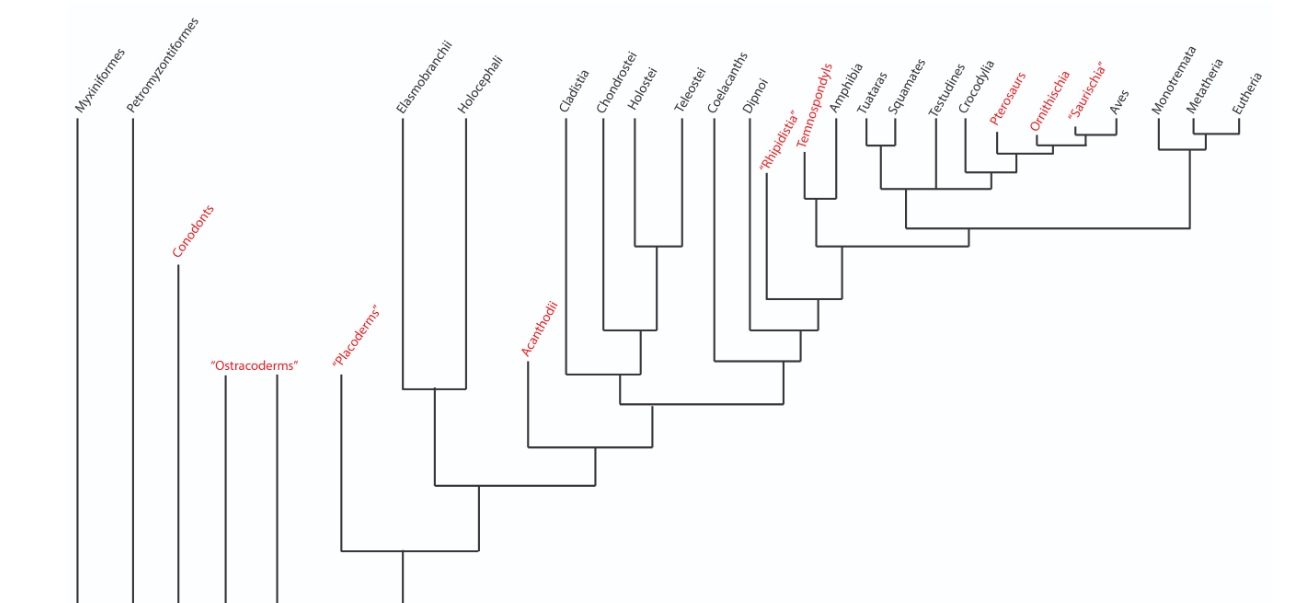
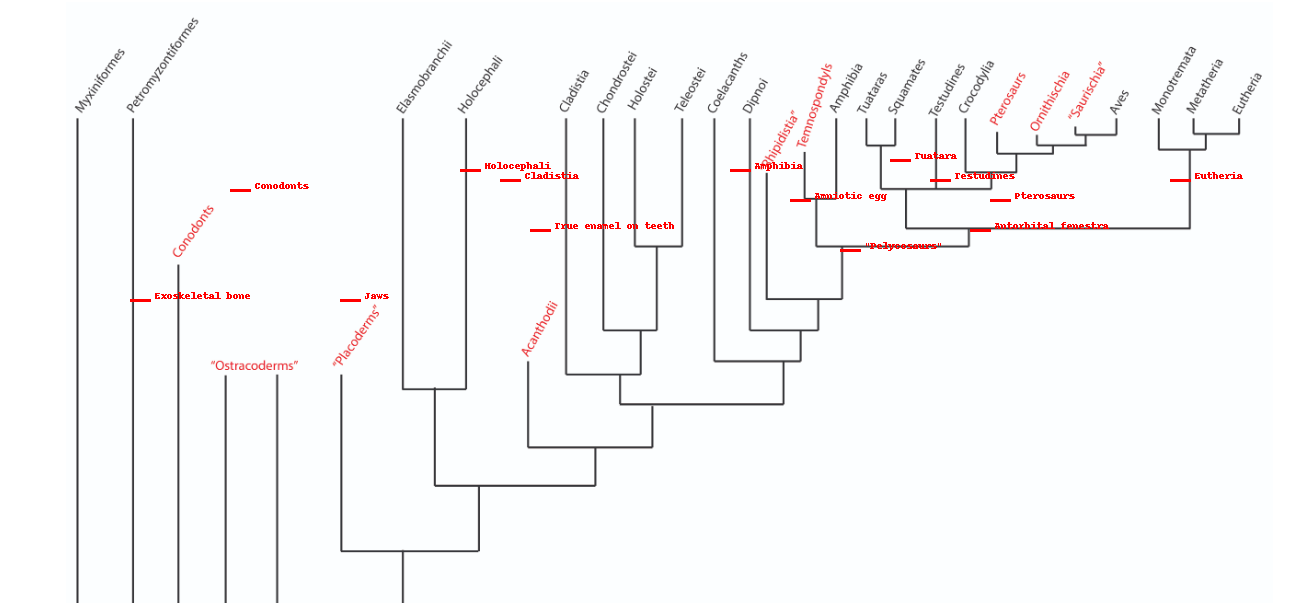
Draw a phylogenetic tree including the following taxa
groups and characteristics
Bonus Round
Amphibia, Amniotic egg, Cladistia, Jaws, Tuatara, Eutheria, Antorbital fenestra, Holocephali, Conodonts, Pterosaurs, Exoskeletal bone, “Pelycosaurs”, Testudines, True enamel on teeth
Summary (From Most Basal to Modern):
Conodonts → Earliest vertebrates, with early tooth-like structures.
Exoskeletal Bone → Early protective feature in vertebrates.
Jaws → Evolution of jawed vertebrates.
Holocephali → Early cartilaginous fish.
Cladistia (Bichirs) → Early ray-finned fish.
Amphibia → First tetrapods transitioning to land.
Amniotic Egg → Evolution of terrestrial vertebrates.
Testudines (Turtles) → Early reptiles with unique shells.
Tuatara → Ancient reptile lineage with distinct characteristics.
Antorbital Fenestra → Feature of archosaurs (dinosaurs, birds, crocs).
Pterosaurs → First flying reptiles.
Pelycosaurs → Early synapsids, leading to mammals.
True Enamel on Teeth → Feature of mammals.
Eutheria (Placental Mammals) → Modern mammals with live birth.