Postmortem Changes
1/75
Earn XP
Description and Tags
Pathology - Lec 10 - Exam 1
Name | Mastery | Learn | Test | Matching | Spaced |
|---|
No study sessions yet.
76 Terms
postmortem
after death
antemortem
before death
perimortem
around death
autopsy
self-examination
necropsy
death examination
postmortem examination (necropsy/autopsy)
exam of the animal after death
What is the goal of postmortem examination?
determine the cause of death/ the extent of disease
Postmortem examination involves the careful process of
dissection, observation, interpretation, & documentation
What is the importance of postmortem examination?
scientific knowledge (physio/ anatomy, etc)
education of students
confirmation of clincial dx
public heath, legal cases, etc
autolysis
decomposition of cells that takes place after somatic death
T/F: Autolysis is the breakdown of the body by endogenous substances/ enzymes.
true
putrefaction
decomposition of organic matter by bacterial or fungal digestion
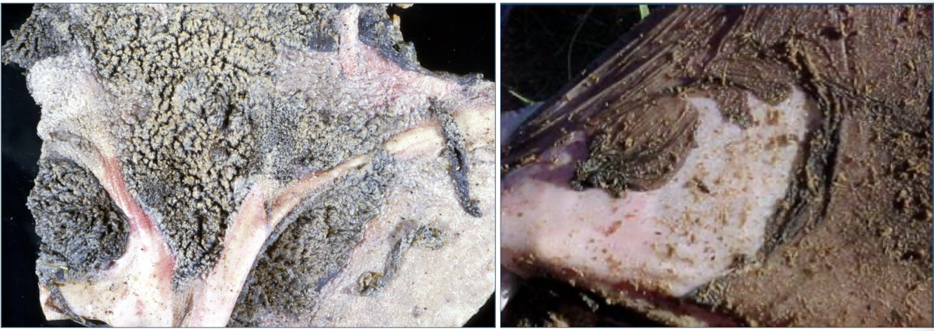
postmortem predication/ scavenging
animals feed on bodies after death
decomposition
process by which dead organic substances are broken down into simpler organic or inorganic matter
Postmorerm degeneration is the result of what?
autolysis and putrefaction
T/F: Postmortem degeneration is evidence of actual lesions.
FALSE
What is the important distinction to make regarding postmortem degeneration?
differentiate antemortem cell death from postmortem cell degeneration
What are some examples of postmortem degeneration?
livor mortis/ hypostatic congestion
postmortem clots
rigor mortis
autolysis & putrefaction
livor mortis/ hypostatic congestion
gravitational pooling of blood on the dependent (down) side of the carcass
What type of animals would you be able to physically see livor mortis?
skin of pale-skinned, sparsely haired animals (pigs)
When does livor mortis begin? What happens when you palpate the carcass?
0.5 - 2 hours after death
blanches on palpation
When does livor mortis become permanent?
once the blood clots → static or “fixed” after 8-12 hours
T/F: Pooled blood will remain on the original down side.
TRUE
What are the 3 main characteristics of a postmortem clot?
smooth shiny surface
lack of lamination
lack of attachment to endothelial surface of vessel
How is a “black currant clot” formed?
blood (RBC) settle at the bottom → buffy coat of leukocytes → clotted serum at top
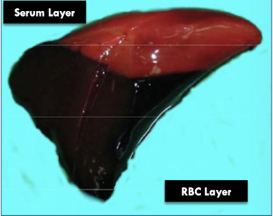
What species would you expect to find a black currant clot in? Why?
horses → high sedimentation rate
What causes an increased sedimentation rate?
inflammation
What causes a decreased sedimentation rate?
anticoagulant rodenticide toxicity
hereditary coagulopathies
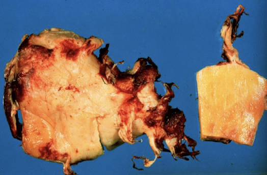
What do “chicken fat clots” look like?
yellow fatty appearance
rigor mortis
generalized contraction of skeletal muscle
T/F: Rigor mortis only affects certain muscles.
FALSE - affects all skeletal muscles
T/F: Smaller muscles are more visible due to rigor mortis.
TRUE
When does rigor mortis start?
1-6 hours after death → can persist for 2+ days
Rigor mortis occurs because muscle relaxation requires what?
ATP
T/F: Intracellular glycogen can provide some postmortem ATP.
TRUE
T/F: Once glycogen stores are depleted, rigor can be reversed by multiple pathways.
FALSE - only reversible via autolysis post depletion
T/F: Rigor mortis requires the complete breakdown of proteins, not just cross-bridges.
TRUE
What 3 things can accelerate rigor mortis?
antemortem exertion
seizures
antemortem hyperthermia
T/F: Autolysis is only caused by endogenous substances/ enzymes.
FALSE - also by aseptic chemical processes
What 2 things contribute to putrefaction?
postmortem bacilli
fermentation
Putrefaction implies the presence of what?
severe softening and gas in the tissue
Putrefaction may suggest what in regards to examination?
samples are not too suitable for microscopic examination
T/F: It is difficult to determine the postmortem interval.
TRUE
Why is it difficult to determine the postmortem interval?
highly variable
limited data in animals → lack of validation
lack of supporting evidence in animals → no last social media post
What factors can cause increased autolysis?
long interval between death & necropsy
high ambient temp
large body size
heavy hair or wool coat
continued fermentation in GI tract (equine/ ruminants)
antemortem hyperthermia
antemortem bacterial infection
What factors can cause reduced autolysis?
short interval between death and necropsy
rapid cooling of carcass postmortem (lower ambient temp, refrigeration)
thin animal (reduced fat & muscle)
high levels of tissue antibiotics
When does autolysis develop in the rumen or intestine? Why?
within 20 mins
exposure to bacteria & digestive enzymes (sloughing of mucosa, pH of rumen drops)
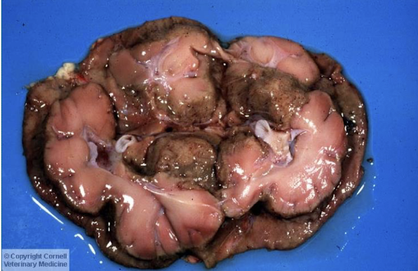
What tissues are quick to autolyze?
brain & spinal cord
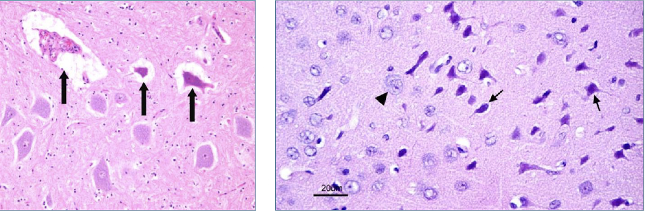
What is a handling artifact to rule out when examining nervous tissue?
dark neurons
T/F: Skeletal muscle retains the ability to contract after somatic death.
TRUE
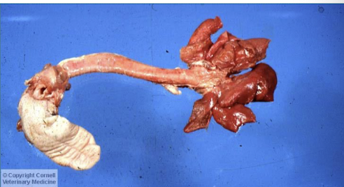
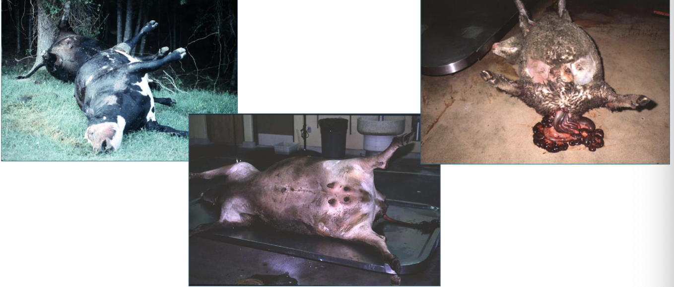
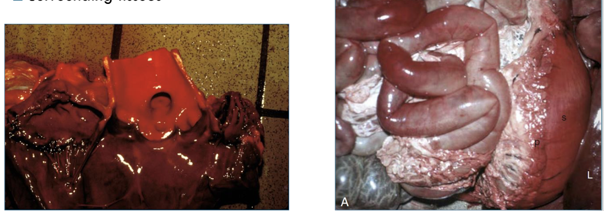
What are 3 examples of postmortem changes?
bloating of carcass
rectal &/or vaginal prolapse
tissue softening, discoloration, & gas bubbles present (putrefaction)
What causes postmortem gas production?
gut bacteria
What is the result of postmortem gas production/ gaseous distension?
bloated appearance → rupture of GI tract or carcass (or diaphragm) → displacement of abdominal viscera → eversion of rectal mucosa through anus (vagina, phimosis, protrusion of tongue)
What causes emphysema in the tissues postmortem?
bacteria traveling to various tissues
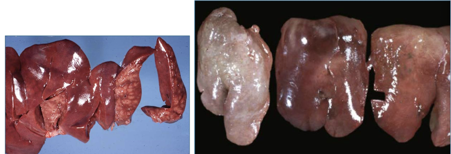
What is the gross appearance of autolysis?
usually diffuse
pallor
tissue soft & pulpy, gas filled
friable (tears easily)
oozes reddish fluid
strong odor (hydrogen sulfide, methyl mercaptan)
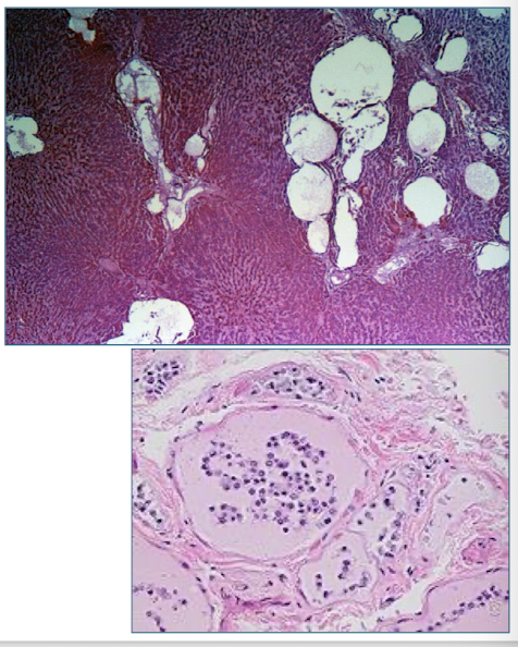
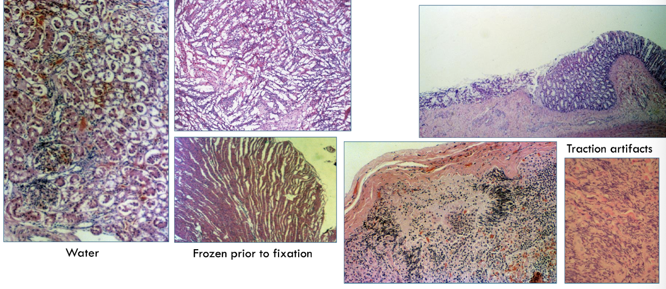
What is the microscopic appearance of autolysis?
tissues pale staining
no inflammatory rx
cadaver bacilli
erythrocytes not preserved
loss of tissue pattern
cells released from basement membrane
What are other possible histological artifacts?
water
frozen prior to fixation
traction artifacts
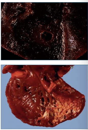
hemoglobin imbibition
reddish discoloration of tissue by hemoglobin from lysed erythrocytes
Where will you normally see hemoglobin imbibition?
intima & endocardium, surrounding tissues
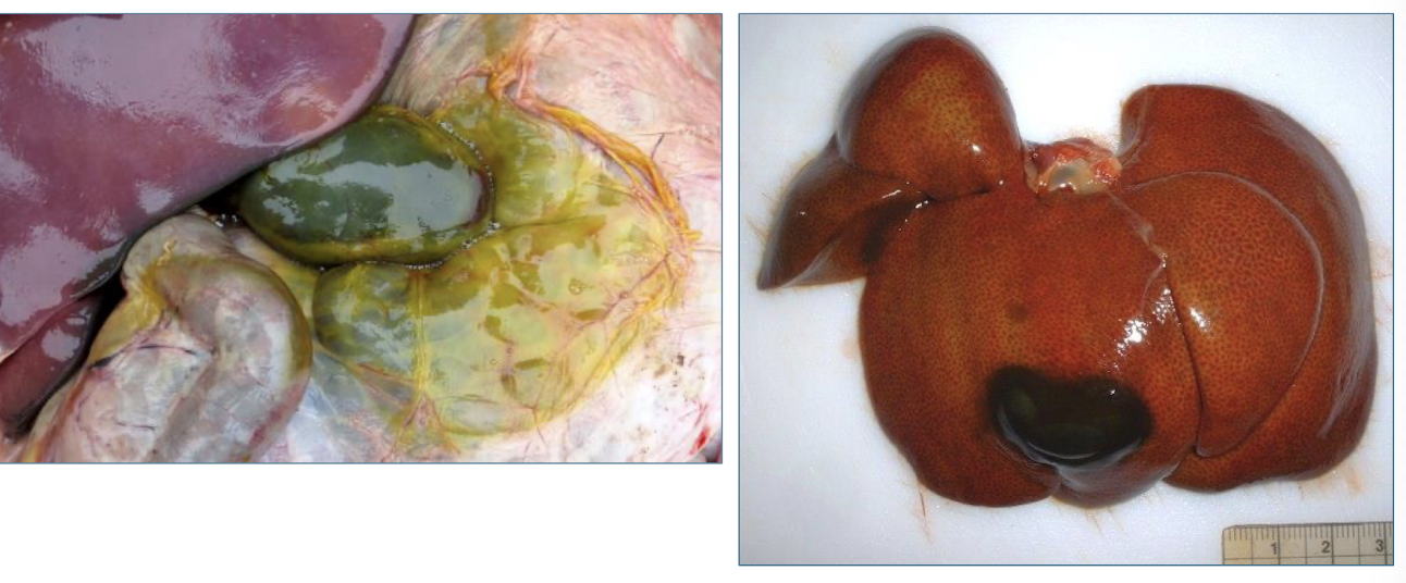
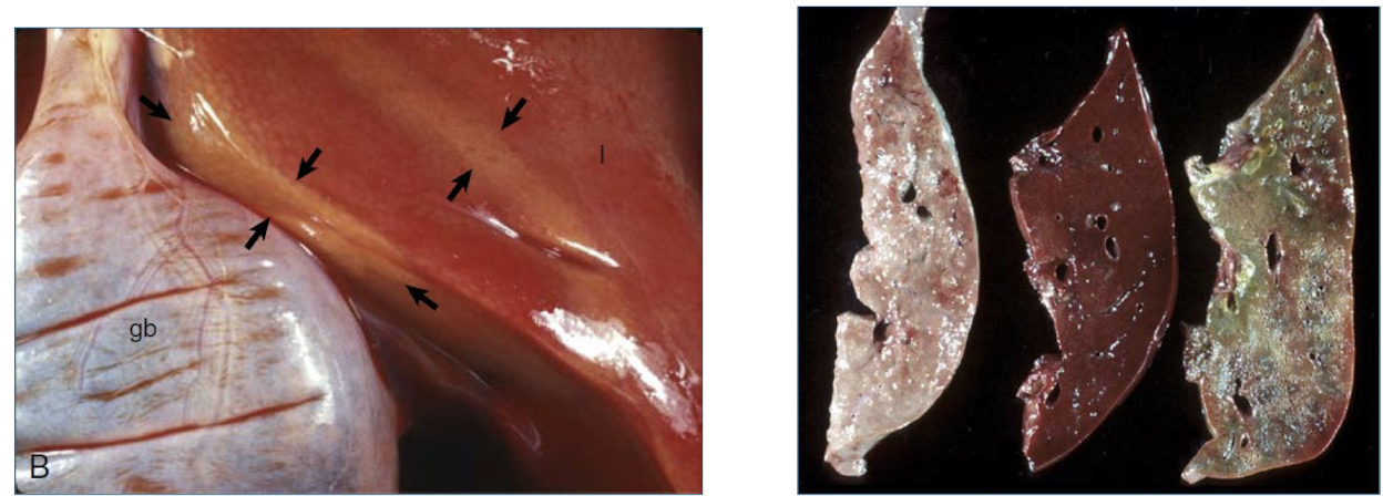
bile imbibition
greenish discoloration from leakage of bile through wall of the gallbladder or bile ducts
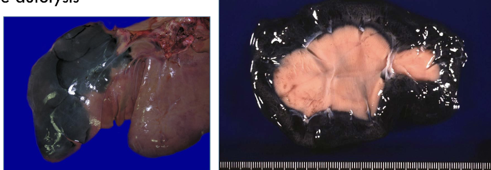
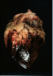
pseudomelanosis
blue-green to black discoloration of tissues by iron sulfide deposits
T/F: Pseudomelanosis deposits are actually melanin.
FALSE - not actually melanin
What reaction causes pseudomelanosis?
rx of hydrogen sulfide from putrefactive bacteria with iron in hemoglobin
What does pseudomelanosis signify?
severe autolysis
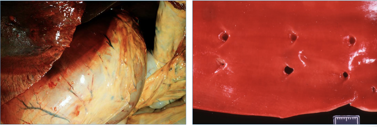
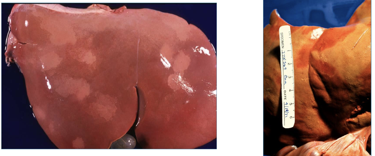
Why do you see pallor?
postmortem pressure on organs can force blood out of tissues → “pale imprints” (rib imprints)
T/F: Pallor can be caused by postemortem bacterial proliferation in the liver.
TRUE - must differentiate from antemortem necrosis
lens artifact
chilling or partial freezing of the carcass can make the lens opaque and white
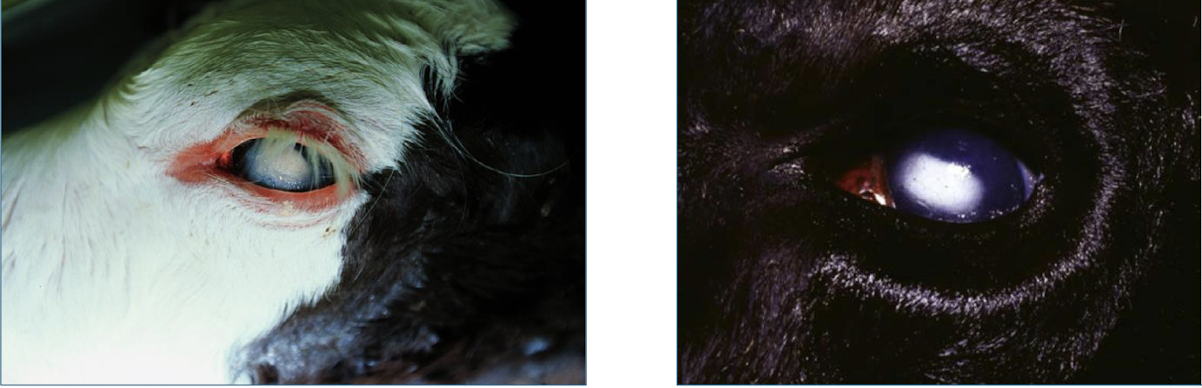
T/F: Lens artifact will not revert back to normal transparency upon warming.
FALSE - but may not after freezing
T/F: Peristalsis continues to occur postmortem.
TRUE
What does postmortem peristalsis cause?
traps blood in ridges
mimics intussusception

perimortem findings
changes associated with the process of dying
Agonal breathing is a perimortem finding. This can cause what?
interstitial edema, tracheal foam
Perimortem hemorrhages are typically seen where?
epicardial & endocardial tissue
What are the artifacts associated with barbituate overdose (euthanasia)?
splenic congestion
pulmonary congestion & edema
discoloration of blood
coagulation of endocardium
What may be seen with electrocution?
petechial hemorrhages
What are the artifacts of resuscitation?
fractured ribs
cutaneous hemorrhage
hemorrhage around injection sites (laceration of heart w/ intracardiac injection)
collapse of lungs if animal was on 100% oxygen (or anesthetic gas)