S&SPU II- Exam 1 Review
 Call Kai
 Learn
Learn Practice Test
 Spaced Repetition
 Match
Match Flashcards
 Knowt Play
1/80

There's no tags or description
Looks like no tags are added yet.
Name | Mastery | Learn | Test | Matching | Spaced | Call with Kai | Chat |
|---|
No analytics yet
Send a link to your students to track their progress
81 Terms
The testes are classified as:
a. endocrine and seminal glands.
b. exocrine and endocrine glands.
c. exocrine and seminal glands.
d. None of these
b
The epididymal head is connected to which portion of the testis?
a. Superior
b. Lateral
c. Inferior
d. Medial
a
Spermatozoa exit the testis through the:
a. efferent ducts.
b. tail of the epididymis.
c. body of the epididymis.
d. head of the epididymis.
a
The penis is composed of:
a. two corpora cavernosa midventrally and one corpus spongiosum dorsolaterally.
b. two corpora spongiosa midventrally and one corpus cavernosum dorsolaterally.
c. two corpora cavernosa dorsolaterally and one corpus spongiosum midventrally.
d. two corpora spongiosa dorsolaterally and one corpus cavernosum midventrally.
c
The main arterial blood supply of the penis includes the:
a. deep artery of the penis, bulbourethral artery, and dorsal artery
b. superficial dorsal artery, deep dorsal artery, and deep artery of the penis.
c. deep artery of the penis, bulbourethral artery, dorsal artery, and pampiniform plexus arteries.
d. deep artery of the penis, dorsal artery, and deep dorsal artery.
a
Testicular parenchyma on ultrasound is:
a. anechoic.
b. mixed solid appearance.
c. heterogeneous.
d. homogeneous.
d
What is the echogenicity of the corpora cavernosa?
a. They cannot be identified on ultrasound.
b. Medium-level echoes
c. High echogenicity
d. Low-level echoes
b
What is the echogenicity of the corpus spongiosum?
a. High echogenicity
b. Low-level echoes
c. It cannot be identified on ultrasound.
d. Medium-level echoes
d
What is the echogenicity of the septum penis?
a. Low-level echoes
b. High echogenicity
c. It cannot be identified on ultrasound.
d. Medium-level echoes
b
What is the echogenicity of the epididymis?
a. Low-level echoes
b. Medium-level echoes
c. It cannot be identified on ultrasound.
d. High echogenicity
b
Which statement is incorrect about the seminal vesicles?
a. They are posterior to the urinary bladder and inferior to the prostate.
b. They help form the ejaculatory duct.
c. They are paired glands.
d. Each gland empties into the distal ductus (vas) deferens.
a
The largest portion of the glandular prostate is the:
a. peripheral zone.
b. periurethral zone.
c. transition zone.
d. central zone.
a
Semen is composed of:
a. 70% alkaline fructose, 13% to 33% sperm, and acidic fluids.
b. 60% alkaline fructose, 13% to 33% alkaline fluid, and sperm.
c. 13% to 33% alkaline fructose, 70% alkaline fluid, and sperm.
d. 60% sperm, 13% to 33% alkaline fluid, and fructose.
b
On a sonogram of the prostate and seminal vesicles, the prostate normally appears:
a. hyperechoic to the seminal vesicles
b. hypoechoic to the seminal vesicles.
c. isoechoic to the seminal vesicles.
d. anechoic to the seminal vesicles.
a
The seminal vesicles join with the ductus deferens to form the ejaculatory ducts.
a. true
b. false
a
Normal serum prostatic specific antigen is less than 4.
a. false
b. true
b
The testes are classified as:
a. endocrine and seminal glands.
b. exocrine and endocrine glands.
c. exocrine and seminal glands.
d. None of these
b
The epididymal head is connected to which portion of the testis?
a. Superior
b. Medial
c. Lateral
d. Inferior
a
The penis is composed of:
a. two corpora cavernosa dorsolaterally and one corpus spongiosum midventrally.
b. two corpora spongiosa dorsolaterally and one corpus cavernosum midventrally.
c. two corpora cavernosa midventrally and one corpus spongiosum dorsolaterally.
d. two corpora spongiosa midventrally and one corpus cavernosum dorsolaterally.
a
The main arterial blood supply of the penis includes the:
a. deep artery of the penis, bulbourethral artery, and dorsal artery
b. deep artery of the penis, bulbourethral artery, dorsal artery, and pampiniform plexus arteries.
c. superficial dorsal artery, deep dorsal artery, and deep artery of the penis.
d. deep artery of the penis, dorsal artery, and deep dorsal artery.
a

Is this a normal or abnormal prostate exam?
a. normal
b. abnormal
b

What is labeled CC in the image?
a. corpus spongiosum
b. deep dorsal vein
c. cavernosal arteries
d. corpus cavernosum
d

What is labeled CS in the image?
a. dorsal vein
b. corpus cavernosum
c. urethra
d. corpus spongiosum
d

What is being scanned in this image?
a. prostate
b. testicles
c. pelvis
d. penile
d
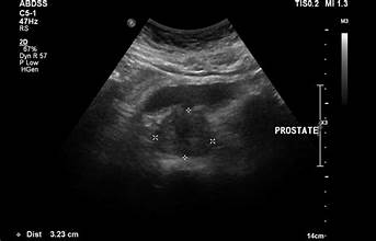
Is this a normal or abnormal prostate image?
a. normal
b. abnormal
a

Pediatric patient comes in ER with abrupt left sided testicular pain. No blood flow is seen in left testicle. What is the probable diagnosis?
a. enlarged lymph nodes
b. epididymo-orchitis.
c. cyst
d. testicular torsion
d

What is the anechoic area seen superior to testicle in the epididymis?
a. solid mass
b. testicular cyst
c. testicular torsion
d. epididymal cyst
d

Is this a normal or abnormal testicular image?
a. normal
b. abnormal
b

What is labeled A?
a. testicular torsion
b. testicle
c. epididymis
d. efferent ducts.
c
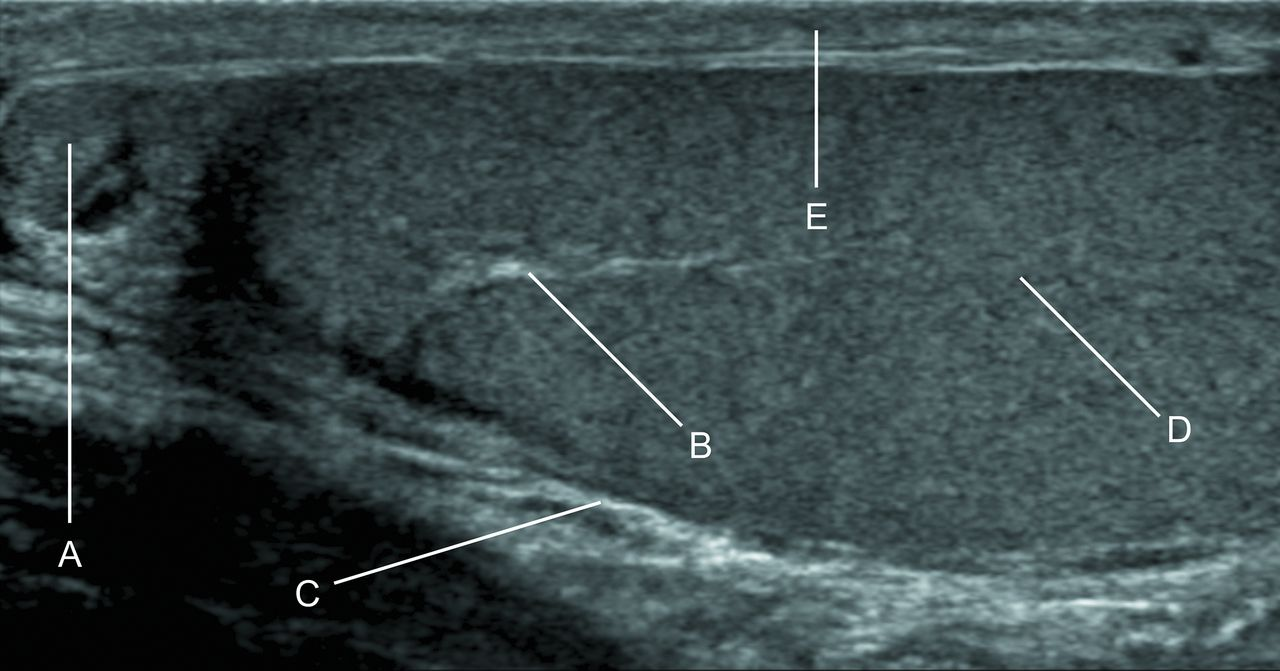
What is labeled B?
mediastinum testis.
epididymal cyst
epididymis
testicular torsion
a
What transducer do you use for scrotal and penile imaging?
high frequency 7.5-15 MHz
What transducer do you use for prostate exams?
curvilinear or transrectal (5-9 MHz)
Ovid, homogeneous echotexture
testes
isoechoic or slightly hypoechoic echotexture
epididymis
surround the testicle; potential space for fluid
tunica vaginalis
visualized superiorly in sagittal plane
spermatic cord
anechoic fluid around testicle
hydrocele
dilated veins >2 mm with Valsalva reflux
varicocele
absent or reduced blood flow: enlarged, hypoechoic testicle
torsion
increased flow, enlarged, heterogeneous
epididymitis/orchitis
hypoechoic intratesticular mass
tumors
patient posittioning for transrectal prostate ultrasound
LLD with knees flexed toward chest
Most prostate cancers occur in which zone?
peripheral
posterior to bladder, superior to prostate
seminal vesicles
seen centrally within prostate
urethra
enlargement of transitional zone
benign prostatic hyperplasia (BPH)
heterogeneous, hypervascular gland
prostatitis
hypoechoic lesion in peripheral zone, often asymetric
prostate cancer
common with aging, appear hyperechoic
calcifications
ventral, surround urethra
corpus spongiosum
surrounds corpora
tunica albuginea
within spongiosum, seen as anechoic tubular structure
urethra
fibrous plaque in tunica; hyperechoic with shadowing
Peyronie’s Disease
Low-flow (ischemic): no arterial flow; High-flow: persistent arterial flow
priapism
Disruption of tunica with hematoma
fracture
Best seen post-void or with injection of contrast
urethral stricture or trauma
Normal testis Doppler findings
symmetric flow, low resistance
Torsion Doppler findings
absent or reduced arterial flow
Epididymitis/Orchitis Doppler findings
hyperemia
The testes are normally __________ in shape and have a __________ echotexture
ovoid; homogeneous
The __________ is located superior and posterior to the testis.
epididymis
The __________ __________ surrounds the testicle and can contain fluid in a hydrocele.
tunica vaginalis
Normal testicular arterial flow is __________ resistance and __________.
low; continuous
Testicular torsion typically presents as __________ or __________ blood flow on
Doppler.
absent; decreased
A varicocele is diagnosed when the pampiniform plexus veins measure over __________
mm and show __________ on Valsalva.
2 mm; reflux
__________ is the most common cause of acute scrotal pain in post-pubertal males.
epididymitis
Seminomas typically appear as __________ (echogenicity) intratesticular masses.
hypoechoic
The patient is typically positioned in the __________ __________ decubitus position for
TRUS.
left lateral
The largest zone of the prostate and most common site of cancer is the __________
zone.
peripheral
The __________ zone is the area that enlarges in benign prostatic hyperplasia (BPH)
transitional
The __________ __________ are located posterior to the bladder and are imaged
during prostate ultrasound.
seminal vesicles
Prostatitis may appear as a __________ and __________ prostate with increased
vascularity.
enlarged; heterogeneous
Prostate cancer often appears as a __________ lesion in the peripheral zone on
ultrasound.
hypoechoic
Normal prostate volume is typically under __________ cc in younger men.
30
The two dorsal erectile structures are the __________ __________.
corpora cavernosa
The __________ __________ surrounds the corpora cavernosa and is disrupted in
penile fracture.
tunica albuginea
__________’s disease is characterized by fibrous plaques in the tunica albuginea.
Peyronie’s
The corpus spongiosum lies __________ to the corpora cavernosa and contains the
__________.
Ventral; urethra
A cavernosal artery peak systolic velocity (PSV) above __________ cm/s is considered
normal in an erectile dysfunction study
30
Low-flow priapism shows __________ or __________ cavernosal artery flow.
absent; minimal
A penile ultrasound is typically performed with the patient in a __________ position.
supine