Biol 240 Unit 1
 Call Kai
 Learn
Learn Practice Test
 Spaced Repetition
 Match
Match Flashcards
 Knowt Play
1/38

There's no tags or description
Looks like no tags are added yet.
Name | Mastery | Learn | Test | Matching | Spaced | Call with Kai | Chat |
|---|
No analytics yet
Send a link to your students to track their progress
39 Terms
3 Major Functions of a Microscope
Magnification
Resolution
Contrast
Magnification
How much larger the image appears compared with the actual object
Example: 400x magnification = image appears 400 times larger
Increasing magnification doesn’t enhance detail
Resolution
What is it
Formula/Determined by
More detail and sharper structures/Ability do discern fine details
Resolution - wavelength/2
Ability to distinguish two very close points as separate objects
Determined by the wavelength of light (or energy) used for illumination. For light microscopy, the limit is ~200nm.
Contrast
What is it?
How is produced?
Telling structures apart. Difference in intensity between the image and the background.
Produced in the specimen by staining with colored dyes that absorb light by using special optical techniques, or by using fluorescent probes.
Difference in appearance (brightness/color) between the object and its background or between different structures
Cells are often naturally transparent, so microscopy techniques/stains are used to increase contrast
Doesn’t necessarily increase resolution
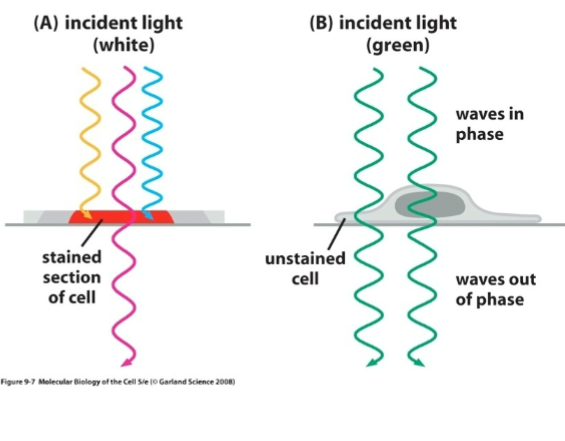
A researcher observes two proteins located extremely close together on a cell membrane. Under the microscope, the region is clearly visible against the surrounding membrane, but the two proteins appear as one single spot rather than two separate spots.
Which property of the microscope most directly limits the researcher’s ability to distinguish the proteins?
Resolution
A student examines an unstained living cell. The nucleus and cytoplasm are within the microscope’s ability to distinguish spatially, but they have nearly identical brightness and therefor are difficult to tell apart.
Which would most directly improve the image?
a. increasing magnification
b. improving contrast
c. improving resolution
d. using a larger image display
Improving contrast
A microscope image is enlarged from 400x to 100x. The image becomes much larger, but previously blurry structures remain blurry.
Which statements could correctly describe what happened?
Magnficiation increased
Resolution necessarily increased
The microscope may heave reached its resolution limit
Making an image larger does not necessarily reveal additional detail
Contrast must have decreased
Magnification increased
Making an image larger does not necessarily reveal additional detail
Two microscopes examine the same pair of organelles, which are 180 nm apart.
Microscope X produces a bright, easily visible image, but the organelles appear as one object
Microscope Y produces a dimmer image, but the two organelles can clearly be distinguished as separate objects
Which microscope has better resolution, and what observation proves it?
Y, because it distinguishes two closely spaced objects
A student says:
“I can’t distinguish the two organelles in this image, so the microscope has poor resolution.”
The professor says this conclusion is not necessarily justified. Which situation would best support the professor?
The organelles are 50nm apart and appear as one continuous object.
The organelles are sufficiently far apart to be resolved, but their signals are almost identical to the surrounding cytoplasm.
Increasing magnification makes the merged organelles appear as a larger merged structure.
The microscope’s resolution limit is 200nm, while the organelles are 100n apart.
The organelles are sufficiently far apart to be resolved, but their signals are almost identical to the surrounding cytoplasm.
A researcher is examining two proteins in a membrane. In the original imgae, she sees one faint patch. After changing the microscopy conditions, she sees one very bright, sharply visible patch. She concludes “The new method improved out ability to resolve the two proteins.”
Is her conclusion justified?
Yes, because the object became easier to see
Yes, because greater contrast necessarily produces greater resolution
No, the increased visibility demonstrated improved contrast, but there is no evidence that the two proteins can now be spatially distinguished.
No, the result demonstrated increased magnification instead.
No, the increased visibility demonstrated improved contrast, but there is no evidence that the two proteins can now be spatially distinguished.
You would need evidence of two separate, distinct structures.
How Scientists Prepare Tissues to be Viewed Under Microscopes
3 Steps:
Process
Function
Fixation - Preserving specimen
Cells/tissues normally start breaking down once they’re removed from an organism
Fixation preserves the cellular structures and essentially locks them in place
Freezing the cell’s structures in their current arrangement / dead tissue / preserving structure, not keeping cells alive
Embedding - Giving tissue support
Tissues get surrounded by something that becomes slid, like paraffin or plastic/resin, to avoid it deforming or falling apart
Soft tissue embedded inside a solid block that allows you to cut extremely thin slices without destroying the organization of the tissue
Sectioning - Cutting it into thin slices
Machine, microtome, takes embedded tissue block and uses a knife to produce extremely thin sections that can be placed onto a microscope slice
A thin section allows us to observe all individual cells-structures inside of it, unlike a thick chunk.
Many microscopy methods need a sufficiently thin slice for illumination or an electron beam to pass through and produce a useful image.
Fix → Embed → Section → View
Preserve → Support → Slice → Microscope

Histological Stains
Why?
Most cell structures don’t naturally look dramatically different form one another, so scientist use stains that selectively bind to particular cellular molecules
Tissue gets treated with different staining to label other cellular/tissue components
Why/How do different things stain differently?
Different cellular structures contain different molecules with different chemical properties
A stain has particular chemical properties that cause it to interact preferentially with certain molecules
Different molecular composition → Different affinity for stains → Different colors → Increased contrast
Adds boundaries, improved contrast
Problem with Staining
Traditional staining usually requires killing the cells
What if you want to watch a cell while its alive?
Differences in Refractive Index
Allows you to generate contrast without killing a cell
Different parts of a cell have different refractive indices
How much a material slows/bends as light passes through it
ie. Cytoplasm, nucleus, organelles, surrounding fluid have slightly different compositions and thus affect light differently
Specialized Microscopes:
Phase-contrast microscopy
Differential Interference Contrast (DIC)
Phase-Contrast microscopy
What
How
Pros
Cons
Phase contrast uses differences in how light travels through cellular structures to create differences in brightness
Invisible difference in light → visible light/dark difference
Pros: Food for transparent cells, lets you watch cells change/move over time
Cons: Can produce halos around structures; less useful for thick specimens, doesn’t tell you what a structure is
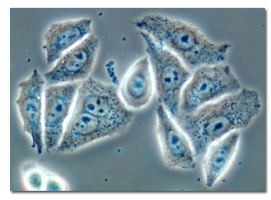
Differential interference Contrast (DIC)
What
How
Pros
Cons
DIC Also exploits differences in how light travels through different parts of the cell, but uses interference between light paths to generate contrast
Creates 3D/Shadowed appearance
Pros: Very sharp boundaries and excellent contrast, useful for fine Cuellar details, 3d appearance
Cons: More complicated/expensive optics, 3D appearance is artificial and may be misleading, doesn’t specifically identify molecules
Distinction of choosing between PC and DIC
What kind of image you want
General observation of living cells
Very clear cell boundaries/edges
Track cells moving/dividing over time
Fine structural/morphological differences
Cheapest/simplest setup
3D like view of cell shape
What kind of image you want
General observation of living cells
Phase contrast
Very clear cell boundaries/edges
DIC
Track cells moving/dividing over time
Phase contrastFine structural/morphological differences
DICCheapest/simplest setup
Phase contrast3D like view of cell shape
DIC
What is the purpose of Florescence Microscopy?
It lets you locate specific molecules of structures inside cells by attaching florescent labels to them and then detecting the light those labels emit
What is the core mechanism in Flurescence?
Flurophores
What are Fluorophores?
How do they work
Explain Excitation/Emission
Relevance to wavelengths
A molecule that can absorb light energy and release some of the energy as light
ie: Specific wavelength of light comes in → Fluorophore absorbs it → Fluorophore becomes excited → Some energy is lost —> Fluorophore emits lower energy light
Excitation = Light IN
Emission = Light OUT
Higher energy = shorter wavelength
Lower energy = longer wavelength
Why does the microscope need different wavelengths?
Brief on how it works
Contextualize it with its background
Takeaway point
To separate what dye is absorbed/reflected
Graph:
Blue curve represents wavelengths that excite the particular dye, green curve represents the light that the dye admits
Shining blue light at specimen → fluorophore absorbs blue → fluorophore emits green → Microscope detects green
Microscope ignores the light used to excite the sample, instead showing the light coming back from the fluorescent molecules
This is also why florescent structures are able to appear brilliantly colored against a dark background
Microscope doesn’t illuminate with light
We shine the appropriate excitation wavelength onto specimen
Only fluorophores capable of absorbing that wavelength become excited and emit their own
TAKEAWAY: Fluorophore absorbs energy and emits a new photon at a different wavelength
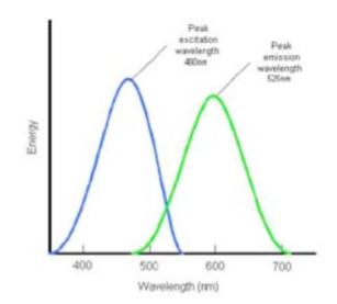
Fluorophores/Colors
How are different colors of fluorescence produced?
The chemical properties of a dye determines its excitation and emission
Different fluorophores have different molecular structures, so they absorb and emit different wavelengths
Label different things with different fluorophores
Color = which florescent label is associated with what cellular target
Molecular Specificity: Colors
Why are colors helpful?
Besides the obvious: Being able to visualize structures
Them being labeled differently in the same cell lets us see their spatial relationships
Does not tell us about whether they specifically interact, only can tell us things like co-localization
Florescence microscopy with Living vs. Fixed Cells
Living cells: Relevance?
Fixed cells: Advantage
Living cells
Fluorescence microscopy can be used on living cells with the appropriate florescent labels
Allows us to watch biological processes change over time
IE. Where proteins move, how mitochondria moves, how ER shapes, what happens during cell division
Dynamic information
Fixed Cells
You’re able to use labeling techniques that may not work as easily in living cells and lets you examine structures very clearly
Cannot examine change, only preserved structural and molecular localization
How does the dye know where to go?
2 ways
Florescent antibodies
Antibody recognizes protein X
Fluorophore attaches to antibody
Antibody binds to protein X
Shine excitation light
Fluorophore emits
You can visualize protein X location
Genetically coded fluorescent proteins (Like Green fluorescent protein)
GFP can be fused to a protein they’re interested in
Protein + GFP, cell produces fluorescent fusion protein and allows researchers to track where protein X goes in living cells
A dye absorbs most strongly around 480 nm and emits around 650 nm.
Which has more energy?
480 nm
Shorter wavelength = Higher energy
A scientists uses two fluorophores
Protein A = Green
Protein B = Red
Some regions contain only green and others only red. What might this mean?
The proteins have different cellular distributions. Some regions may overlap which suggests co-localization, but doesn’t prove interaction.
A researcher wants to observe mitochondria moving around a cell for 30 minutes. Would a fixed-cell technique work?
No.
Light Path of a Florescence Microscope: Entire Path
Why is it set up in a certain way
How its set up: Orderly
Every mechanic
Every process from mechanic
Complete path of light
Microscope needs to control which wavelengths travel where
Light Source → Excitation filter
Chooses light going INNNNNN
Selects/filters the wavelength/range of wavelengths capable of exciting our fluorophore
Dichroic Mirror
Treats different wavelengths differently
Reflects certain wavelengths while transmitting others to the objective lens
Objective
Magnifies and Focuses light onto the specimen
(Recall we filter what light does in)
Collects the emitted fluorescent light coming back from the specimen
Light travels through objective in both directions
Going down = Excitation light → Objective → Specimen
Coming back = Specimen fluorescence → Objective → Specimen
Fluorophore
Shorter wavelength excitation light absorbs
Fluorophore excited
Energy lost
Longer wavelength emission light released
Emission Filter
After light comes back through the Dichroic Mirror, the filter selects wavelengths that we want our detector to receive
Helps mitigate stray excitation light, unwanted wavelengths, background light
Extra specificity
CLEANS UP FROM THE FLUOROPHORE
Detector
Appropriate fluorescent light emissions reach the detector which measure the fluorescence and allows an image to be produced
Fluorophore present → Emission → Detector receive signal → Bright
Little/no emission = little/no signal → dark
Immunofluorescence consists of
Fluorescent protein tagging consists of
Immunofluorescence: Antibodies and Fluorescent marks
Fluorescent Protein Tagging: Genetically encoded fluorescent proteins like GFP
What is an antibody
Structure
Mechanism
Production
Structure
2 Heavy chains + 2 Light chains
Mechanism
Have high affinity to particular antigens
Production
Can be produced in animals like rabbits, mice, sheep, goats, and humans
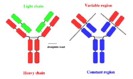
Antibody Structures: Variable Regions
Location
Importance
The tips of the Y
Determine what the antibody recognizes and binds to
Antibody Structures: Constant
Location
Importance
Everything but the Tips
Not really relevant
Antigen
Molecule/Structure recognized by an antibody
Ex: Tubulin, Golgi protein
How Variable Regions Work
Antibody recognizes protein
Antibody binds protein
Fluorescent marker becomes associated with that antibody
Fluorescence appears when antibody is accumulated
We infer the location of the protein
Antibody = Specificity ; Fluorophore = Visibility
Antibodies and Antigens: Detailed Example
Part 1: Antibodies
Inject protein A into a rabbit (Antigen A)
Rabbits immune system recognizes and generates antibodies against protein/Antigen A
Researchers collect blood from the rabbit and separate out the serum containing antibodies
Serum contains tons of different antibodies, not just the one we want
We want to isolate the protein A antibodies
Attach Protein A to beads
Pour the rabbit serum through
Antibodies that don’t recognize protein A don’t bind
Antibodies that do recognize protein A bind strongly and stay stuck in the column
Disrupt the interaction and release the antibodies by using high salt
Collect a purified serum enriched for antibodies that recognize antigen
Antibodies and Antigens: Detailed Example
Part 1: Primary/Secondary Antibodies and Immunocytochemistry/Immunofluorescence
Primary antibody recognizes biological target
In our previous explained example: Rabbit antibody directed against Antigen/Protein A = Primary Antibody
Primary antibody binds directly to Antigen A
Secondary Antibody recognizes the Primary antibody
Marker-coupled antibody that recognizes rabbit antibodies
Secondary fluorescence reveals antigen