H.A chapter 14 pt 2
1/38
There's no tags or description
Looks like no tags are added yet.
Name | Mastery | Learn | Test | Matching | Spaced |
|---|
No study sessions yet.
39 Terms
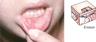
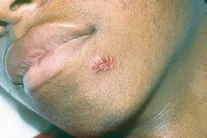
Erosion
Loss of superficial epidermis that does not extend to the dermis.
It is a depressed, moist area. Examples include rupture vesicle, scratch mark, and aphthous ulcer (aphthous stomatitis, commonly called a canker sore, pictured below).
Erosion image
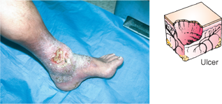
Ulcer
Skin loss extending past epidermis, with necrotic tissue loss.
Bleeding and scarring are possible.
Examples include stasis ulcer of venous insufficiency (stasis dermatitis with venous stasis ulcer, pictured below) and pressure injury.
Ulcer image
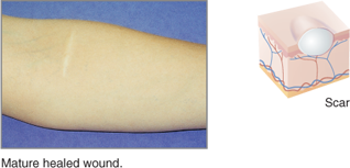
SCAR (CICATRIX)
healing of wound or lesion that represents replacement by connective tissue of the injured tissue.
Young scars are red or purple, whereas mature scars
white or glistening. Examples include healed wound and healed surgical incision.
Scar Cictrix image
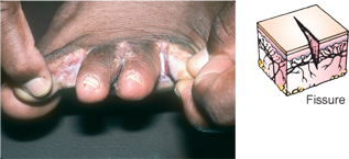
Fissure
Linear crack in the skin that may extend to the dermis and may be painful.
Examples include chapped lips or hands and athlete’s foot.
Interdigital tinea pedis with fissures and maceration is pictured below.
Fissure image
Vascular Skin Lesions
Vascular skin lesions are associated with bleeding, aging, circulatory conditions, diabetes, pregnancy, and hepatic disease, among other problems.
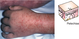
Petechia
tiny pinpoint bleeding spots.
Definition: Tiny pinpoint spots (≤2 mm) caused by small capillary bleeding.
Appearance: Red, purple, or brown dots; flat and don’t blanch (don’t turn white when pressed).
Causes: Platelet problems (low platelet count, clotting disorders), trauma (like tight tourniquet, coughing, vomiting).
Petechia image
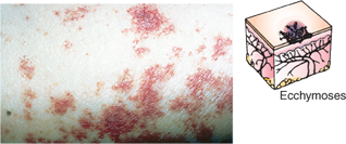
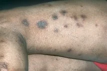
Ecchymosis
larger bruise-like bleeding under skin.
Definition: Larger area of bleeding under the skin, >1 cm.
Appearance: What we usually call a “bruise.” Color changes over time (red → purple/blue → yellow/green → brown).
Causes: Trauma (most common), bleeding/clotting disorders, medications (like anticoagulants).
Ecchymosis image
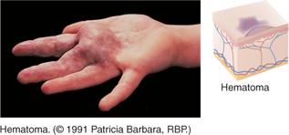
Hematoma
Definition: A collection (pool) of blood under the skin, tissue, or organ due to vessel rupture.
Appearance: Swollen, raised, painful lump; can be firm or fluctuant depending on size.
Causes: More significant trauma, surgery, or vessel injury.
Hematoma image
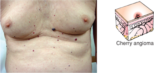
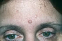
Cherry Angioma
Papular and round, red or purple lesion found on the trunk or extremities.
It may blanch with pressure. It is a normal age-related skin alteration and usually not clinically significant.
Cherry Angioma image
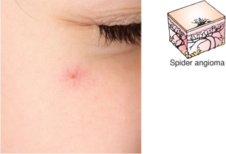
Spider Angioma
Red arteriole lesions with a central body and thin branches
Found on face, neck, arms, and upper chest
Rare below the waist
Associated with liver disease, pregnancy, and vitamin B deficiency
Spider Angioma image
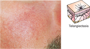
Telangiectasis
Blush or red lesion with varying shape (spider-like or linear)
Does not blanch with pressure
Cause/Pathophysiology: Due to superficial dilation of venules vessels and capillaries.
Common Locations: Legs, anterior chest.
Associated Conditions: States of increased venous pressure (e.g., varicosities, venous hypertension).
Telangiectasis image
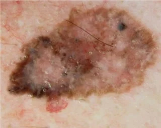
Malignant melanoma
can be deadly if not discovered and treated early, which is one reason why professional health assessment and skin self-assessment can be life-saving procedures.
ABCDE
A for asymmetrical;
B for borders that are irregular (uneven or notched);
C for color variations;
D for diameter greater than 1/4 in. or 6 mm; and E for evolution (changes over time)
Basal Cell Carcinoma
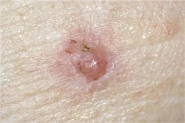
Squamous cell Carcinoma
Most dangerous
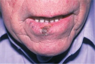
Melanoma image
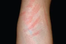
Configurations of skin lesions
Describing lesions by shape, distribution, or configuration is one way to communicate specific characteristics that can help identify causes and treatments.
Linear configuration example dermatographism
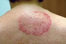
Annular configuration example tiena corporis
Clustered configuration, lesions grouped together. An example is herpes simplex
Discrete configuration individual distinct lesions. An example is multiple nevi
Nummular configuration, coin-shaped lesions. An example is nummular eczema
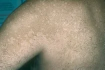
Confluent configuration
Smaller lesions run together to form larger lesions. Example is tiena versicolor
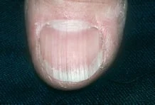
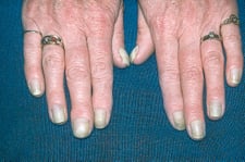
Longitudinal ridging parallel ridges running lengthwise. May be seen in the elderly and some people with no know etiology
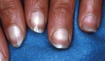
Half-and-half nails. Nails that are white on the upper proximal half and pink on the distal half
May be seen the chronic renal disease
CKD
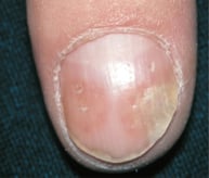
Pitting seen with psoriasis
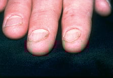
Koilonychia,
Spoon-shaped nails that may be seen with trauma to cuticles or nail folds or in iron deficiency anemia, or endocrine or cardiac disease
iron-deficiency anemia
Yellow nail syndrome
Nails grow slow and are curved
May be seen in AIDs and respiratory syndromes
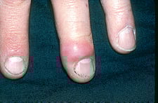
Paronychia, local infection