Pulse Anatomy Exam 2
 Call Kai
 Learn
Learn Practice Test
 Spaced Repetition
 Match
Match Flashcards
 Knowt Play
1/376

There's no tags or description
Looks like no tags are added yet.
Name | Mastery | Learn | Test | Matching | Spaced | Call with Kai | Chat |
|---|
No analytics yet
Send a link to your students to track their progress
377 Terms
the abdominal cavity extends superiorly from the _______ _______ to the _______ ______ inferiorly
abdominal diaphragm, pelvic inlet
what is found in the abdominal cavity
much of the gastrointestinal tract, parts of the urogenital organs, and all associated arteries, veins, and nerves
what is the hard boundary that separates the abdominal and thoracic cavity
the diaphragm
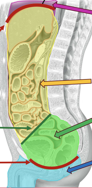
what is the purple
thoracic cavity
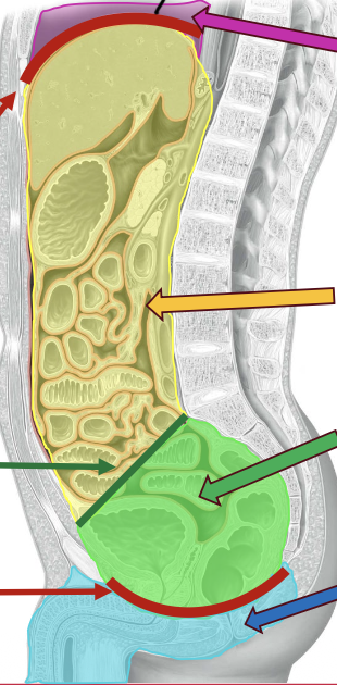
what is the yellow
abdominal cavity
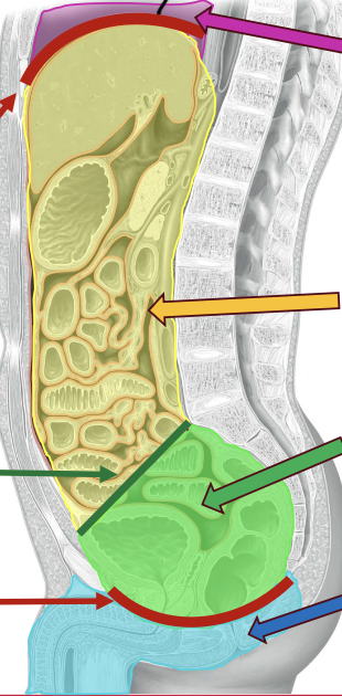
what is the green
pelvic cavity
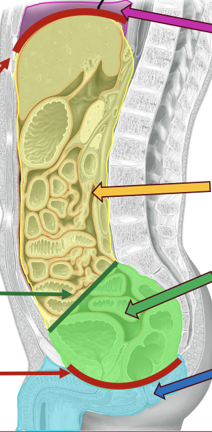
what is the red on the top
abdominal diaphragm
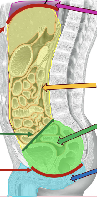
what is the red on the bottom
pelvic diaphragm
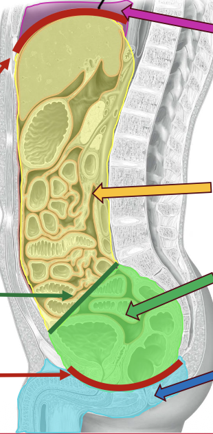
what is the blue
perineum
is the abdominal cavity a hard boundary?
no
what are the functions of the abdomen?
contains the major elements of the GI system, spleen, and parts of the urinary system, assists in breathing, and changes in intra-abdominal pressure (pressure in the abdomen) assist in voiding the bladder/rectum, and child birth
the primary openings for structures of the thorax to pass through are:
vena caval foramen (T8 level) for the inferior vena cava and some branches of R. phrenic nerve, esophageual hiatus (T10 level) for the esophagus anterior from the L. vagus nerve and posterior to the vagal trunks from R. vagus nerve, and the aortic hiatus (T12 level) for the aorta, azygous vein, and thoracic duct
what is the mnemonic for the primary openings for the structures of the thorax to pass through
I Ate 10 Eggs At 12 (inferior vena cava T8, esophagus (T10), and aorta (T12)
the abdominal surface topography divides the abdomen into ____ quadrants and ___ regions
4, 9
what are the 9 region divide of the abdomen
right hypochondriac region, epigastric region, left hypochondriac region, right lumbar region, umbilical region, left lumbar region, right iliac region, hypogastric region, and left iliac region
what are the 4 quadrant divide of the abdomen
right upper quadrant, left upper quadrant, right lower quadrant, and left lower quadrant
the anterior abdominal wall is composed of layers. from superficial to deep, they are:
skin, superficial fascia of (camper’s fascia and scarpa’s fascia, in that order), muscles, transversalis fascia, extraperitoneal fat, and parietal peritoneum
camper’s fascia is the
fatty layer
scarpa’s fascia is the
membranous layer
what are the muscles of the anterior abdominal wall from superficial to deep:
external oblique, internal oblique, and transversus abdominus
the muscle fibers of the anterior abdominal wall begin
laterally, pass anteriorly and terminate as an aponeurosis
what is an aponeuosis
flattened tendon for muscle attachment
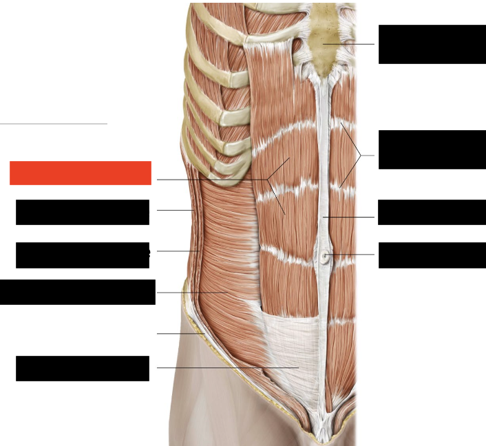
what is the red box
rectus abdominus
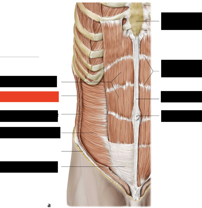
what is the red box
external oblique
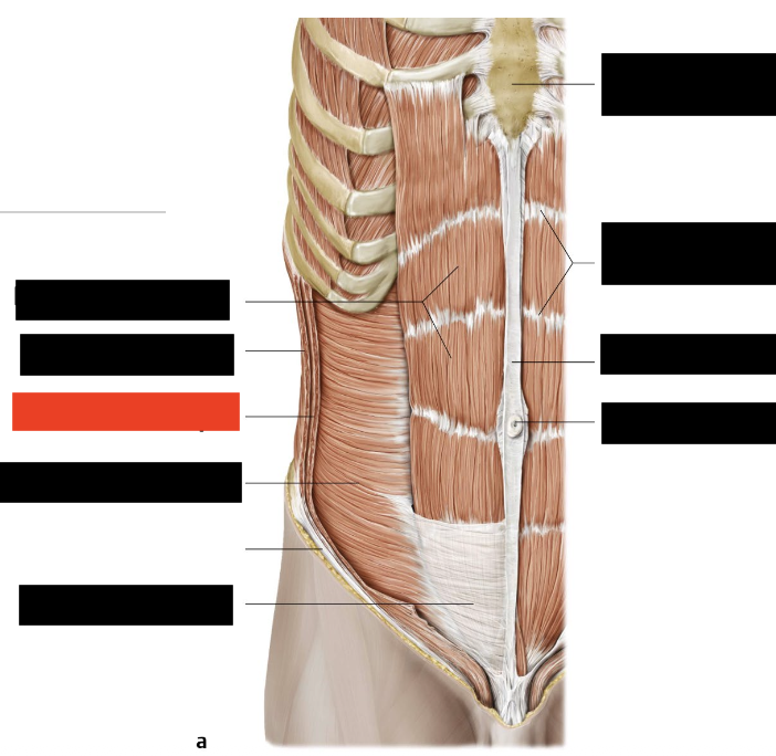
what is the red box
internal oblique
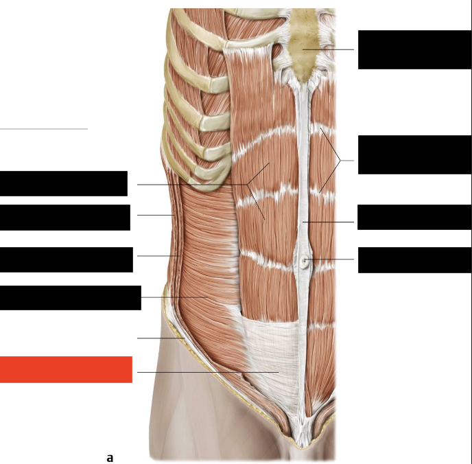
what is the red box
rectus sheath
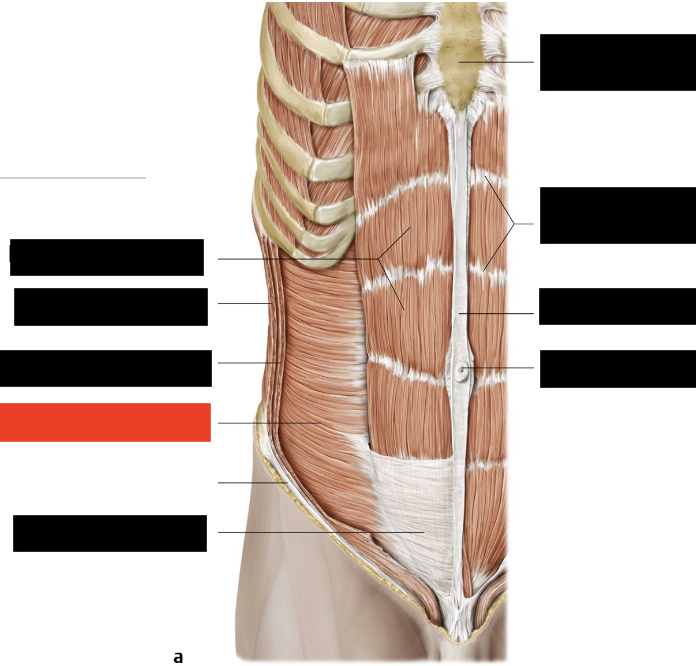
what is the red box
transversus abdominus
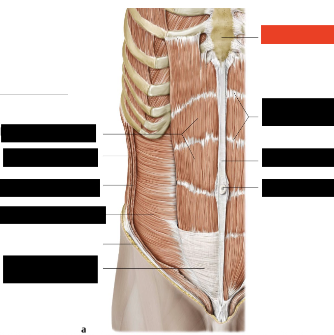
what is the red box
sternum
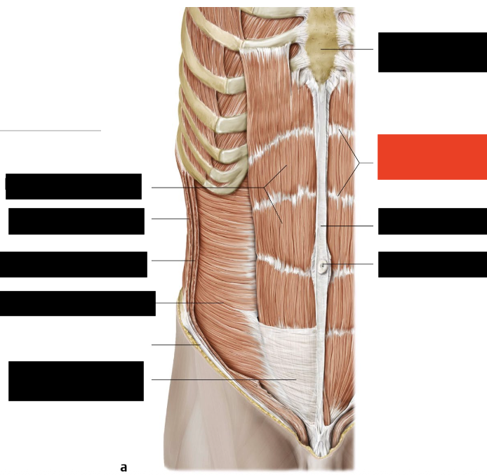
what is the red box
tendinous intersections
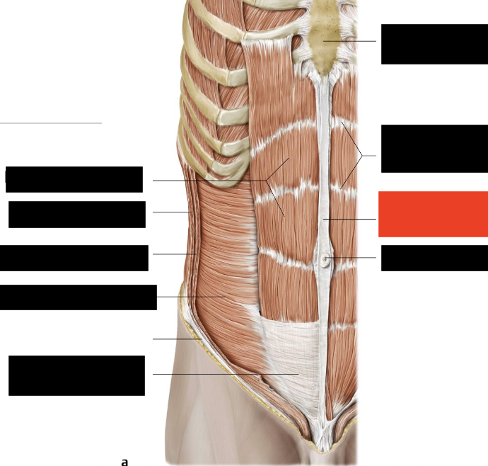
what is the red box
linea alba
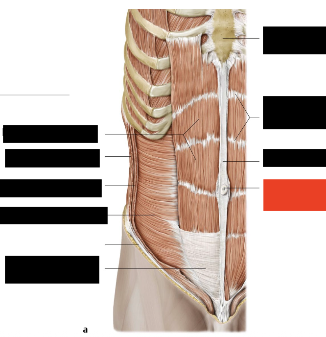
what is the red box
umbilicus
what are the muscles that run lateral?
external oblique, internal oblique, and transversus abdominus
what are the muscles that run medial?
rectus abdominus and rectus sheath
what is the rectus abdominus enclosed in
the rectus sheath
what is the rectus sheath composed of?
the fused aponeuroses of the lateral abdominal wall muscles
what muscle has no proper tendon, but has an aponeurosis that spans across the abdomen from the midline
external oblique
what is the fiber orientation for external oblique muscles
supero-lateral to infero-medial (hands in your pockets)
what is the action of the external oblique muscle
compresses and supports the abdominal viscera and flexes (bending forward) and rotates the trunk (moving side to side)
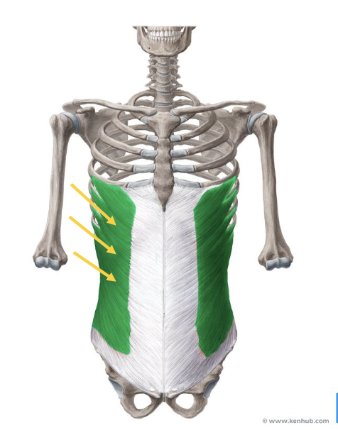
what is the green
external oblique muscle
what muscle has no proper tendon, but has an aponeurosis that spans across the abdomen from the midline
internal oblique
what is the fiber orientation of the internal oblique?
infero-lateral to the supero-medial (hands on your chin)
what is the action of the internal oblique
compresses and supports the abdominal viscera, flexes and rotates the trunk
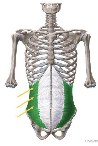
what is the green?
internal oblique muscle
what is the deepest of the abdominal wall muscles and has no proper tendon, but has an aponeurosis that spans from across the abdomen from the midline
transversus abdominis
what is the fiber orientation of the transversus abdominis
transverse/horizontal
what is the action of the transversus abdominis muscle
compresses and supports the abdominal viscera, flexes and rotates the trunk
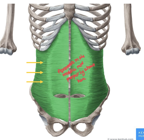
what is the green
transversus abdominis
which muscle is the only vertically oriented muscle of the abdominal wall and has paired muscles separated by linea alba
rectus abdominis
with the rectus abdominis muscle, each one has multiple bellies separated by
tendinous intersections
what is the action of the rectus abdominis
flexes trunk and tenses abdominal wall
the aponeuroses of the other muscles create the ______ _____ which surrounds the rectus abdominis muscles
rectus sheath
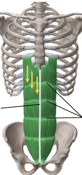
what is the green
rectus abdominis, and the bellies (white lines between the little pouches) are the tendinous intersections
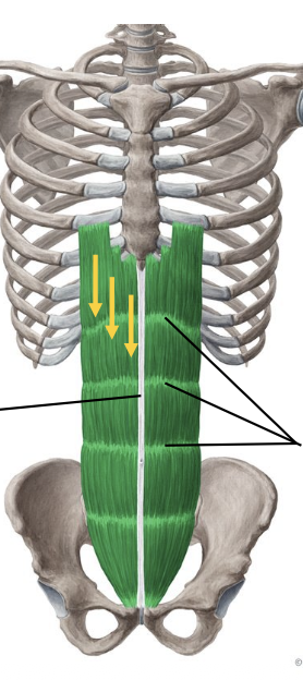
the grey in the middle is the
linea alba
what lines run along lateral edges of rectus abdominis and is the site of union where aponeuroses of external and internal obliques muscles meet the rectus sheath
semilunar lines
aponeuroses of the external and internal oblique muscles merge together at the _____ _____ along the midline of the two parallel rectus abdominus muscles
linea alba
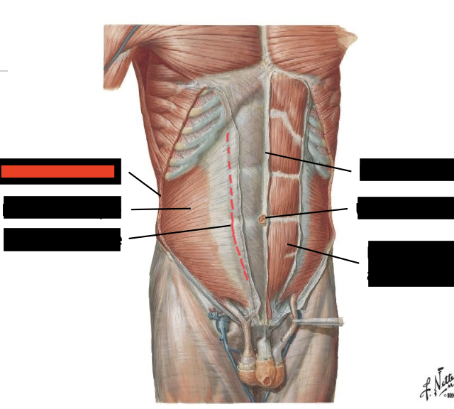
what is the red box?
external oblique
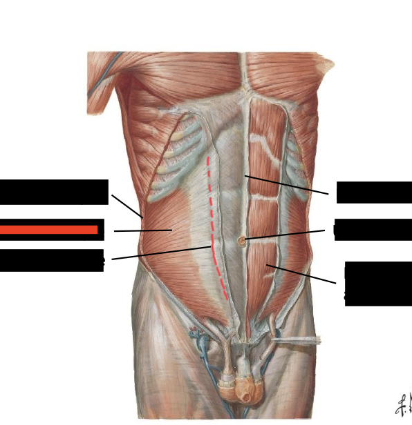
what is the red box?
internal oblique
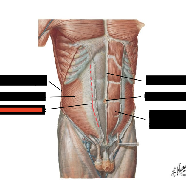
what is the red box?
semilunar line
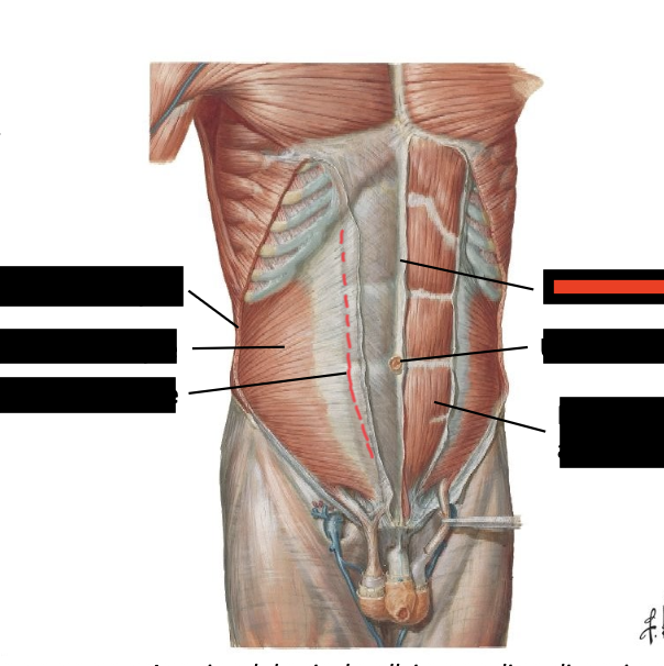
what is the red box?
linea alba
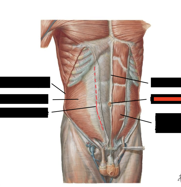
what is the red box?
umbilicus
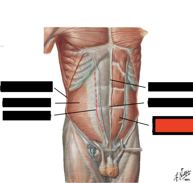
what is the red box?
rectus abdominis
rectus abdominis muscles are enclosed in _______ and ______ which are in other words the _____ ______
aponeuroses, fascia, rectus sheath
above the arcuate line is the
anterior and posterior rectus sheath
the anterior rectus sheath is composed of above the arcuate line
external and internal aponeurosis (above the umbilicus)
the posterior rectus sheath is composed of above the arcuate line
internal and transversus aponeurosis
what is the transition that occurs midway between umbilicus and the pubic symphysis. it is visible on the deep surface of anterior abdominal wall; place where the anterior rectus sheath disappears
arcuate line
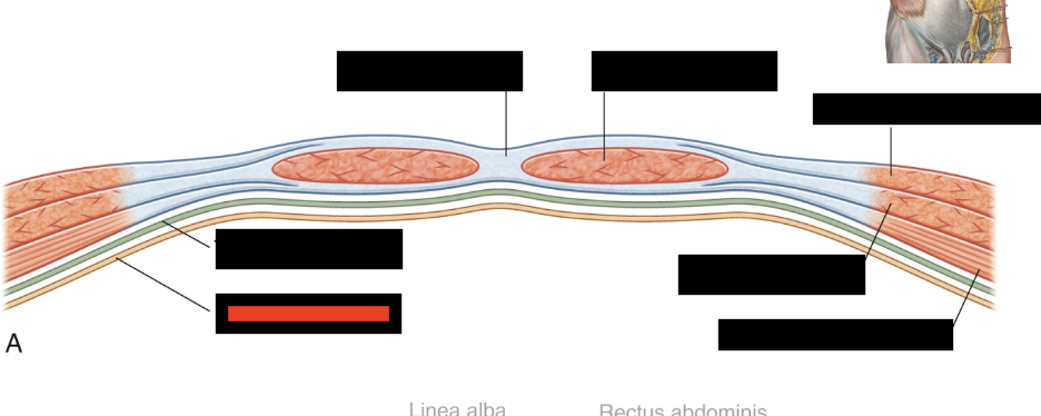
what is the red box
parietal peritoneum
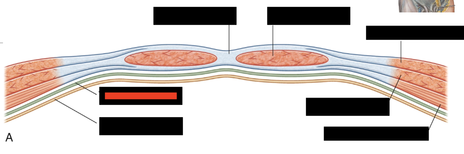
what is the red box
transversalis fascia
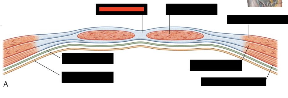
what is the red box
linea alba
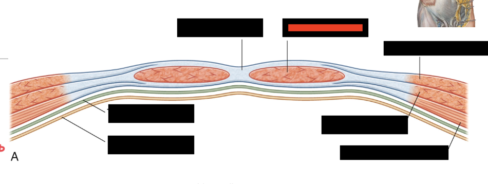
what is the red box
rectus abdominis
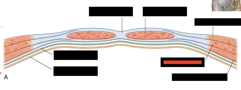
what is the red box
internal oblique
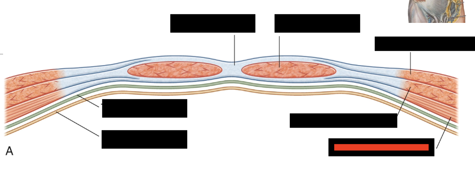
what is the red box
transversus abdominis
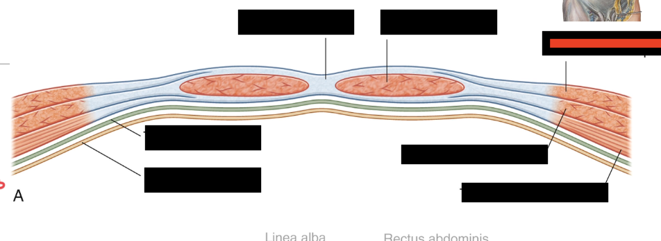
what is the red box
external oblique
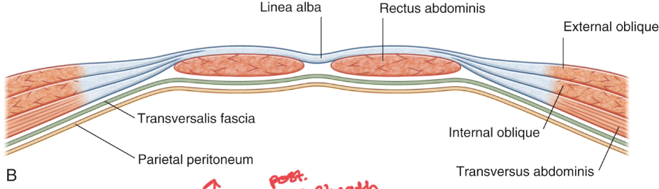
below the arcuate line there is
no posterior rectus sheath, only transversalis fascia and peritoneum present posterior to rectus abdominus
what is in the anterior rectus sheath below the arcuate line
external aponeurosis, internal aponeurosis, and transversus aponeurosis
what is the posterior rectus sheath below the arcuate line
there is none; it is what is below the belly button and since this area is soft, it is prone to hernia’s
a condition in which part of an organ (or fat) is abnormally displaced and protrudes through the cavity in which it normally resides
hernia
what are the 3 conditions of hernias?
a point of weakness (gap in muscle for ex), pressure, and something to protrude
what is common in newborns as a result of a defect in the linea alba. fat or sometimes intestines push through the layers of the anterior abdominal wall at near the umbilicus. can be a problem if the tissue is trapped (i.e. bowel becomes incarcerated, or lack of fully fused linea alba)
paraumbilical hernia
the arterial supply of the superior part of the anterior abdominal wall is supplied by
musculophrenic arteries and superior epigastric arteries (both branches of the internal thoracic artery)
the arterial supply of the interior part of the anterior abdominal wall is supplied by
inferior epigastric arteries (branches of the external iliac arteries)
both of the superior and inferior epigastric arteries are _____ to the anterior rectus sheath, but ________ to the rectus abdominis muscles
deep, superficial
if there is a blockage in the superior epigastric artery, will you still get blood and why?
yes because the external iliac artery branches blood from the inferior portion
what are the layers of the anterolateral abdominal wall
skin, superficial fascia (campers fascia and scarpas fascia), external abdominal oblique msucle, internal oblique muscle, transversus abdominis muscle, transversalis fascia, extraperitoneal fat, parietal peritoneum
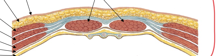
is this above or below the arcuate line? how do you know?
below because there is no post rectus sheath
in the inguinal region, what is the band of connective tissue that extends from the anterior superior iliac spine (ASIS) to the pubic tubercle?
inguinal ligament
what is formed by the free inferior border of the aponeurosis of the external oblique muscle
inguinal ligament
the inguinal ligament demarcates the division between the
abdomen and the superior thigh
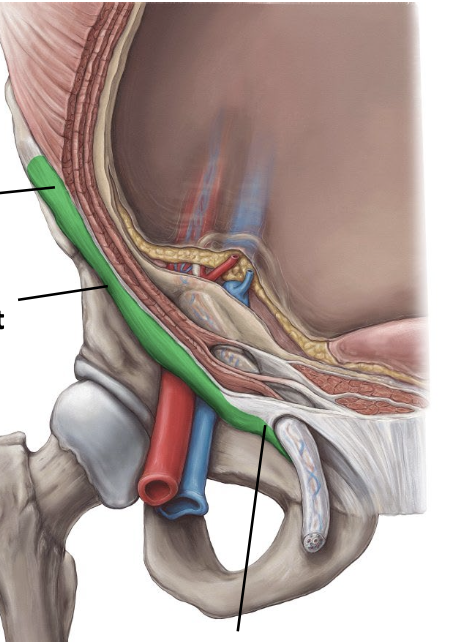
what is the green, second line of the left
inguinal ligament
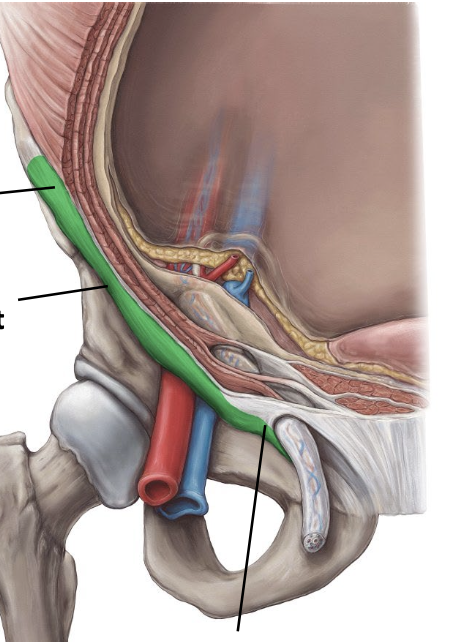
what is the green, first line of the left
ASIS
in the inguinal region, what is the short passage that allows structures to pass from inside the abdomen to the external genitalia
inguinal canal
the inguinal canal is situated
superior and parallel to the medial half of the inguinal ligament
in males, what is found in the inguinal canal
spermatic cord
in the inguinal canal of females, what is found
round ligament of the uterus
the beginning and end of the inguinal canal is marked by two openings which are
deep inguinal ring (inside ab. wall /entrance), and superficial inguinal ring (outside ab. wall/exit)
when does the inguinal canal form
during the development of female or male gonands
from deep to superficial, the inguinal canal passes through/what is the pathway?
deep inguinal ring in transversalis fascia, transversus abdominis muscle, internal oblique muscle, superficial inguinal ring in the external oblique aponeurosis
in the internal abdominal wall, the umbilical folds are
raised ridges of parietal peritoneum covering underlying structures
when is the umbilical folds function?
during fetal development and some are still functional
what are the 5 umbilical folds
median umbilical fold (unpaired), medial umbilical folds (paired), lateral umbilical folds (paired)