Nervous System
1/30
There's no tags or description
Looks like no tags are added yet.
Name | Mastery | Learn | Test | Matching | Spaced | Call with Kai |
|---|
No study sessions yet.
31 Terms
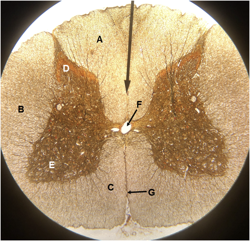
In the image above, identify structures A-G. Question H: the cell bodies of what type of neurons are located in structure E?
A. posterior (dorsal) white column
B. lateral white column
C. anterior (ventral) white column
D. posterior (dorsal) horn
E. anterior (ventral) horn
F. central canal
G. anterior (ventral) median fissure
H. somatic motor
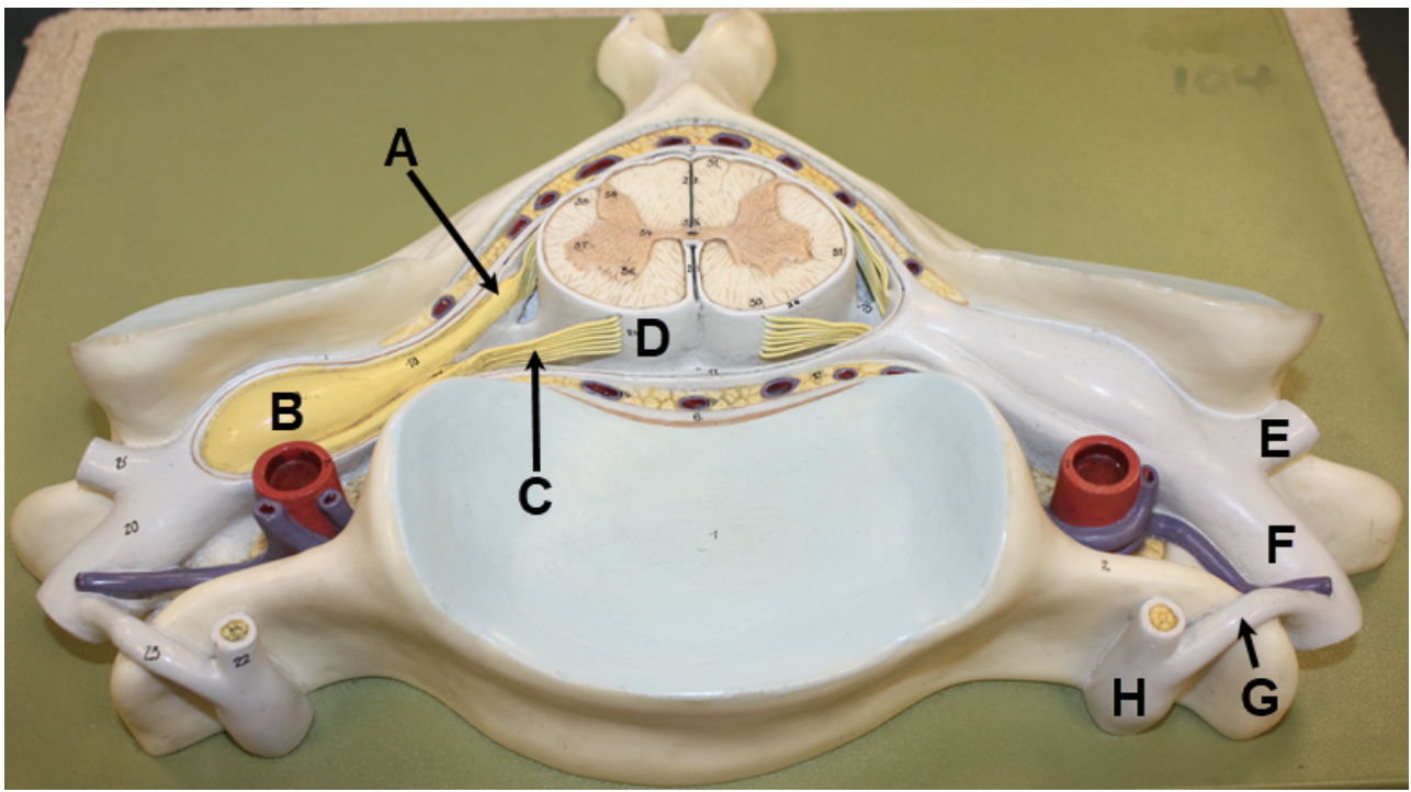
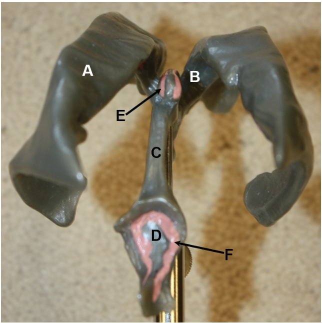
In the image above, identify structures A-C, meningeal layer D, and structures E-H. Question I: the cell bodies of what type of neurons are located in structure B? (Hint for structure G: this model only shows one structure here, but there are actually two.)
A. posterior (dorsal) root
B. spinal ganglion (posterior root ganglion or dorsal root ganglion)
C. anterior (ventral) root
D. pia mater
E. posterior (dorsal) ramus
F. anterior (ventral) ramus
G. rami communicantes (although this model only shows one structure to represent them, so ramus communicans is also an acceptable answer)
H. sympathetic ganglion (autonomic ganglion)
I. sensory (both somatic sensory and visceral sensory)
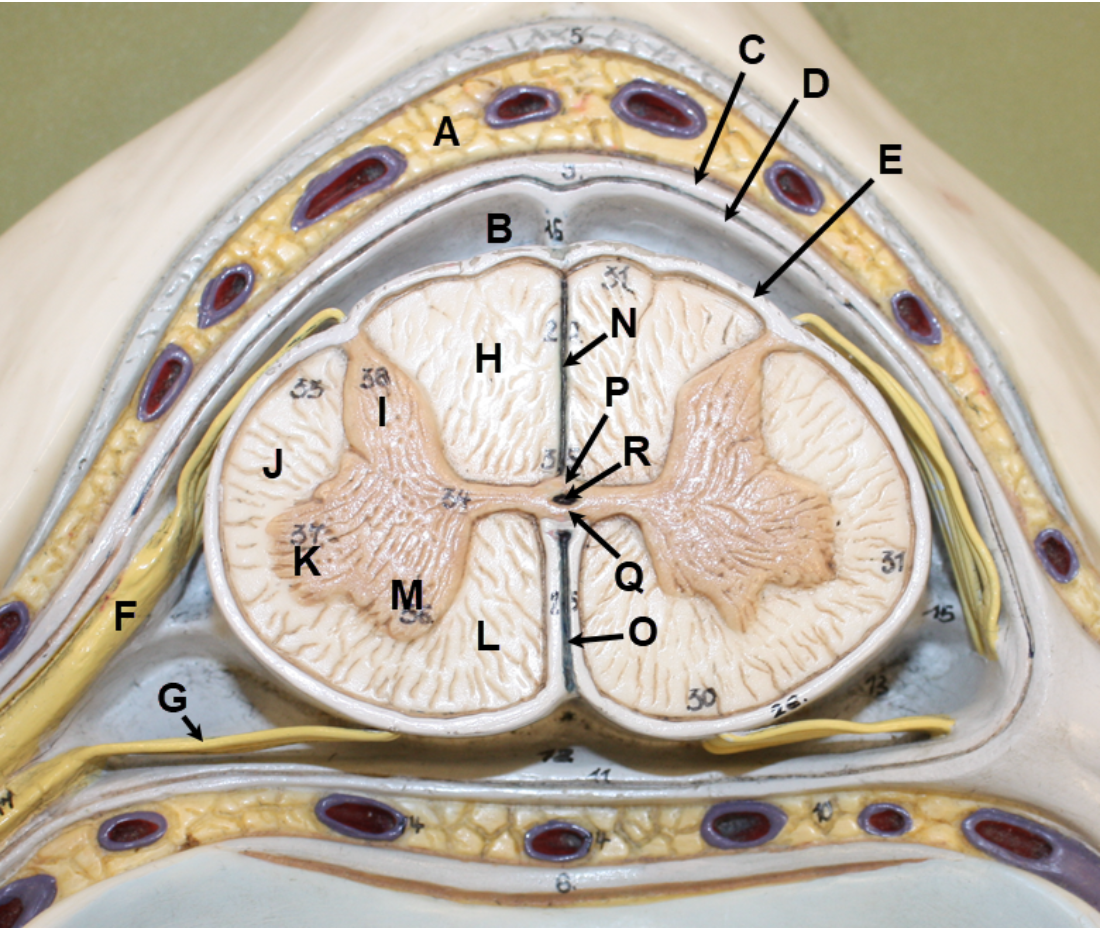
In the image above, identify spaces A and B, meningeal layers C-E, and structures F-R. Question S: the cell bodies of what type of neurons are located in structure K? Question T: the cell bodies of what type of neurons are located in structure M?
A. epidural space
B. subarachnoid space
C. dura mater
D. arachnoid mater
E. pia mater
F. posterior (dorsal) root
G. anterior (ventral) root
H. posterior (dorsal) white column
I. posterior (dorsal) horn
J. lateral white column
K. lateral horn
L. anterior (ventral) white column
M. anterior (ventral) horn
N. posterior (dorsal) median sulcus
O. anterior (ventral) median fissure
P. posterior (dorsal) gray commissure
Q. anterior (ventral) gray commissure
R. central canal
S. visceral motor
T. somatic motor
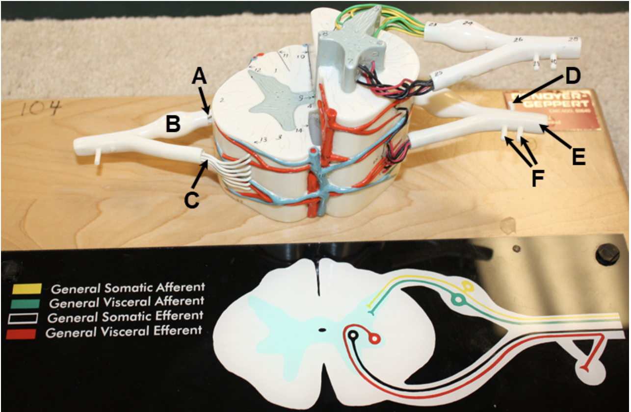
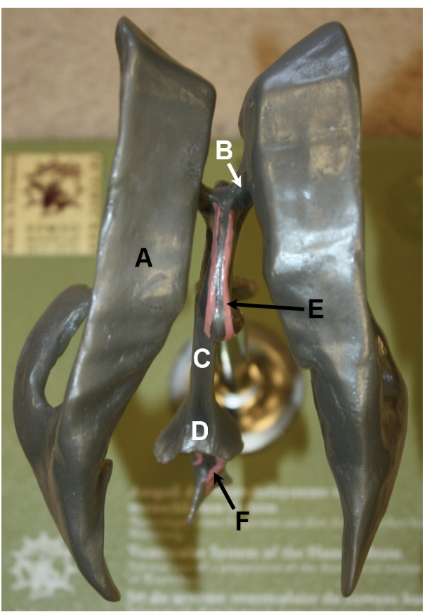
In the image above, identify structures A-F. Question G: the cell bodies of what type of neurons are located in structure B?
A. posterior (dorsal) root
B. spinal ganglion (posterior root ganglion or dorsal root ganglion)
C. anterior (ventral) root
D. posterior (dorsal) ramus
E. anterior (ventral) ramus
F. rami communicantes
G. sensory (both somatic sensory and visceral sensory)
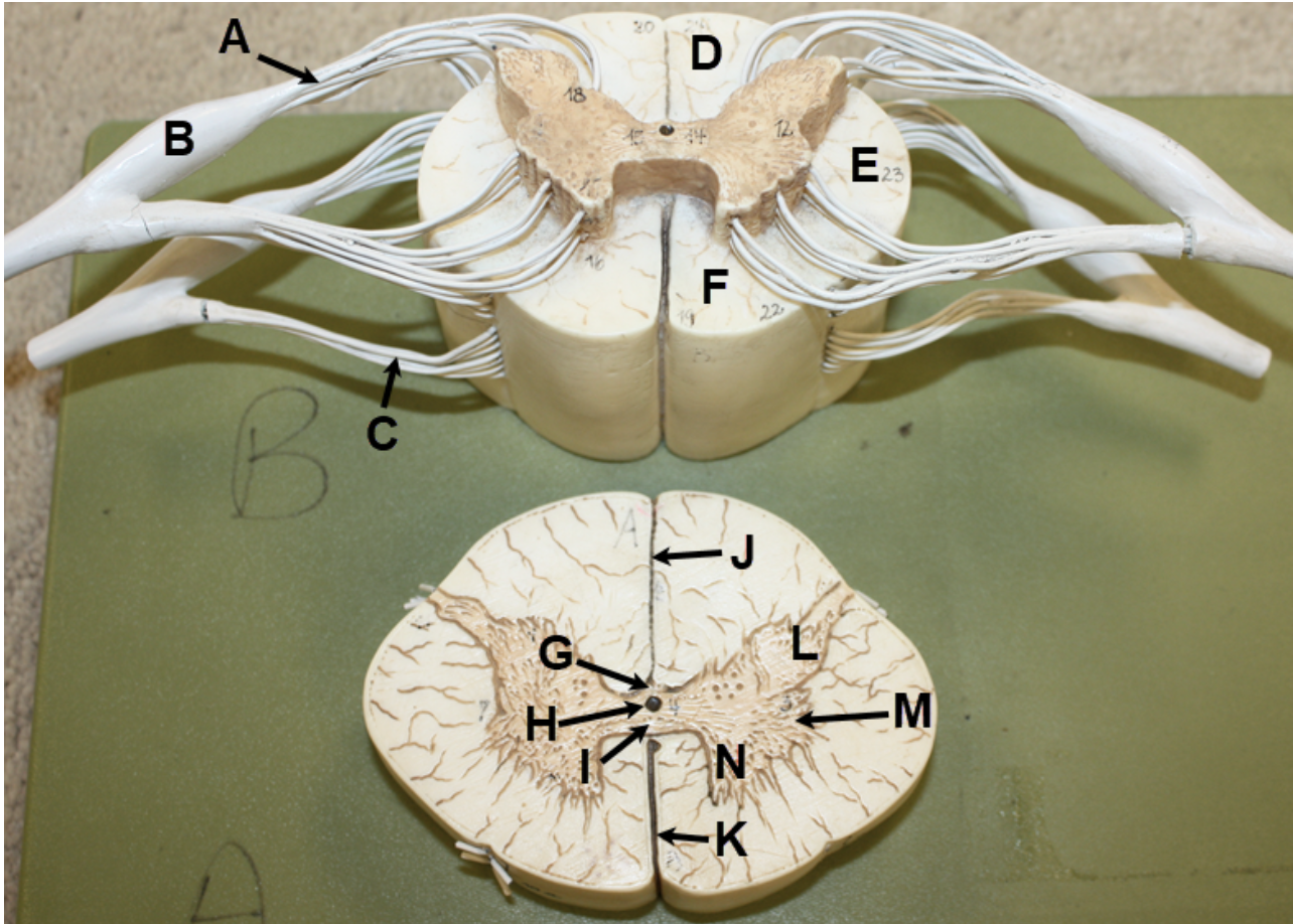
In the image above, identify structures A-L. Question M: the cell bodies of what type of neurons are located in structure D? Question N: the cell bodies of what type of neurons are located in structure F?
A. posterior (dorsal) white column
B. posterior (dorsal) horn
C. lateral white column
D. lateral horn
E. anterior (ventral) white column
F. anterior (ventral) horn
G. posterior (dorsal) root
H. anterior (ventral) root
I. posterior (dorsal) gray commissure
J. anterior (ventral) gray commissure
K. anterior (ventral) white commissure
L. central canal
M. visceral motor
N. somatic motor
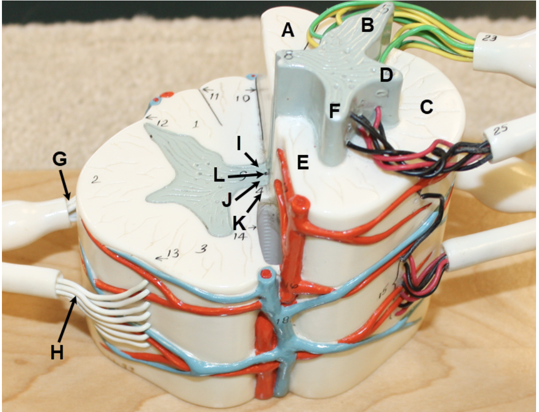
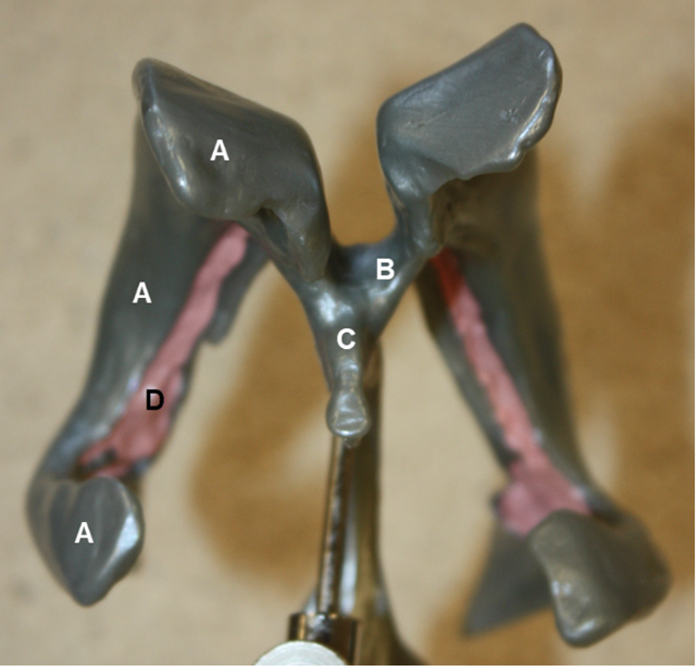
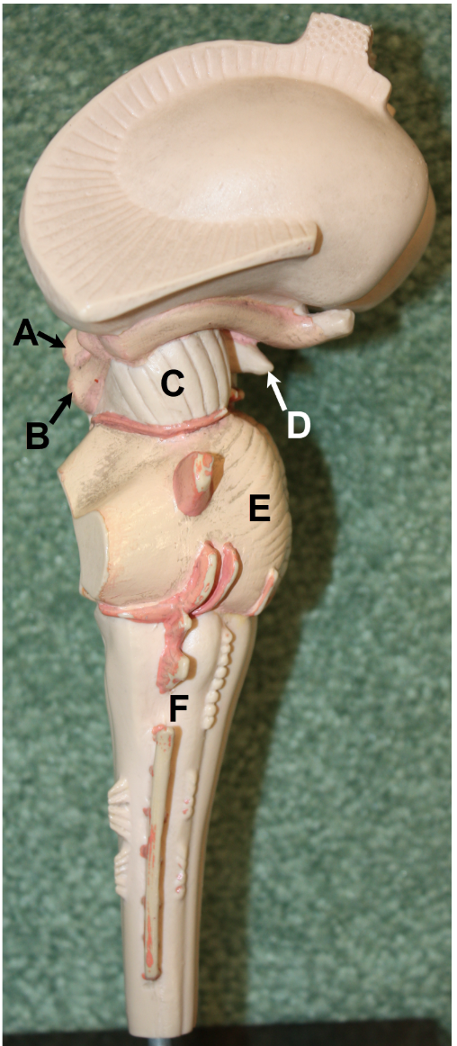
In the image above, identify structures A-N. Question O: the cell bodies of what type of neurons are located in structure B? Question P: the cell bodies of what type of neurons are located in structure M? Question Q: the cell bodies of what type of neurons are located in structure N?
A. posterior (dorsal) root
B. spinal ganglion (posterior root ganglion or dorsal root ganglion)
C. anterior (ventral) root
D. posterior (dorsal) white column
E. lateral white column
F. anterior (ventral) white column
G. posterior (dorsal) gray commissure
H. central canal
I. anterior (ventral) gray commissure
J. posterior (dorsal) median sulcus
K. anterior (ventral) median fissure
L. posterior (dorsal) horn
M. lateral horn
N. anterior (ventral) horn
O. sensory (both somatic sensory and visceral sensory)
P. visceral motor
Q. somatic motor
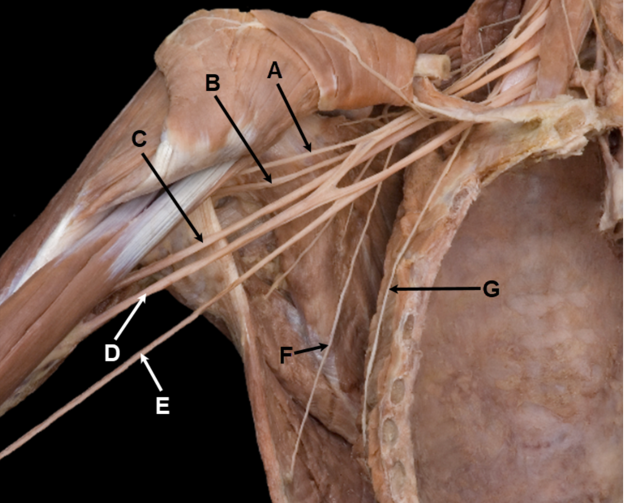
In the image above, identify nerves A-G.
A. musculocutaneous nerve
B. axillary nerve
C. radial nerve
D. median nerve
E. ulnar nerve
F. thoracodorsal nerve
G. long thoracic nerve
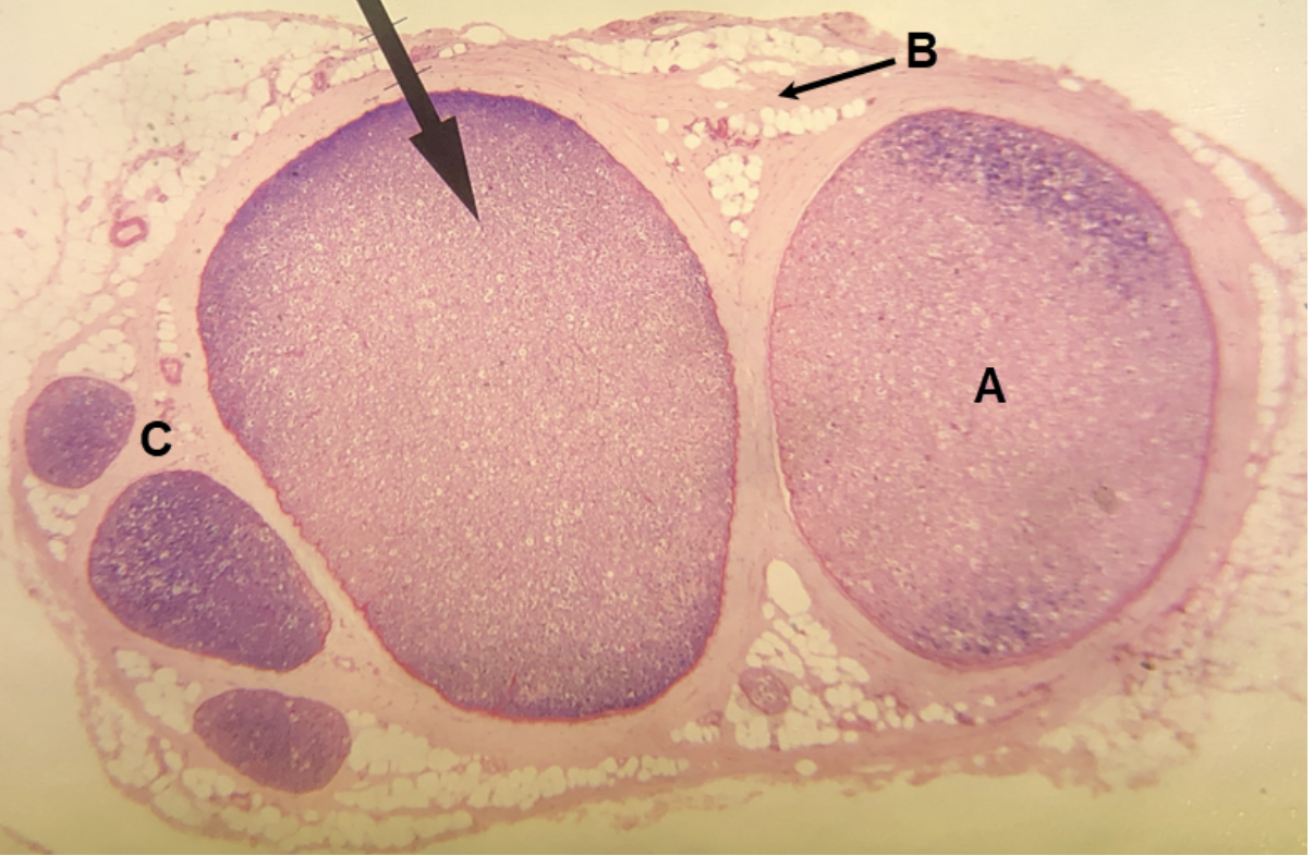
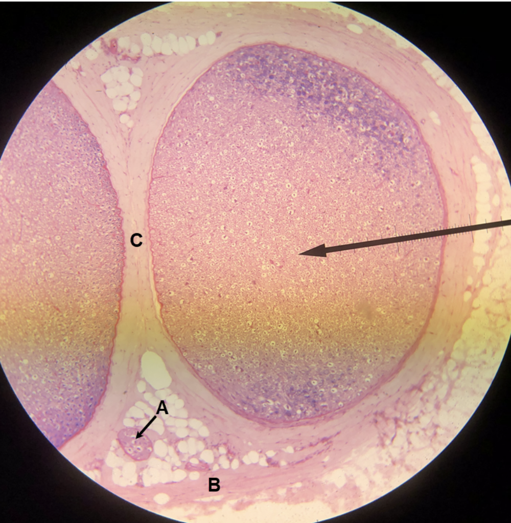
In the image above, identify structures A-C.
A. fascicle
B. epineurium
C. perineurium

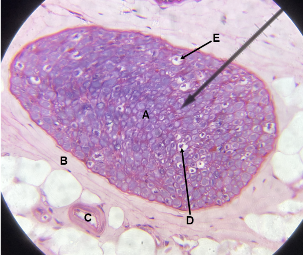
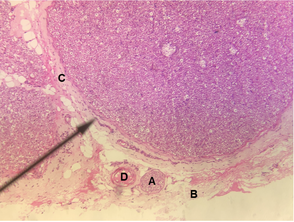
In the image above, identify structures A-D.
A. fascicle
B. epineurium
C. perineurium
D. vasa nervorum
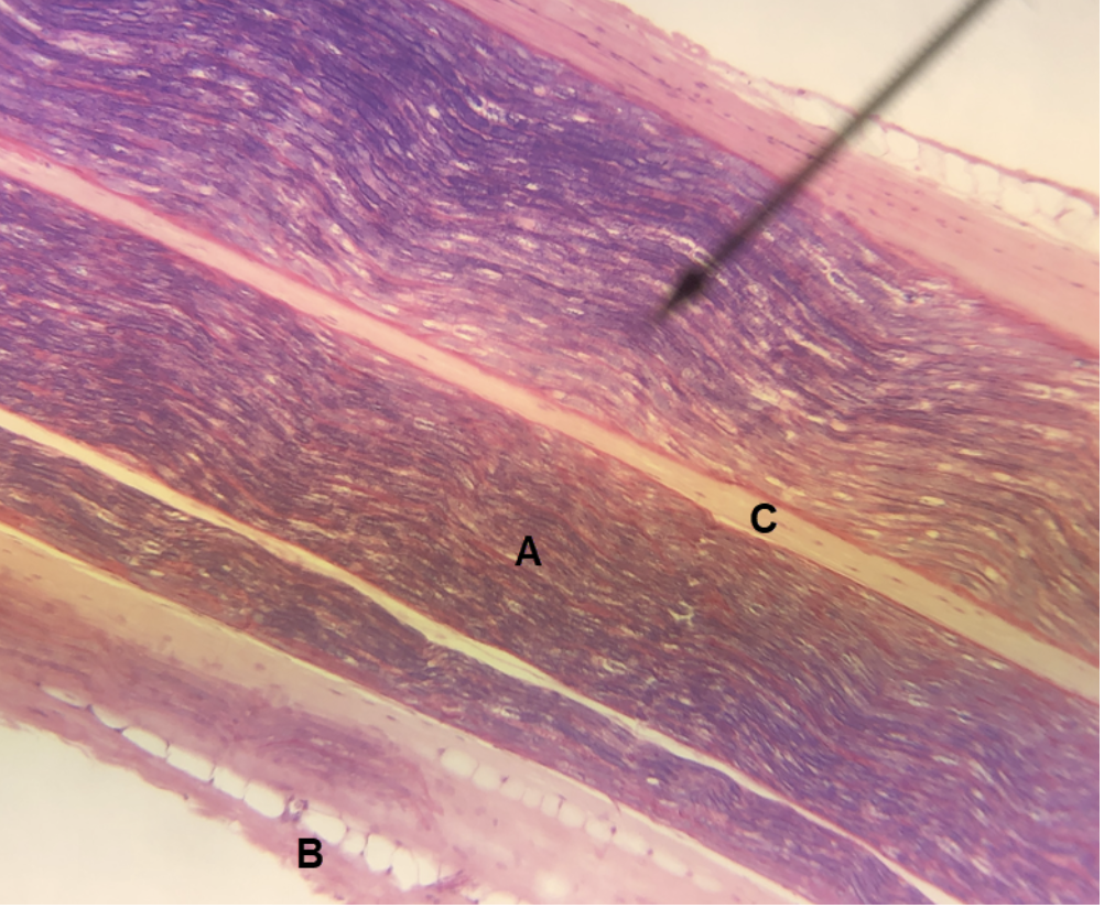
In the image above, identify structures A-C.
A. fascicle
B. epineurium
C. perineurium
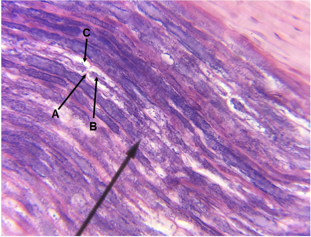
In the image above, identify structures A-E. (Hint: D is purple, E is white.)
A. fascicle
B. perineurium
C. vasa nervorum
D. axon
E. Schwann cell (myelin)
In the image above, identify structures A-C.
A. fascicle
B. epineurium
C. perineurium
In the image above, identify structures A-C. (Hint: B is purple, C is white.)
A. node (node of Ranvier)
B. axon
C. Schwann cell (myelin)
In the image above, identify structures A-C. (Hint: B is purple, C is white.)
A. node (node of Ranvier)
B. axon
C. Schwann cell (myelin)

In the image above, identify structures A-C.
A. fascicle
B. epineurium
C. perineurium
In the image above, identify structures A-D.
A. fascicle
B. epineurium
C. perineurium
D. vasa nervorum
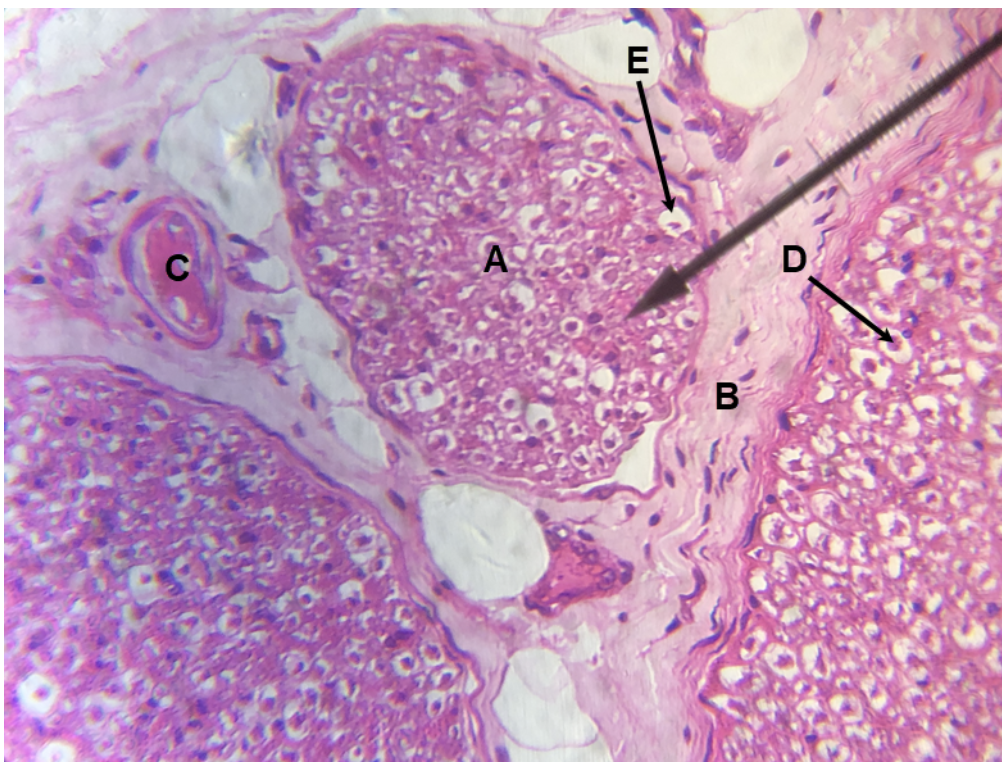
In the image above, identify structures A-E. (Hint: D is purple, E is white.)
A. fascicle
B. perineurium
C. vasa nervorum
D. axon
E. Schwann cell (myelin)
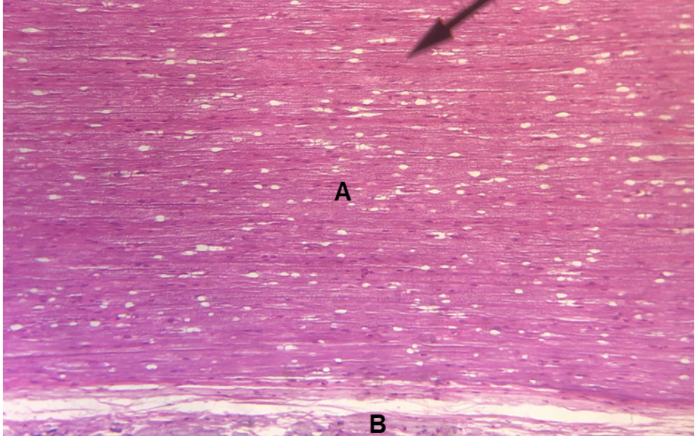
In the image above, identify structures A and B.
A. fascicle
B. perineurium
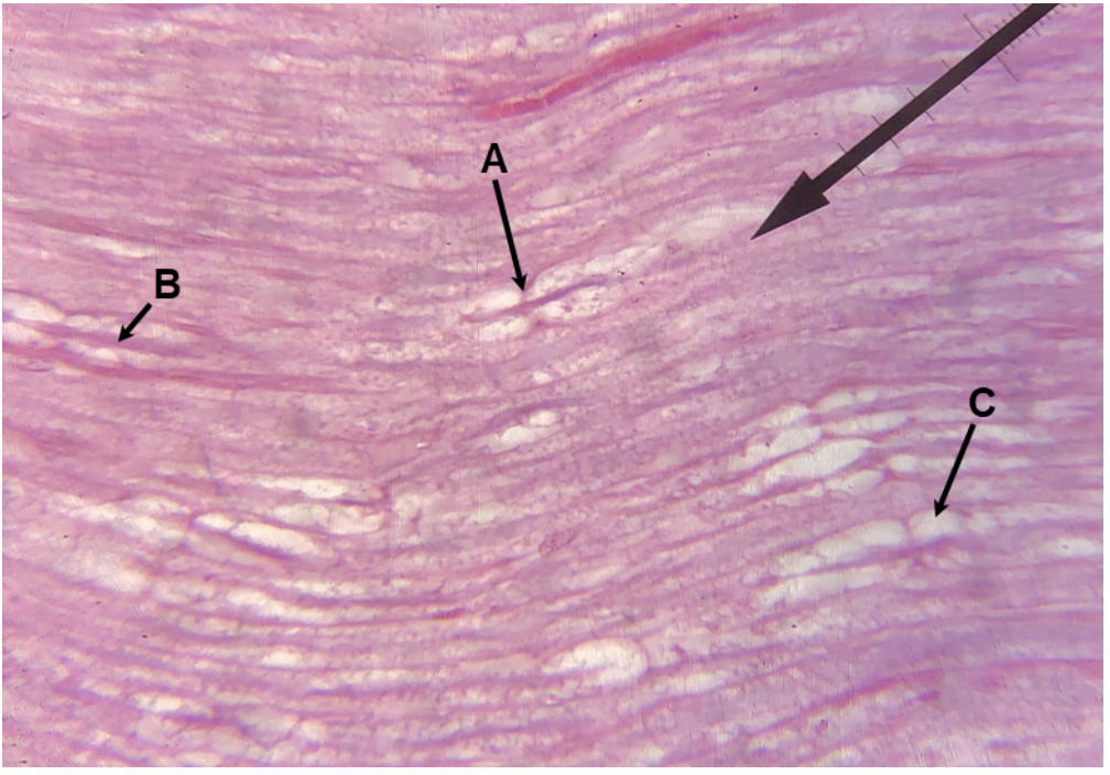
In the image above, identify structures A-C. (Hint: B is purple, C is white.)
A. node (node of Ranvier)
B. axon
C. Schwann cell (myelin)

In the image above (ventricular system), identify hollow structures A-E, and pink structures F-G.
A. lateral ventricle
B. interventricular foramen
C. third ventricle
D. cerebral aqueduct
E. fourth ventricle
F. choroid plexus of third ventricle
G. choroid plexus of fourth ventricle
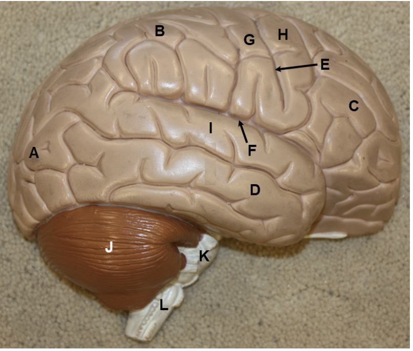
In the image above, identify lobes A-D, specific grooves E-F, specific ridges G-I, and structures J-L.
A. occipital lobe
B. parietal lobe
C. frontal lobe
D. temporal lobe
E. central sulcus
F. lateral sulcus (lateral fissure)
G. postcentral gyrus
H. precentral gyrus
I. superior temporal gyrus
J. cerebellum
K. pons
L. medulla oblongata
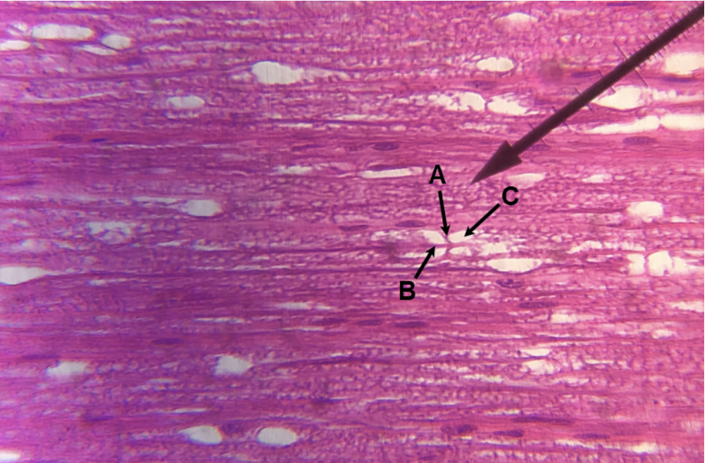
In the image above (brainstem/diencephalon region), identify structures A-E.
A. infundibulum
B. cerebral peduncle
C. mammillary body
D. pons
E. pyramids
In the image above, identify dural sinuses A-C, dural fold D, structures E-W, and white structure X. For Y, identify the specific brown structure that surrounds X.
A. superior sagittal sinus
B. straight sinus
C. confluence of sinuses
D. tentorium cerebelli
E. optic chiasm
F. hypothalamus
G. anterior commissure
H. corpus callosum
I. septum pellucidum
J. fornix
K. choroid plexus of third ventricle
L. interthalamic adhesion
M. pineal gland
N. thalamus
O. infundibulum
P. pituitary gland
Q. mammillary body
R. cerebral peduncle
S. posterior commissure
T. pons
U. medulla oblongata
V. fourth ventricle
W. corpora quadrigemina (the superior and inferior colliculi are not distinct from each other on this model)
X. arbor vitae
Y. cerebellar cortex
In the image above (ventricular system), identify hollow structures A-C, and pink structure D.
A. lateral ventricle
B. interventricular foramen
C. third ventricle
D. choroid plexus of lateral ventricle
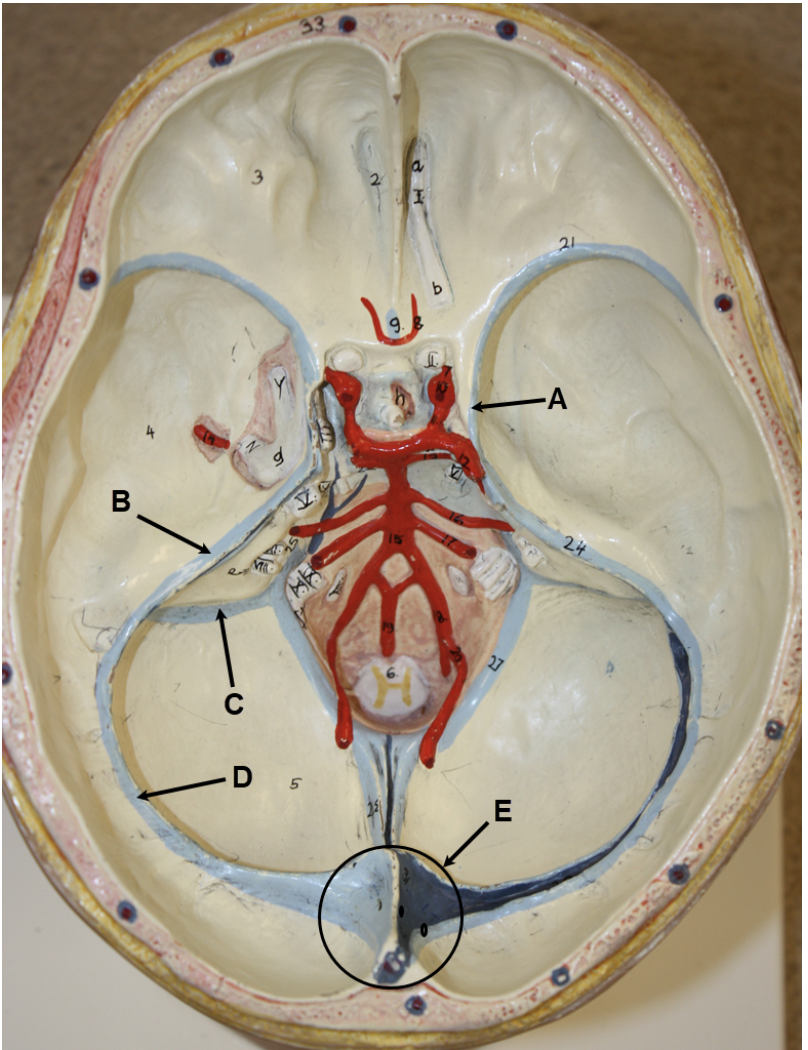
In the image above (superior view of cranium with calvaria and brain removed), identify dural sinuses A-E.
A. cavernous sinus
B. superior petrosal sinus
C. sigmoid sinus
D. transverse sinus
E. confluence of sinuses
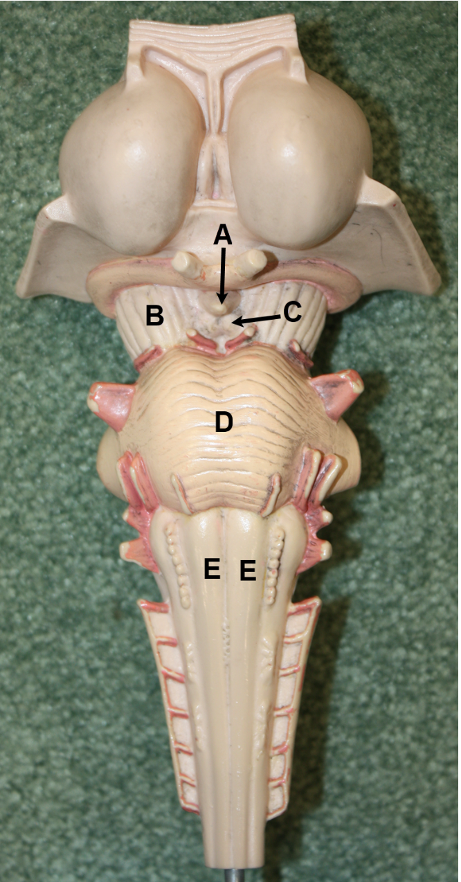
In the image above (brainstem/diencephalon region), identify structures A-G.
A. third ventricle
B. thalamus
C. pineal gland
D. superior colliculus
E. inferior colliculus
F. pons
G. medulla oblongata

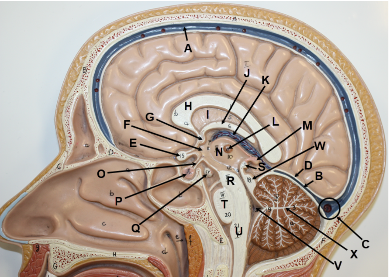
In the image above, identify structures A-R, and white structure S. For T, identify the specific brown structure that surrounds S.
A. anterior commissure
B. septum pellucidum
C. corpus callosum
D. fornix
E. choroid plexus of third ventricle
F. interthalamic adhesion
G. pineal gland
H. corpora quadrigemina (the superior and inferior colliculi are not very distinct from each other on this model)
I. cerebral aqueduct
J. fourth ventricle
K. hypothalamus
L. posterior commissure
M. optic nerve (CN II)
N. optic chiasm
O. mammillary body
P. cerebral peduncle
Q. pons
R. medulla oblongata
S. arbor vitae
T. cerebellar cortex
In the image above (ventricular system), identify hollow structures A-D, and pink structures E-F.
A. lateral ventricle
B. interventricular foramen
C. cerebral aqueduct
D. fourth ventricle
E. choroid plexus of third ventricle
F. choroid plexus of fourth ventricle

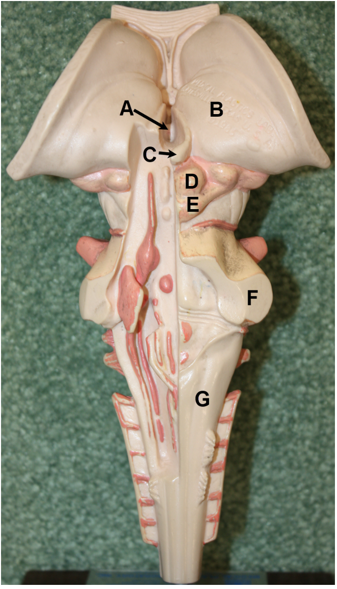
In the image above, identify lobes A-B, and structures C-G. Hint #1: use Martini et al. Fig. 14-18 (11th Ed.) or Fig. 14-19 (10th Ed.) if you need help. Hint #2: structure C has been cut at its base.
A. frontal lobe
B. temporal lobe
C. infundibulum
D. mammillary body
E. cerebral peduncle
F. pons
G. medulla oblongata
In the image above (brainstem/diencephalon region), identify structures A-F.
A. superior colliculus
B. inferior colliculus
C. cerebral peduncle
D. infundibulum
E. pons
F. medulla oblongata
In the image above (ventricular system), identify hollow structures A-D, and pink structures E-F.
A. lateral ventricle
B. interventricular foramen
C. cerebral aqueduct
D. fourth ventricle
E. choroid plexus of third ventricle
F. choroid plexus of fourth ventricle