Lecture 8: development of the pharyngeal apparatus
 Call Kai
 Learn
Learn Practice Test
 Spaced Repetition
 Match
Match Flashcards
 Knowt Play
1/27

There's no tags or description
Looks like no tags are added yet.
Name | Mastery | Learn | Test | Matching | Spaced | Call with Kai | Chat |
|---|
No analytics yet
Send a link to your students to track their progress
28 Terms
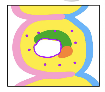
Pharyngeal Arches
a series of tissue bulges that appear on the ventro-lateral side of the embryonic head
first pharyngeal arch
arch gives rise to the maxillary arch and mandibular arch
Pharyngeal cleft
separates pharyngeal arches on the outside
indentation between pharyngeal arches on their external surfaces
lined by the ectoderm
4 pairs
Pharyngeal pouch
Indentation between pharyngeal arches on their internal surfaces
Lined by endoderm
4 pairs
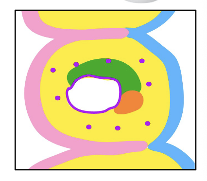
Neural crest-derived mesenchyme
the yellow
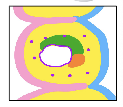
Mesoderm mesenchyme
The green
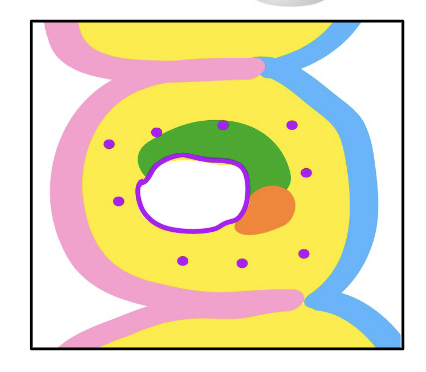
Pharyngeal arch artery
purple dots
part of the vascular endothelium
make up the aortic arch artery, and aortic arch
embedded in the mesenchyme of the pharyngeal arches
derived from splanchnic mesoderm
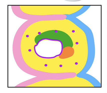
Angioblasts
the purple border with the white in the middle
part of the vascular endothelium
Precursor of blood vessels
from paraxial mesoderm and splanchnic mesoderm
Nerves
Orange
Pharyngeal Endoderm
component of the pharyngeal arch
wall of the pharyngeal gut
covers the internal surface of the pharyngeal arches, and lines the pharyngeal pouches
Surface Ectoderm
component of the pharyngeal arch
covers the external surface of the pharyngeal arches, and lines the pharyngeal clefts
Neural Crest-Derived Mesenchyme
called ectomesenchyme or mesctoderm to distinguish it from the mesoderm mesenchyme
Contains cranial neural crest cells (NCC) and Cardiac NCC
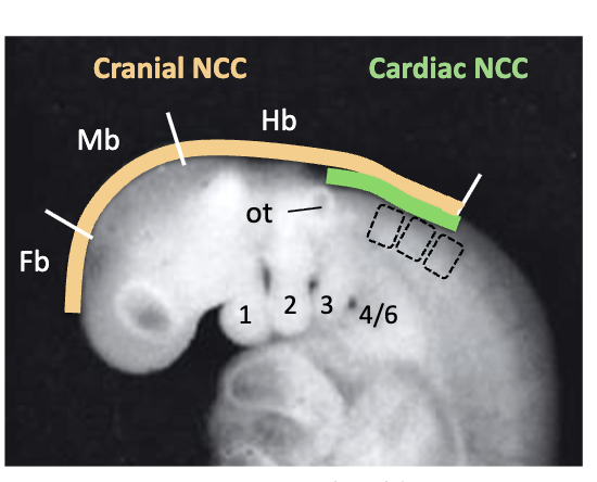
Cranial NCC (neural crest cells)
from forebrain, midbrain, and hindbrain
Only those in the midbrain and hindbrain enter the pharyngeal arches
becomes skeleton and connective tissue
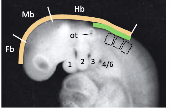
Cardiac NCC (neural crest cells)
part of the neural crest-derived mesenchyme
from mid-otic placode to the level 3rd somite (2nd pharyngeal cleft - 6th arch)
Contributes to heart and blood vessels
Rhombomeres
the embryonic hindbrain is organized into 8 segments called what?
R1, R2
which rhombomeres that are the only ones of the eight that are in the midbrain go to the 1st pharyngeal arch
R4
Rhombomere that goes to the 2nd pharyngeal arch, part of the hindbrain
R6, R7
Rhombomeres that go to the 3rd, 4th, and 6th pharyngeal arches
R3, R5
Which two rhombomeres produce only very few cardiac neural crest cels, which joins the streams from neighboring R
paraxial, splanchnic
The mesoderm mesenchyme is mostly from blank mesoderm, and some blank mesoderm
Mesoderm core
at the center of each pharyngeal arch, mostly becomes skeletal muscles of the face and neck
part of the mesoderm mesenchyme
Second heart field
part of the mesoderm mesenchym
splanchnic mesoderm
at the junction of the pharyngeal arches and the heart
becomes part of the heart (cardiac muscles)
1st pharyngeal arch
which arch gives rise to Trigeminal (cranial nerve 5)
2nd pharyngeal arch
which arch gives rise to Facial (cranial nerve 7)
3rd pharyngeal arch
which arch gives rise to Glossopharyngeal (cranial nerve 9)
4th and 6th pharyngeal arch
which arch gives rise to vagus (cranial nerve 10)
Pharyngeal Ectoderm
Regulates development of the neural crest mesenchyme
Fibroblast growth factor (FGF) and bone morphogenetic protein (BMP)
The major signaling molecules produced by the ectoderm.