Chapter 25&49 SG
5.0(1)
Studied by 56 peopleCard Sorting
1/32
Last updated 2:35 PM on 11/28/22
Name | Mastery | Learn | Test | Matching | Spaced | Call with Kai |
|---|
No analytics yet
Send a link to your students to track their progress
33 Terms
1
New cards
What is an ekg/ecg?
The process by which a graphic pattern is created from the electrical impulses generated within the heart as it pumps.
2
New cards
In order, what is the pathway of electrical impulses through the heart?
SA Node, AV Node, Bundle of his, Purkinje fibers
3
New cards
What is known as the pacemaker of the heart and why?
The SA node because it triggers a sequence of electrical events in the heart
4
New cards
What is depolarization?
the orderly passage of electrical current sequentially through the heart muscle, changing it, cell by cell, from the resting polarized state to the depolarized state
5
New cards
What are leads?
a graphical description of the electrical activity of the heart and it is created by analyzing several electrodes.
6
New cards
What is an artifact?
electrocardiographic alterations, not related to cardiac electrical activity.
7
New cards
Poor skin preparation, dangling wire, or breathing movement creates what type of artifact?
Wandering Baseline
8
New cards
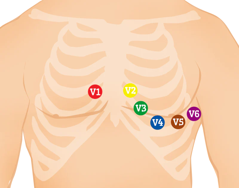
What are the specific locations of the chest leads
V1: 4th intercostal space (ICS), RIGHT margin of the sternum
V2: 4th ICS along the LEFT margin of the sternum
V4: 5th ICS, midclavicular line;
V3: midway between V2 and V4;
V5: 5th ICS, anterior axillary line;
V6: 5th ICS, mid-axillary line
V2: 4th ICS along the LEFT margin of the sternum
V4: 5th ICS, midclavicular line;
V3: midway between V2 and V4;
V5: 5th ICS, anterior axillary line;
V6: 5th ICS, mid-axillary line
9
New cards
What is stress testing?
an exercise stress test, shows how your heart works during physical activity.
10
New cards
What is a holter monitor?
a small, wearable device that records the heart's rhythm. It's used to detect or determine the risk of irregular heartbeats (arrhythmias).
11
New cards
What is forced vital capacity?
(FVC) is the total amount of air exhaled during the FEV test.
12
New cards
What are the letters of the deflections on an ecg tracing?
P wave (atrial depolarization )
QRS complex (ventricular depolarization)
T wave (ventricular repolarization).
U wave ( Purkinje repolarization),
QRS complex (ventricular depolarization)
T wave (ventricular repolarization).
U wave ( Purkinje repolarization),
13
New cards
What does the P wave represent?
The P wave represents the electrical depolarization of the atria.
14
New cards
What does the QRS complex on an ecg represent?
ventricular depolarization.
15
New cards
What portion of the limbs should the electrodes be placed on?
On the fleshy part of the limbs
16
New cards
What position for an ecg should be used if the patient is uncomfortable lying flat?
Semi-Fowler's
17
New cards
What causes somatic tremor?
caused by patient movement, even shivering or chewing gum
18
New cards
What are the peak flow zones, and what do they indicate?
Green zone: Your asthma is well-controlled.
Yellow zone: Your asthma is getting worse or is poorly controlled
Red zone: Your asthma is severe. It requires emergency care.
Yellow zone: Your asthma is getting worse or is poorly controlled
Red zone: Your asthma is severe. It requires emergency care.
19
New cards
When is pressure in the arteries the greatest?
when blood is pumped out of the heart into the arteries.
20
New cards
Pericardium
Membrane that encloses the heart.
21
New cards
Myocardium
Middle layer of the heart, is the thickest layer of the wall and is primarily made up of cardiac muscle, which makes it the working layer of the heart.
22
New cards
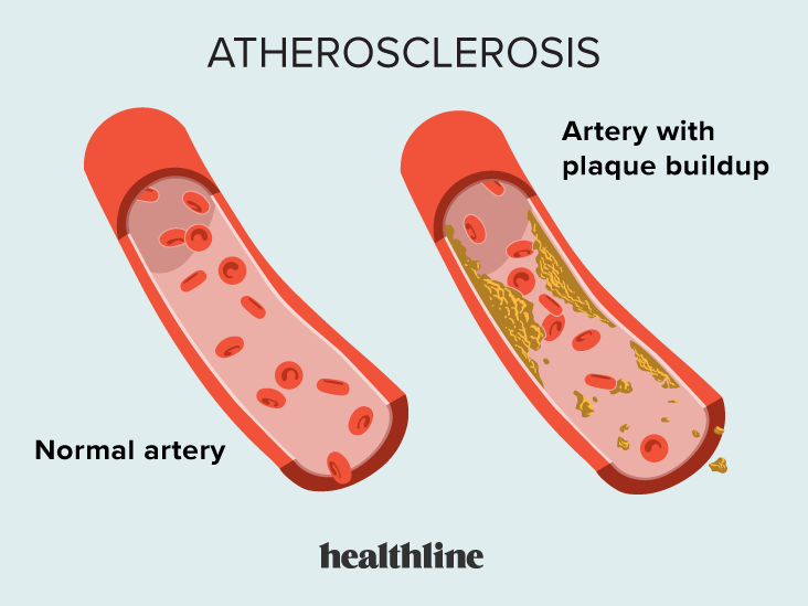
Atherosclerosis
A disease of the arteries characterized by the deposition of plaques of fatty material on their inner walls.
23
New cards
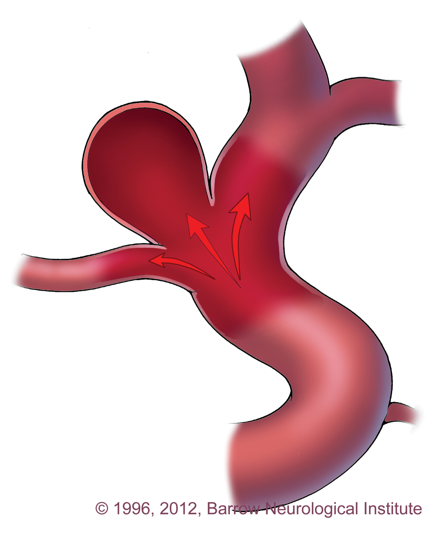
Aneurysm
a bulge in a blood vessel caused by a weakness in the blood vessel wall, usually where it branches
24
New cards
Where is the bicuspid valve located?
Located between the left atrium and left ventricle.
25
New cards
Where is the aortic semilunar valve located and what does it do?
Is located between the left ventricle and the aorta. It prevents blood from flowing back into the left ventricle
26
New cards
What do capillaries connect?
They are the connecting vessel between the arterioles of the arterial system and venules of the venous system.
27
New cards
Increased cardiac output causes what?
It causes an increase in blood pressure
28
New cards
How does vasoconstriction affect blood pressure?
-If blood pressure falls too low, the muscular walls of the arteries can constrict to increase blood pressure.
29
New cards
What are symptoms of a pulmonary embolism?
Shortness of breath, Chest pain, and Cough, which may contain blood. Leg pain or swelling. Back pain, Excessive sweating. Lightheadedness, dizziness, Bluish lips or nails.
30
New cards
What type of chest pain occurs during movement?
Costochondritis causes chest pain, felt at the front of the chest.
31
New cards
What might chest pain that increase when bending over be?
People with heartburn or a pulmonary embolism may experience worsening chest pain when bending forward.
32
New cards
What is the purpose of the chordae tendineae?
a group of tough, tendinous strands in the heart. Play a vital role in holding the atrioventricular valves in place while the heart is pumping blood.
33
New cards
Identify the blood flow through the heart, and name the structures?
1)body
2) inferior/superior vena cava
3) right atrium
4) tricuspid valve
5) right ventricle
6) pulmonary arteries lungs
8) pulmonary veins
9) left atrium
10) mitral or bicuspid valve
11) left ventricle
12) aortic valve
13) aorta
2) inferior/superior vena cava
3) right atrium
4) tricuspid valve
5) right ventricle
6) pulmonary arteries lungs
8) pulmonary veins
9) left atrium
10) mitral or bicuspid valve
11) left ventricle
12) aortic valve
13) aorta