Structure and function of microorganisms
1/51
There's no tags or description
Looks like no tags are added yet.
Name | Mastery | Learn | Test | Matching | Spaced |
|---|
No study sessions yet.
52 Terms
why should we study microbiology? 3
microbes cause most common dental diseases
we need to understand infection to enable effective treatment options and preventions
by understanding the basic processes of microbes we can develop future treatments
what common dental infections are caused by microorganisms?
caries and periodontal diseases
what is the definition of microbiology?
the biology of organisms that are too small to be seen by the naked eye
what are some examples of microbes? 4
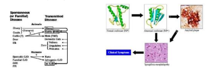
• TSEs “scrapie-like” agents (Transmissable Spongiform Encephalopathies.
Viruses.
Bacteria.
Eukaryotic microbes: fungi and protozoa.
are protozoa eukaryotic or prokaryotic?
eukaryotic
what are TSEs?
TSEs “scrapie-like” agents (Transmissable Spongiform Encephalopathies)
‘infective’ proteins
Examples – Kuru, Scrapie, Creutzfeld-Jacob, Scrapie
Cause sponge-like lesions in the brain - by modulating the conformation of a particular protein - normal conformation vs misfolded proteins - eg amyloid plaques - may become insoluble - causes destruction of cells - abnormally folded protein
this may cause a further rection which causes normally folded proteins into abnormally folded proteins - infective nature
bacteria
bacteria come in a wide variety of forms / shapes/ sizes
they are all much smaller than a mammalian cell - some examples but BNL very small
viruses
even smaller than a bacteria - similar ratio that bacteria have with animal cells
differentiation between microbes?
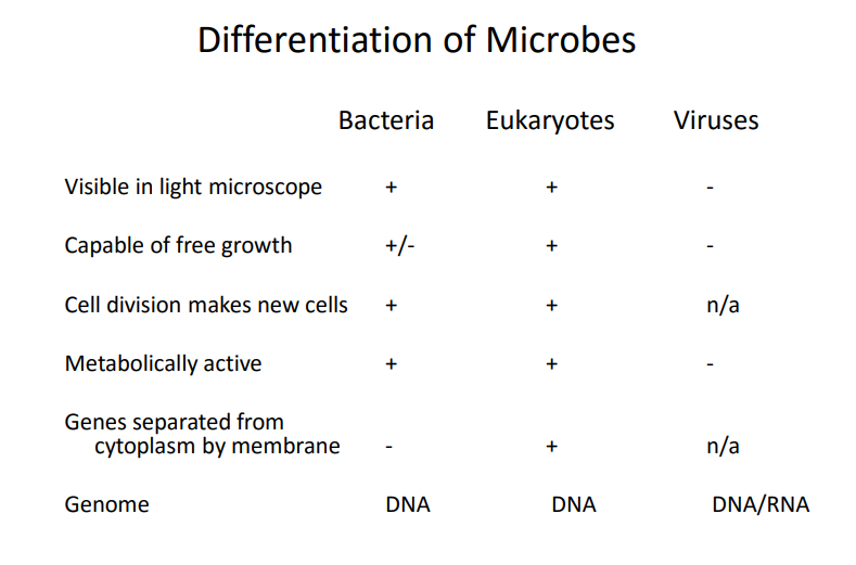
which microbes are visible in a light microscope?
bacteria and eukaryotes
which microbes are capable of free growth? - can grow on their own
eukaryotes and some bacteria
ALL viruses need other cells to grow in - obligate intracellular paracites
cell division makes new cells?
both bacteria and eukaryotes
viruses don’t
which microbes are metabolically active?
bacteria and eukaryotes - viruses AREN’T (reliant on host)
which microbes have genes separated from cytoplasm via a membrane?
only eukaryotes - compartmentalisation , n/a for viruses
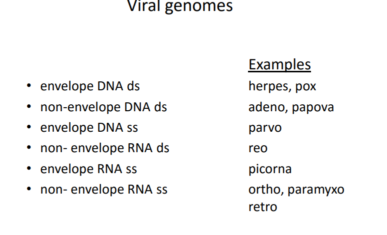
different genomes in different microbes
bacteria have DNA, eukaryotic have DNA and viruses have DNA/RNA
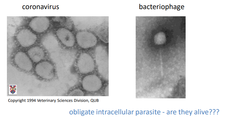
What are viruses?
infect bacteria, plants and animals
small - only “visible” in electron microscope
10 to 200 nm
are they alive????
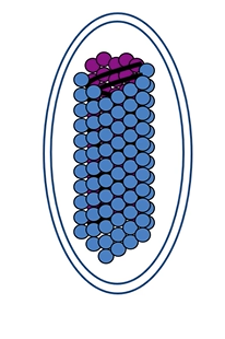
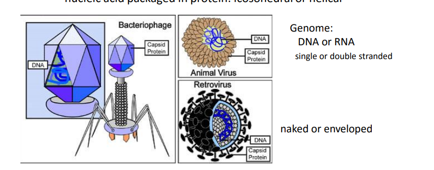
Viral structure
nucleic acid packaged in protein- nuclear capsid : icosohedral - geometic or helical
genome - DNA/RNA
nuclear capsid can be naked or enveloped (by host cell membrane/protein)
Viral replication
virus comes into contact with the animal cell
entry into the cell - recognition b/w virus and surface of the cell, triggers a fusion event + gains entry
after gaining entry, the virus will then uncoated - releases its genome into the nucleoplasm but mainly cytoplasm
uses host cells machinery to replicate its genome via RNA or DNA -
at the same time it will transcribe its genome to start producing viral proteins - nuclear capsules or immunosuppresant molecules - or shifting machinery down a different route
all proteins assemble and package new viruses - bud off cell or explodes
mechanisms of viral replication - reverse transcription
viral genome - RNA is converted to DNA
mechanisms of viral replication - integration
reverse transcribed DNA can insert itself into hosts genome
viral genome examples
all have capsids
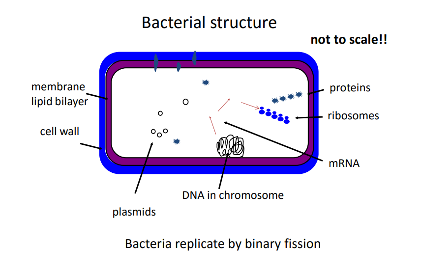
bacterial structure
living organisms - metabolically active, reproduce , binary fission
plasmids code for a variety of things - antimicrobial resistance, virulence factors
some proteins that are produced become membrane associated proteins
bacterial cell walls
can be quite complex - gram negative bacteria
simple - gram positive bacteria
structural support
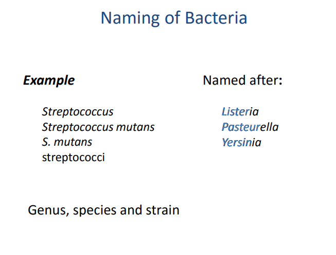
naming bacteria
genus and species are in italics
genus is in capitals, species in lowercase
genus can be shortened , say original first
plural of the genus - all lowercase and plural suffix
named after famous microbiologists
Identification and Taxonomy
Range of complexity: from groups of bacteria to individual isolates
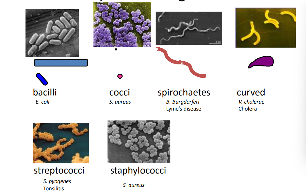
shape and size
arrangement of growing bacteria
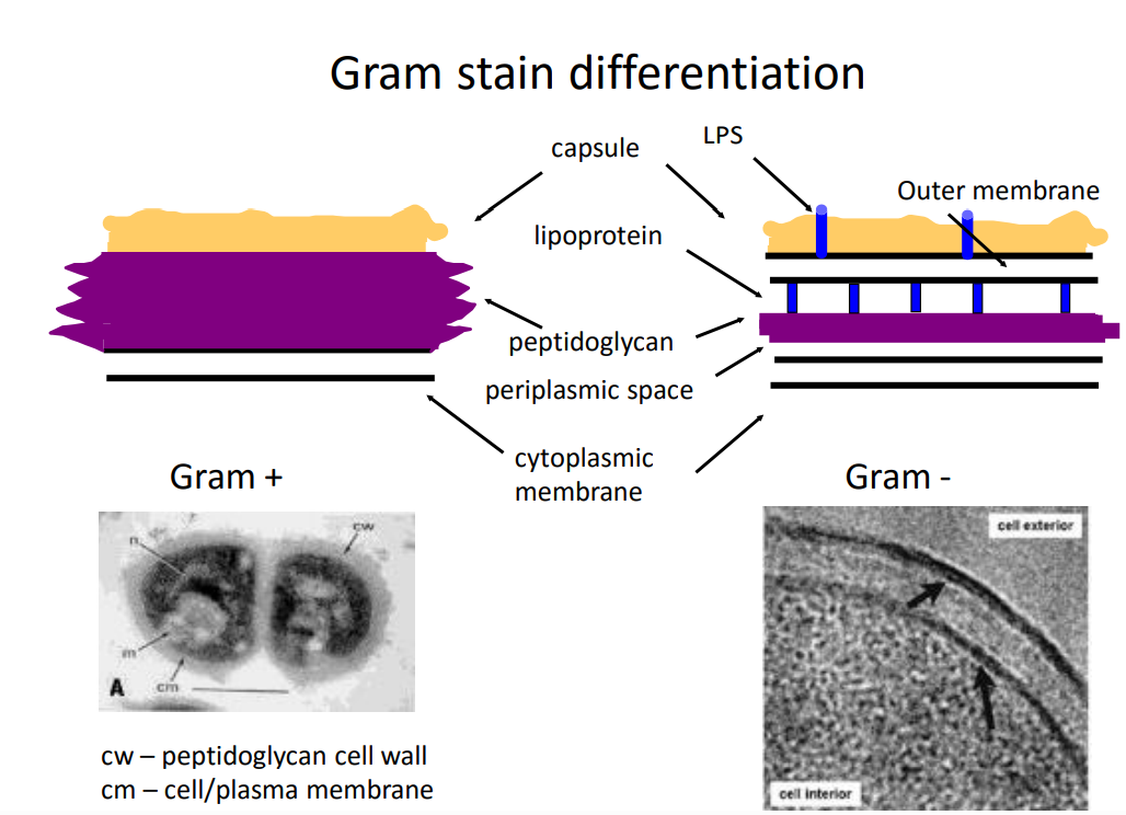
Gram stain
culture requirements - acidogenic or aciduric
biochemical reactions
antigenic structure
nucleic acid technologies
typing
Shape and arrangement
arrangement - streptococci (lines) vs staphylococci (clusters)
Gram stain differentiation
gram positive has a thick peptidoglycan layer
gram negative has a space between the peptidoglycan layer and the cytoplasmic membrane - periplasmic space , thinner pdl layer
both have a glycocalyx exterior structure - slime layer or capsule like layer - support and evading immune responses
gram negative also have an outer membrane and lipoproteins that connect the two layers
gram negative bacteria has a compound called lipopolysaccride on the outer membrane - triggers septic shock - immunogenic
colony characteristics - of bacteria
haemolysis
surface texture
colony morphology
size indicator dyes
different nutrients
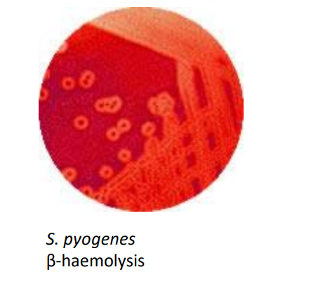
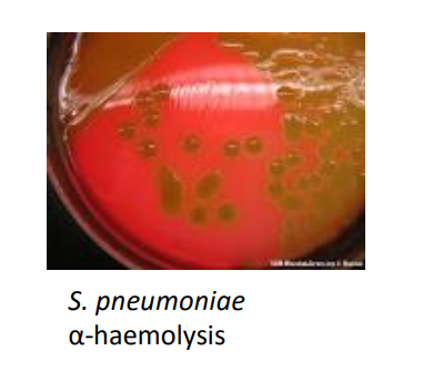
haemolysis types
agar plate that contains whole blood cells
alpha, beta and gamma haemolysis - breaking down blood cells to release its components
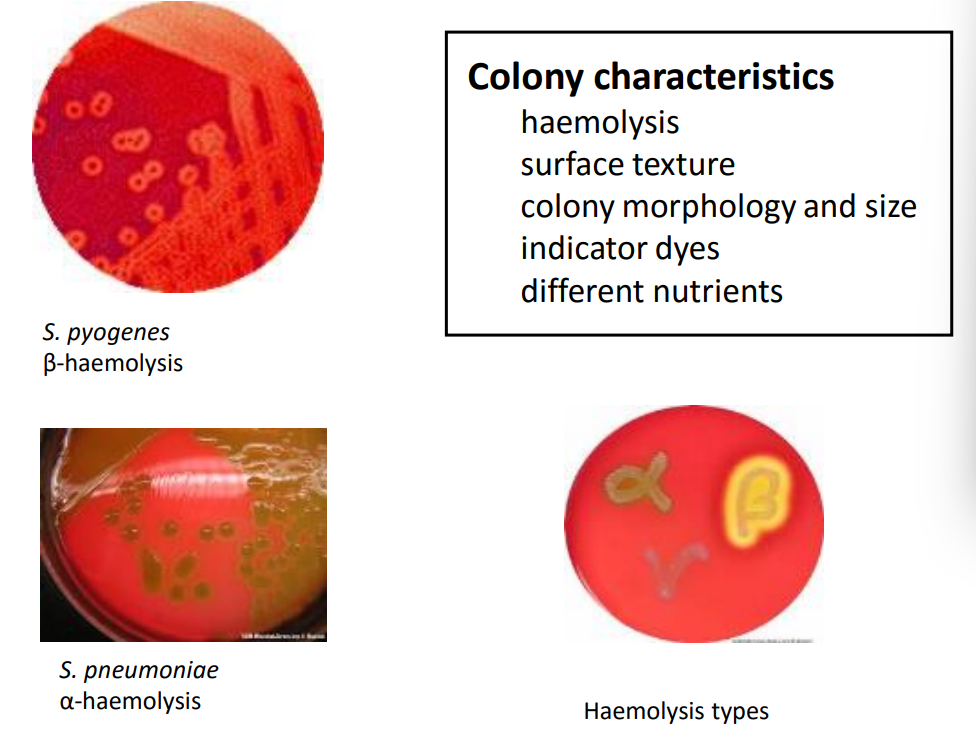
beta haemolysis
clear zone where the bacteria has broken down the red blood cells/haemoglobin
alpha haemolysis
no clear zone but there is a greenish tinge - haemoglobin breakdown a la bile
Biochemical Tests to distinguish bacteria
sugar fermentation profiles - gas? - acid?
enzyme profiles eg coagulase
single test not enough - use loads of tests - metabolite profile on a structure - mass spec vs database
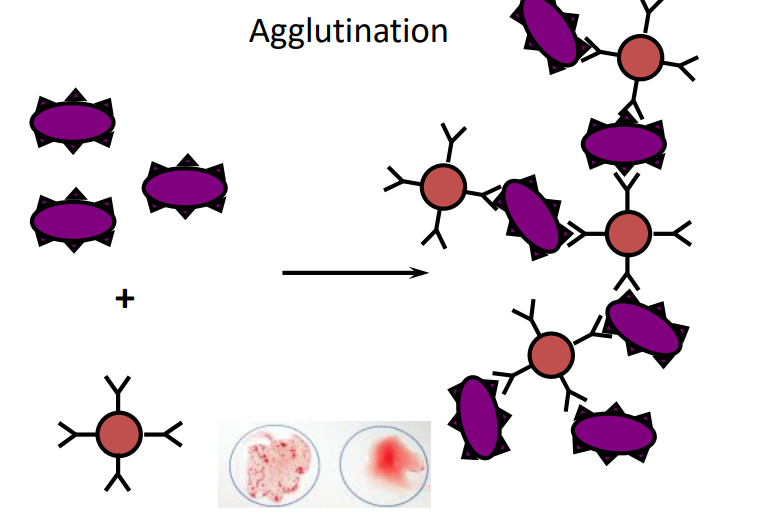
Antigenic structure
based on specificity of antibody-protein interactions proteins on bacterial surface unique to that bacterium
antigen is unique to species and even strain
identifying the antibodies via agglutination
mixed with red blood cells coated in antibodies - they agglutinate - large immune complex - clustering effect
Typing - defining it based on a type
• serotyping using antisera - see previous card
• phage typing using phage (bacterial viruses) that recognise surface proteins
• genetic typing using sequence properties of DNA
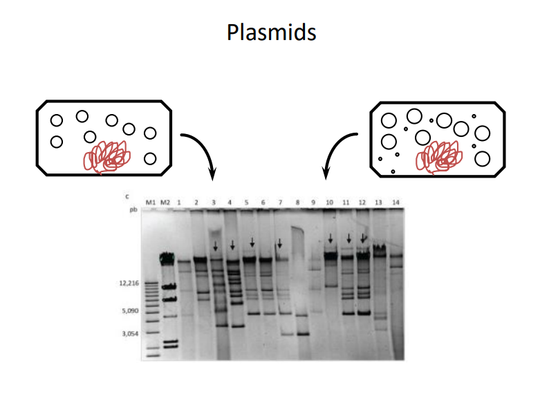
Genetic typing
All characteristics encoded by DNA
plasmid profile – Salmonella
sequence and RFLP - fully sequence the organism - restriction enzyme fragment polymorphism - endonuclease
probes - match to sequence
polymerase chain reaction (PCR)useful for all pathogens
plasma profiling
size separation via electrophoresis
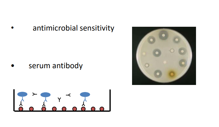
Other analyses
antimicrobial sensitivity
serum antibody - ELISA plate
Prokaryotic vs Eukaryotic - general differences
no internal membranes
rigid cell wall
70s ribosomes
vs
DNA encased in nucleus
mitochondria - energy metabolism
Some have no rigid cell wall
larger than bacteria
80s ribosomes
Fungi and protozoa
fungi: single or multi-cellular digest food with extracellular enzymes some dimorphic such as: Candida albicans – yeast forms or hyphae
protozoa single cellular - may have chloroplast - amoeba
Fungi
Yeasts – Grow as single cells, but can be dimorphic (grow as single cells and as hyphae – e.g. Candida spp, Cryptococcus spp
Moulds – Grow as hyphae - furry (filamentous) only – e.g. Aspergillus spp, Penicillium spp, Fusarium spp
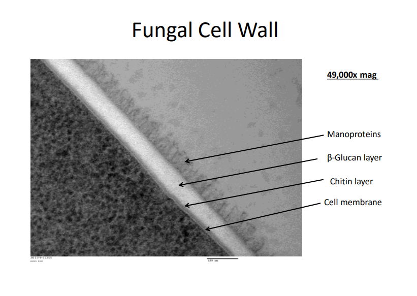
the fungal cell wall
chitin -
ergosterol instead of cholesterol
glucan filaments
mannoproteins - high degree of mannose sugars
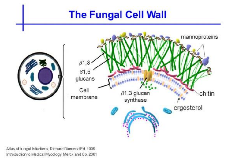
fungal cell wall - continued
glucan layer is thickest
history of microbiology
louis pasteur - germ theory
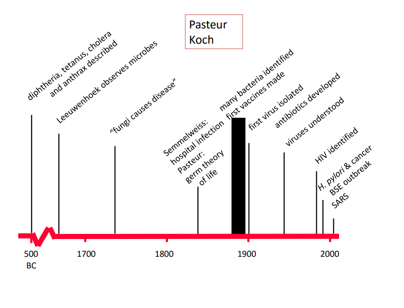
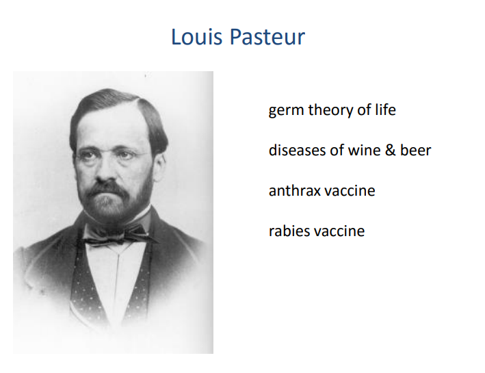
germ theory - were alive and can cause disease but not all of them
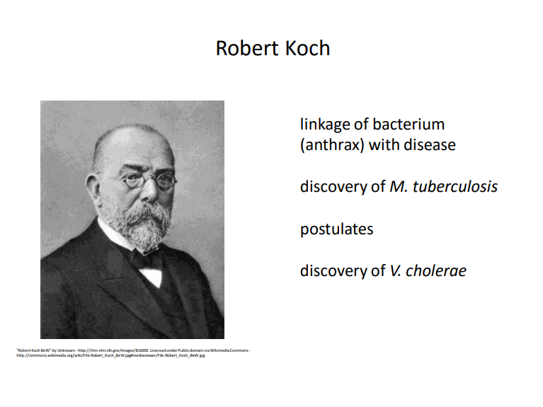
Koch’s Postulates
The bacteria must be present in every case of the disease.
The bacteria must be isolated from the host with the disease and grown in pure culture.
The specific disease must be reproduced when a pure culture of the bacteria is inoculated into a healthy susceptible host.
some nuance in later decades
Microbiology and disease
pathogenic bacteria – cause diseass
commensals, normal flora – harmless, “good bacteria”
competitive exclusion bad stuff, eg Salmonella – nutrient competition – pH – immune system
synthesis of nutrients
Commensals or opportunistic pathogens
• antibiotics overuse – Salmonella – C. difficile - disrupts microflora
HIV
– TB
– Candida - in the gut - thrush when HIV positive
trauma – S. aureus - abrasions
opportunistic bacteria
• commensals, normal flora - “good bacteria”
Pathobionts - candida, samonella
pathogenic bacteria •opportunistic bacteria -
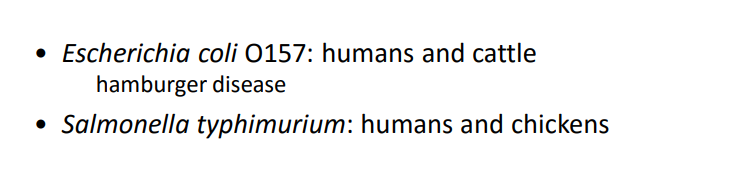
Host parasite relationship
disease state is complex and unstable - we can cure diseases
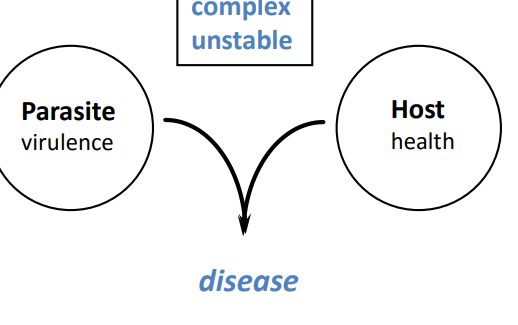
Host parasite relationship
cattle don’t react badly to E-coli