histology practical
1/24
There's no tags or description
Looks like no tags are added yet.
Name | Mastery | Learn | Test | Matching | Spaced |
|---|
No study sessions yet.
25 Terms

What is the organ shown in the diagram with respiratory epithelium and hyaline cartilage?
Trachea.
What type of epithelium lines the trachea?
Pseudostratified columnar ciliated epithelium.
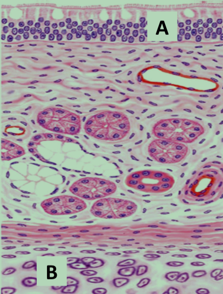
What structure is labeled A in the trachea diagram?
Respiratory epithelium.

What structure is labeled B in the trachea diagram?
Hyaline cartilage.
What type of cartilage is found in the trachea?
Hyaline cartilage.
How is tracheal cartilage arranged?
In rings.
What is an identifying histological feature of the trachea?
Pseudostratified ciliated columnar epithelium.
Rings of hyaline cartilage
What organ is identified by pseudostratified epithelium and hyaline cartilage rings?
Trachea.
Identify
Trachea

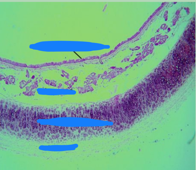
What structure is shown in the lung diagram with folded mucosa and cartilage plates?
Lobar bronchus.
What type of mucosa is present in lobar bronchus?
Folded mucosa.
What type of muscle layer is present in lobar bronchus?
Circular smooth muscle layer.
What type of cartilage is present in lobar bronchus?
Hyaline cartilage plates.
Are cartilage rings or plates found in lobar bronchus?
Plates.
What glands are present in the adventitia of lobar bronchus?
Mucus glands.
What lymphoid structure may be present in lobar bronchus?
Lymphatic nodules.
What organ contains lobar bronchi and alveoli?
Lung.
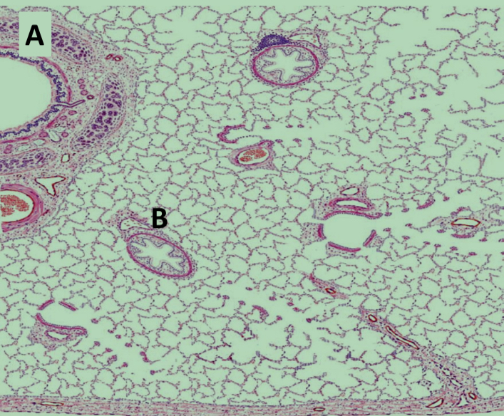
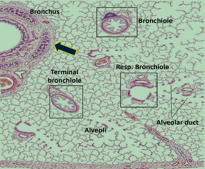
What is structure B in the lung diagram showing bronchioles?
Terminal bronchiole.

What is structure A in the same lung diagram?
Lobar bronchus.
What type of epithelium lines terminal bronchioles?
Simple cuboidal epithelium.
Are terminal bronchioles ciliated?
Yes.
Which two cells line the epithelium of terminal bronchioles?
Ciliated cuboidal cells and Clara cells.

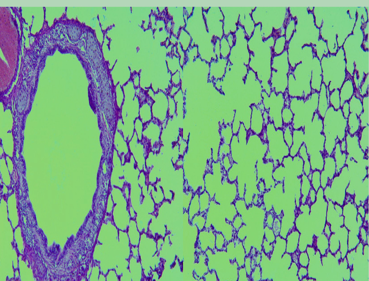
What organ is identified in the slide showing lobar bronchus and alveoli?
Lung.

What are the identifying points of lung histology?
Lobar bronchus with hyaline cartilage plates and lung alveoli.