UNIT 4. ERYTHROCYTE PRODUCTION
 Call Kai
 Learn
Learn Practice Test
 Spaced Repetition
 Match
Match Flashcards
 Knowt Play
1/126

There's no tags or description
Looks like no tags are added yet.
Name | Mastery | Learn | Test | Matching | Spaced | Call with Kai | Chat |
|---|
No analytics yet
Send a link to your students to track their progress
127 Terms
Mature erythrocytes
nonnucleated, biconcave discs with a central pallor that occupies one-third of the cell
120 days
erythrocytes life span
Oxygen transport: transports or carries oxygen from the lungs to the tissues where oxygen is released
erythrocytes main function
CO2 transport: transports carbon dioxide from the tissues to the lungs
buffers the pH of the blood
erythrocytes 2ndary function
IL 3
GM-CSF
Kit Ligand
BFU-E matures into CFU-E under the influence of what factors/substance?
Erythropoietin
CFU-E matures into RBC Precursor Cells (Pronormoblast) under the influence of _
Pluripotent HSC → Multipotent Progenitors (MPP) → Common Myeloid Progenitor (CMP) → BFU-E → CFU-E → RBC Precursor cells → Mature RBC
Enumerate the sequence of erythropoiesis
Burst-Forming Unit-Erythroid
Colony-Forming Unit-Erythroid
Enumerate the Erythroid Progenitors
BFU-E
earliest committed progenitor of the erythrocytic lineage
gives rise to large colonies
CFU-E
erythroid progenitors that gives rise to smaller colonies
1 week
BFU-E → CFU-E takes up to _
1 week
CFU-E → pronormoblast takes up to _
6 to 7 days
Pronormoblast → mature RBC takes up to _
18-21 days
BFU-E → mature RBC takes up to a total of _
CFU-E
more sensitive to EPO because of its many EPOR in its surface
EPO
major hormone for stimulating RBC production
targets cells containing EPOR
cell division (proliferation) and maturation
Normoblastic proliferation occurs through _ and _
Pronormoblast → Basophilic normoblast → Polychromatic normoblast → Orthochromic normoblast → Polychromatic erythrocyte
Enumerate the sequence of ERYTHROID PRECURSORS
Erythrocytes
term used for mature RBCs and erythroid cells without nucleus
Erythroblasts/Normoblasts
term used for immature RBCs; nucleated precursors in the BM; developing nucleated cells with normal appearance
Pronormoblast
Basophilic normoblast
Polychromatic (polychromatophilic) normoblast
Orthochromic normoblast
Polychromatic (polychromatophilic) erythrocyte
Erythrocyte
Enumerate the Erythroid precursors in sequence using the NORMOBLASTIC NOMENCLATURE
Rubriblast
Prorubricyte
Rubricyte
Metarubricyte
Polychromatic (polychromatophilic) erythrocyte
Erythrocyte
Enumerate the Erythroid precursors in sequence using the RUBRIBLASTIC NOMENCLATURE
Proerythroblast
Basophilic erythroblast
Polychromatic (polychromatophilic) erythroblast
Orthochromic erythroblast
Polychromatic (polychromatophilic) erythrocyte
Erythrocyte
Enumerate the Erythroid precursors in sequence using the ERYTHROBLASTIC NOMENCLATURE
Polychromatic normoblast
the last stage capable of cell division (proliferation)
“polychromatophilic erythrocyte”
“diffusely basophilic erythrocyte
These are terms sometimes used for reticulocytes (peripheral blood film)
Diameter of the cell decreases
Diameter of nucleus decreases more rapidly than the cell ; N:C ratio decreases
Nuclear chromatin pattern → coarser, clumped, condensed (raspberry-like appearance)
Nucleoli disappear → cessation of protein synthesis
Cytoplasm changes from blue to gray-blue (Basophilic) to salmon pink (Acidophilic/Eosinophilic)
ENUMERATE CRITERIA USED IN THE IDENTIFICATION OF ERYTHROID PRECURSORS
raspberry-like appearance
As erythroid precursors mature, their nuclear chromatin pattern become coarser, clumped, condensed, resembling a _
protein synthesis
Disappearance of nucleoli is attributed to the cessation of _
condensation of nuclear chromatin
Decrease in nuclear size and the N:C ratio is attributed to the _
Blueness or basophilia
due to its acidic components which attract basic stain (methylene blue)
Directly proportional
↑ Basophila ↑ Ribosomal RNA
Relationship between basophilia and the amount of ribosomal RNA, ribosomes, and other organelles
Pinkness or eosinophilia/acidophilia
due to its basic components which attract acid stain (eosin)
Directly proportional
↑ Eosinophila ↑ Hemoglobin production
Relationship between Eosinophila and Hgb production
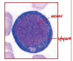
Rubriblast (Pronormoblast)
the earliest recognizable erythroid precursor
12-19 µm (largest)
Rubriblast (Pronormoblast) size
8:1
Rubriblast (Pronormoblast) N:C ratio
Rubriblast (Pronormoblast)
Contains 1 or 2 nucleoli
Large, round nucleus
Rubriblast (Pronormoblast)
Chromatin is purple red and has a fine pattern / open
Rubriblast (Pronormoblast)
Deep blue cytoplasm (due to ribosomes-RNA activity)
without granules
Rubriblast (Pronormoblast)
High RNA activity (needed for production of CHONs required for Hgb synthesis)
Rubriblast (Pronormoblast)
Globin production & Heme synthesis begins
Rubriblast (Pronormoblast)
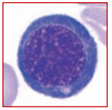
12-17 µm
Prorubricyte (Basophilic Normoblast) size
6:1
Prorubricyte (Basophilic Normoblast) N:C ratio
Prorubricyte (Basophilic Normoblast)
Nucleoli may be present in the early stage but disappears later
Prorubricyte (Basophilic Normoblast)
Nuclear chromatin is deep purple red and begins to condense → “Patially clumped/condensed”
Prorubricyte (Basophilic Normoblast)
Deeper, richer blue cytoplasm = Protein synthesis begins
Prorubricyte (Basophilic Normoblast)
Start of hemoglobin synthesis (Hgb pigmentation not yet evident)
Eosinophila is being masked by the high amount of ribosomal RNA & ribosomes in the cytoplasm
Why is Hgb pigmentation (eosinphilia) not yet evident in Prorubricytes even though Hgb synthesis have alr started?
Prorubricyte has a coarser chromatin and absence of nucleoli
Most helpful criteria in comparing Prorubricyte with rubriblast
Prorubricyte (Basophilic Normoblast)
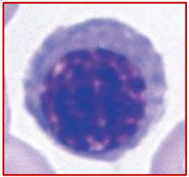
11-15 µm
Rubricyte (Polychromatophilic normoblast) size
4:1
Rubricyte (Polychromatophilic normoblast) N:C ratio
Rubricyte (Polychromatophilic normoblast)
No nucleoli are present
Increased clumping of the chromatin → “Moderately clumped/condensed”
murky gray-blue from Pink (Hgb) + Blue (RNA)
Rubricyte (Polychromatophilic normoblast) cytoplasm color
Rubricyte (Polychromatophilic normoblast)
Hemoglobin synthesis increases (Hgb pigmentation becomes evident)
Rubricyte (Polychromatophilic normoblast)
Last stage capable of mitosis
Progressive ↓ in DNA synthesis
Rubricyte (Polychromatophilic normoblast)
Metarubricyte (Orthochromic normoblast or nucleated RBC/NRBC)
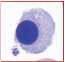
8-12 µm
Metarubricyte (Orthochromic normoblast) size
1:2
Metarubricyte (Orthochromic normoblast) N:C ratio
Metarubricyte (Orthochromic normoblast)
Chromatin pattern is tightly/completely condensed → Pyknotic nucleus
Pyknotic nucleus
dense/compact mass of degenerated chromatin is called _
Metarubricyte (Orthochromic normoblast)
nucleus becomes inactive
synthetic activity and cell division ability is lost
Metarubricyte (Orthochromic normoblast)
where nucleus explusion/extrusion occurs
Metarubricyte (Orthochromic normoblast)
Reddish pink with slightly bluish color of cytplasm
Metarubricyte (Orthochromic normoblast)
Last nucleated stage
Hgb synthesis continues
Metarubricyte (Orthochromic normoblast)
Hgb synthesis is nearing completion
RNA begins to degrade
vimentin
Nucleus expulsion is initiated when _, a protein which hold organelles in proper location in the cytoplasm, is lost by the cell.
myosin
In the pinching process of nucleus expulsion, _ contracts to push the nucleus outside the cell.
Pyrenocyte
pyknotic/enveloped extruded nucleus is called _
Phosphatidylserine (phospholipid)
the pyrenocyte is covered by the cell membrane which contains _ acting as “eat me flag”
Macrophages
bone marrow
_ in the _ engulfs the pyrenocyte
7-10 µm
Reticulocyte (Polychromatophilic erythrocyte) size
No N:C ratio because it is anucleate
Reticulocyte (Polychromatophilic erythrocyte) N:C ratio
Reticulocyte (Polychromatophilic erythrocyte)
Cell is anuclear
Diffuse reticulum
Reticulocyte (Polychromatophilic erythrocyte)
Various degrees of polychromasia (mixed pink and blue staining)
pink is more dominant with bluish tints
Polychromatophelia
Increased Polychromatic erythrocytes in the peripheral blood
Reticulocyte (Polychromatophilic erythrocyte)
End-stage of Hgb synthesis
Reticulocyte (Polychromatophilic erythrocyte)
residual RNA & ribosomes are completely lost at the end of this stage
endoribonuclease
In reticulocytes, _ digests the ribosomes
Diffusely Basophilic Erythrocyte
term used for Polychromatophilic erythrocyte when bluish tint is scattered/spread in the cytoplasm
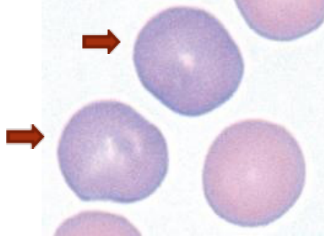
Wright’s stain
Diffusely Basophilic Erythrocyte is demonstrated by _
Reticulocyte
term used for Polychromatophilic erythrocyte when bluish tint appear as small, blue, granulofilamentous strands due to the precipitation of residual RNA with the stain
reticulum
small blue strands found in reticulocytes are called _
Supravital stain
Reticulocytes are demonstrated using _, which stains cells alive
New Methylene Blue (NMB)
Brilliant Cresyl Blue (BCB)
Examples of Supravital Stains
Punctate basophila
occurs when residual RNA appears as distinct blue dots all over the cell’s cytoplasm
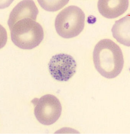
Wright’s stain
Punctate basophila is demonstrated using _
lead poisoning
Punctate basophila is manifested in events like _
1-2 or 2-3 days
Reticulocyte (Polychromatophilic erythrocyte) resides in the BM for __ prior to its release in PB
spleen
After residing in the BM, reticulocytes circulate in the _ to undergo needed changes before their eventual release to the PB
pitting & membrane polishing → BICONCAVE DISCOID MATURE RBC
What happens to the reticulocytes as their circulate in the spleen
1 day
Reticulocyte (Polychromatophilic erythrocyte) stays in the PB for about _ before becoming mature RBC
Erythrocyte
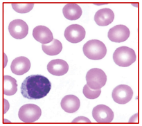
6-8 µm
Erythrocyte size
Erythrocyte
Salmon-pink with central pale area cytoplasm
Erythrocyte
cannot divide (anucleate
No mitochondria
Biconcave disc (discocyte)
Hemoglobin
main component of erythrocytes
oxygen-carrying component
Anaerobic glycolysis (Embden–Meyerhof–Parnas (EMP) Pathway);
plasma
Since erythrocytes have no mitochondria, energy production is done through _, which uses glucose from _