Anatomy CA2
1/58
There's no tags or description
Looks like no tags are added yet.
Name | Mastery | Learn | Test | Matching | Spaced |
|---|
No study sessions yet.
59 Terms
where are the 10 new cranial pairs divided into
the medulla, pons, and the midbrain
What does the brainstem contain
The brainstem contains the 10 true cranial pairs
Where is the Brainstem located
It is located under the cerebrum and is continuos with the spinal cord
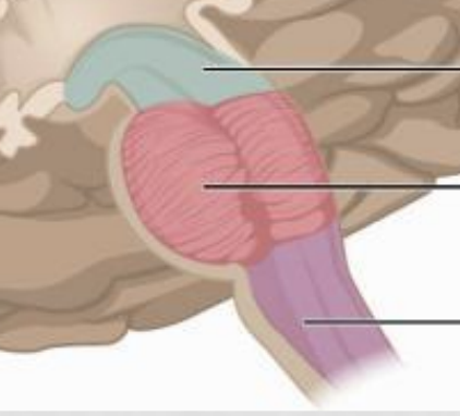
Label the parts
Midbrain
Pons
Medulla
What si the difference in grey matter between the brainstem and spinal cord?
In the brain stem the grey matter is broken up into defined nuclei
What happens to the central canal in the brainstem?
The central canal greatly expands in the brainstem to create a ventricle
Do nerves emerge often in the brainstem?
No nerve emergence doesn’t happen as frequently in the brain stem
Where does the Medulla come from?
The medulla comes from the upper rootlet of the first cervical spinal nerve (C1) to the pons
What nuclei is present in the medulla?
Glossopharyngeal (IX) - Vagus (X) - Accessory (XI) - Hypoglossal (XII)
What does pons look like it is conencting?
The right and left cerebrum hemispheres
What are the nuclei present in pons?
Trigeminal (V) - Adbucens (VI) - Facials (VII) - Vestibulocochlear (VIII)
What is the midbrain?
The midbrain is a short part of the brainstem that connects the Pons to the Cerebrum
Where are the Oculomotor and Trochlear nuclei located at?
In the Midbrain
Where is the fourth canal expanded to?
The central canal is expanded to the fourth ventricle of the brainstem
Where is the fourth ventricle located at
It is located in the upper medulla and pons
Whata are choroid plexus
They are vascular fringes that are projected into the cavity of the fourth ventricle
What is the Posterior wall of the infra temporal fossa?
Styloid apparatus
What is the Anterior wall of the Infra temporal fossa?
Posterior surface of the maxilla
What is the roof of the Infra temporal fossa?
Greater wing of sphenoid
What is the lateral wall of the infra temporal fossa?
ramus of the mandible
What is the Medial wall of the infra temporal fossa?
lateral pterygoid plate
What is the floor of the infra temporal fossa
medial pterygoid muscle
What are the pterygoid muscles that are in the infra temporal fossa
Lateral pterygoid and Medial Pterygoid
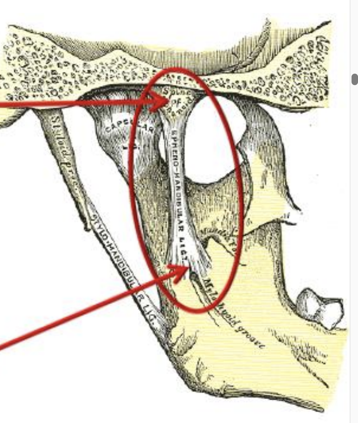
What is this and what are the arrows pointing at
This is the sphenomandibular ligament
1- spine of sphenoid
2- Lingual and adjacent margin of the mandibular foramen
What nerves are in the Anterior trunk of the mandibular nerve?
Buccal nerve
Masseteric nerve
Deep temporal nerve
Nerves of lateral pterygoid
What nerves are in the posterior trunk of the Mandibular nerve
Auriculotemporal nerve
Superficial temporal branches
Lingual nerve
Inferior alveolar nerve and mental branches
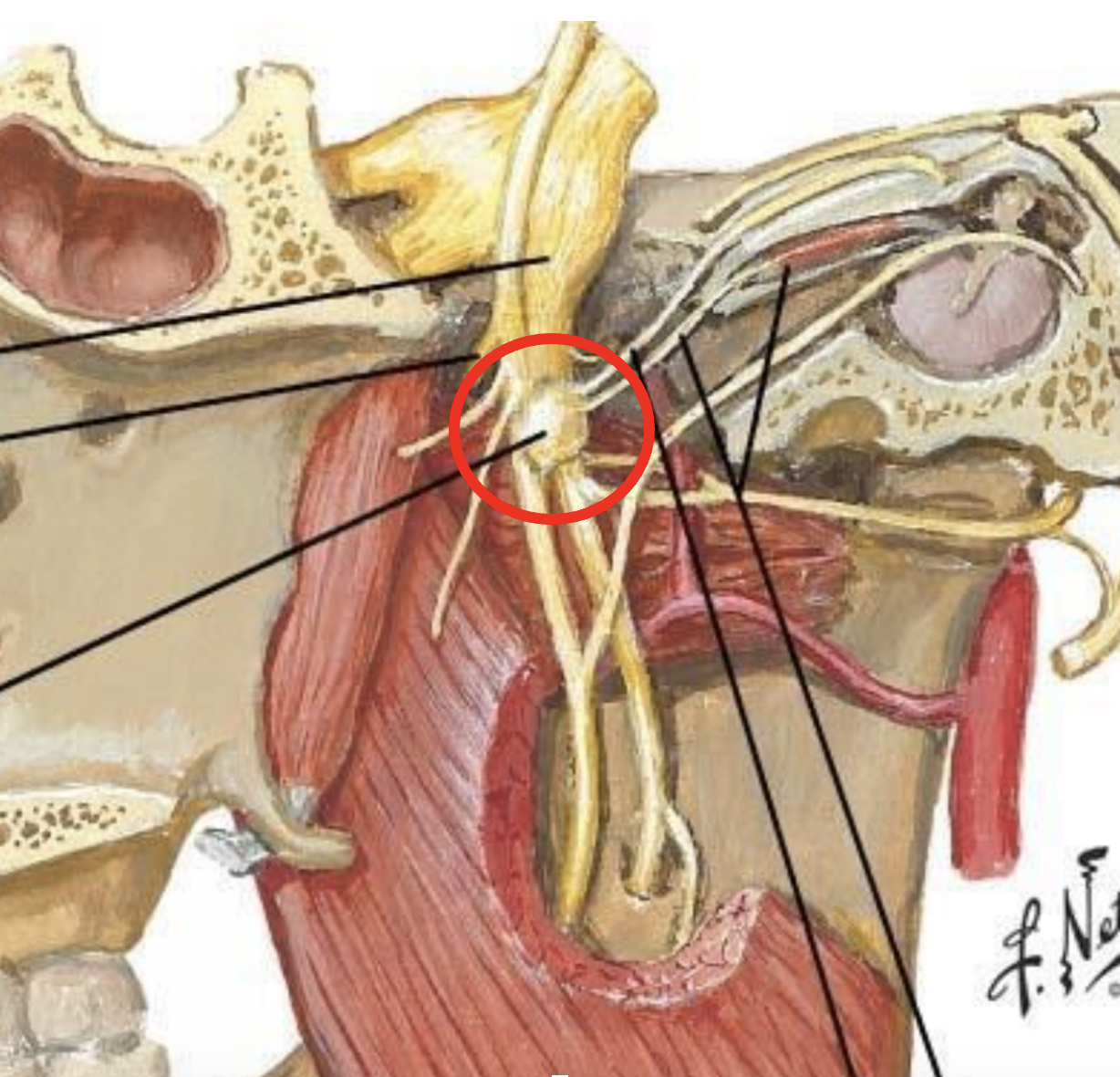
What is the red arrow pointing at
Otic ganglion
What are the 5 arteries and branches in the maxillary artery?
1) Articular and anterior tympanic arteries
2) Middle and accessory maningeal arteries
3) Inferior alveolar artery and mental branch
4) Deep temporal, masseteric, buccal branch
5) Lingual branch
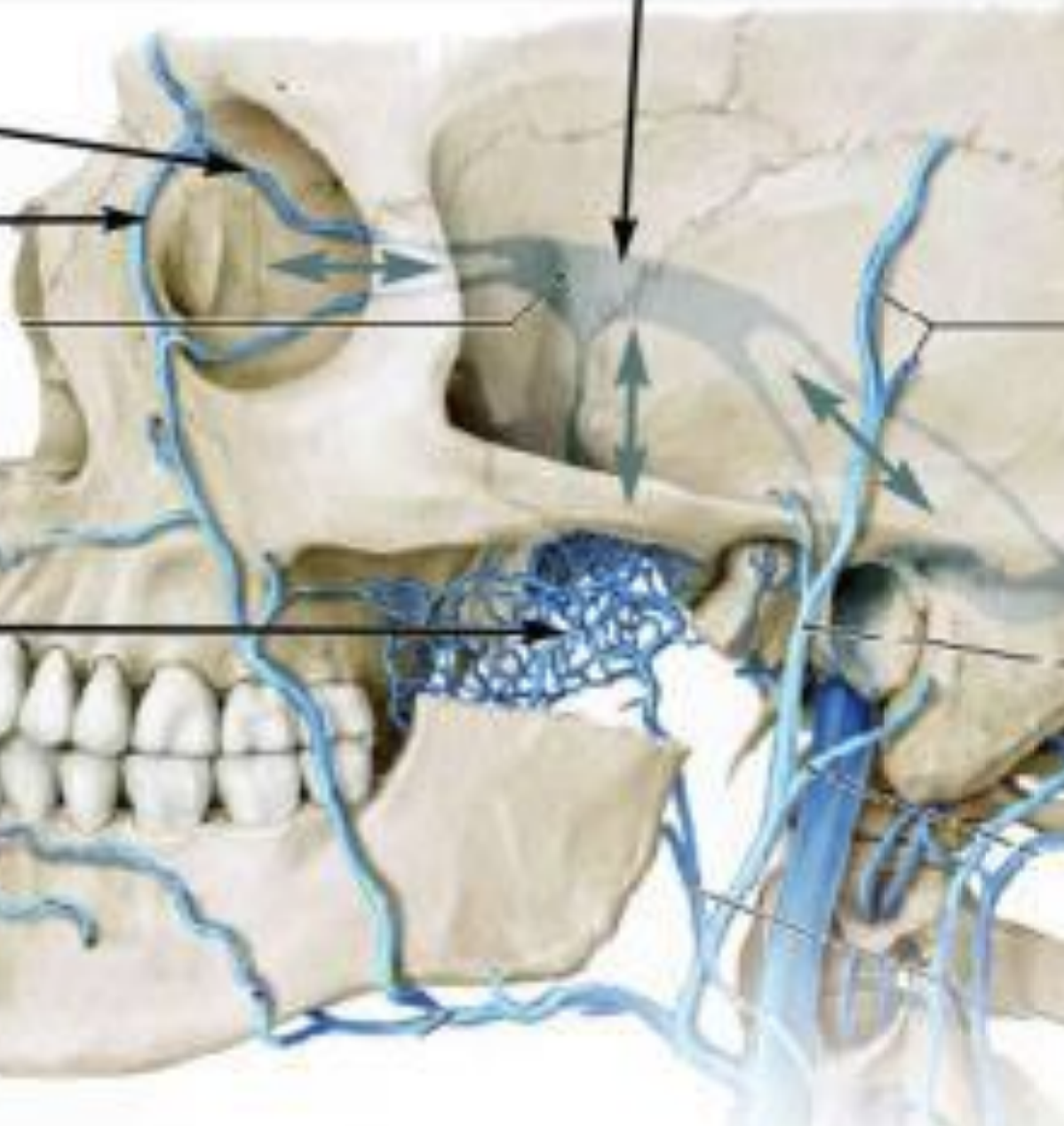
What is this and what are the arrows starting from top to bottom
Pterygoid plexus
1- Cavernous sinus
2- Ophthalmic vein
3- Angular vein
4- Pterygoid plexus
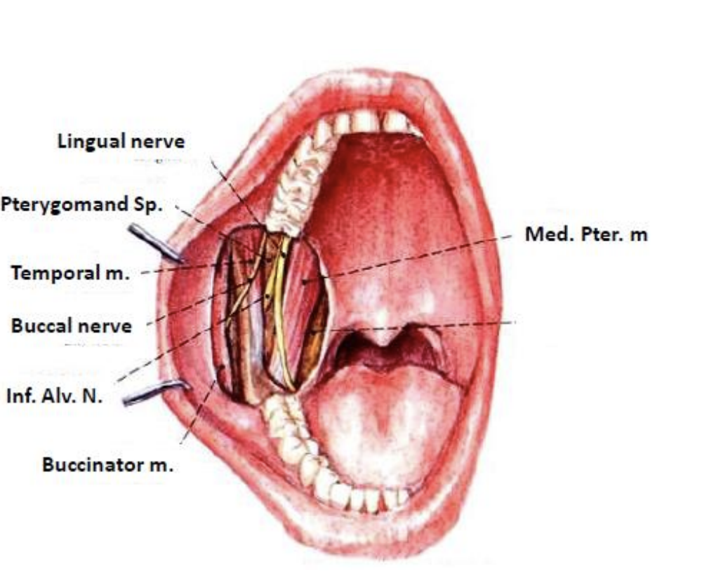
What is this
pterygomandibular space
What are the 3 muscles found in the styloid apparatus?
Stylopharyngeal muscle - styloglossal muscle - stylomandibular muscle
What are the 2 ligaments found in the styloid apparatus
Sylomandibular ligament - sylohoid ligament
What does the pterygomaxillary fissure look like?
An inverted triangle
What are the 4 nerves in the pteygoidpalatine ganglion?
1- greater palatine nerve
2- Lesser palatine nerve
3-nasal nerve
4-nasopalatine nerve
What 4 branches are in the maxillary artery?
Palatine branch, Nasal branch, pharyngeal branch, posterior superior alveolar branch
What artery is in the maxillary artery
The infraorbital artery
What canal is in the Maxillary artery
The artery of the pterygoid canal
What is the anatomical structure of the orbit
The roof, the lateral wall, the medial wall, and the floor
What is in the roof of the orbit
The orbital part of the frontal bone, and the lesser wing of sphenoid
What is in the lateral wall fo the orbit?
The orbital surface of the zygomatic bone, and the greater wing of sphenoid
What is in the medial wall of the orbit
frontal process of the maxilla, the lacrimal bone, orbital plate of ethmoidal laberynth, body of sphenoid
What is in the floor of orbit
orbital plate of palatine bone, and the upper surface of the body of maxilla / orbital surface
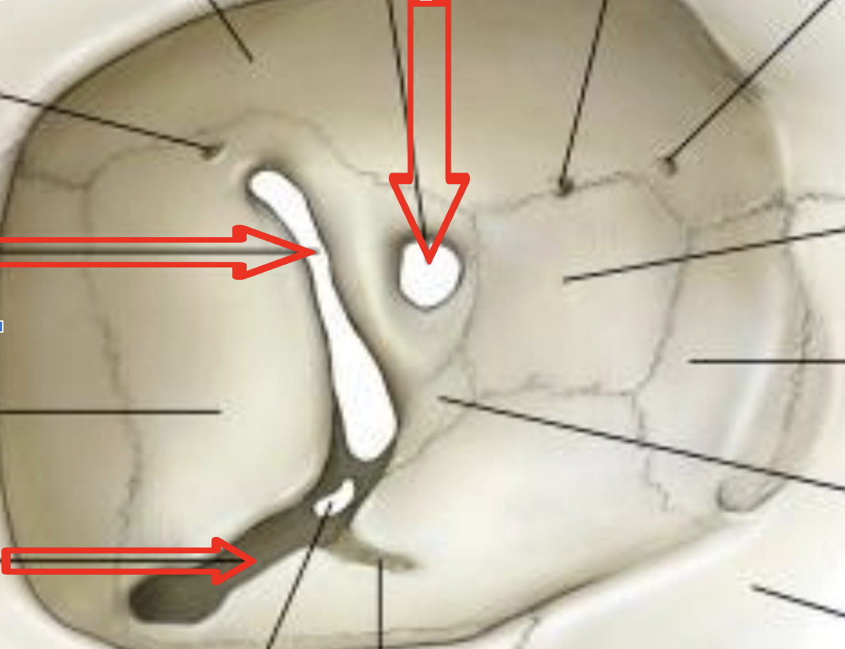
What are the red arrows pointing at from top to bottom
The optic canal
superior orbital
Inferior orbital
What nerve is in the optic canal
Optic nerve (II)
What nerve is present in the inferior orbital fissure
The ophthalmic branch of the trigeminal nerve (V)
What nerves are present in the superior orbital fissure?
The oculomotor nerve (III) and trochlear (IV) nerves
The ophthalmic branch of the trigeminal nerve (V)
Ophthalmic vein
What are the contents of the orbit?
eyeball and optic nerve
The extraocular muscles
vessels and nerves
How is the the eyeball divided into 3 parts
The fibrous layer, the vascular layer, and the inner layer
What is in the fibrous layer
cornea and sclera
What is in the vascular layer
choroid
ciliary body
iris
What is in the inner layer
retina
what is in the extraocular muscle
recti - Superior oblique - inferior oblique - levator plapebrae superioris
What does the recti include of
medial, lateral, inferior, and superior
What movable things protects the eyelid
Movable flaps
What muscles move the upper eyelid?
The levator palpebral superiors
orbicularis oculi
What are the contents of the eye lids
Tarsal plates covered by the skin
Tarsal glands
Conjunctiva
Eyelashes
What is the conjunctiva?
The conjunctiva is a transparent membrane that covers the anterior aspect of the sclera