Exam 2 Study Guide
0.0(0)
Card Sorting
1/131
Last updated 7:36 PM on 10/10/22
Name | Mastery | Learn | Test | Matching | Spaced | Call with Kai |
|---|
No analytics yet
Send a link to your students to track their progress
132 Terms
1
New cards
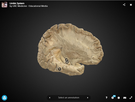
pathway tract
The ________ for visual information starts with the first group of bipolar sensory neurons in the retina (back of eye) Optic nerve exits the posterior of the eye.
2
New cards
Tract
________ is of major importance to speech production.
3
New cards
joint flexes
Rubrospinal: Modulates flexor tone (the amount of tension present in our muscles when ________) in upper extremities.
4
New cards
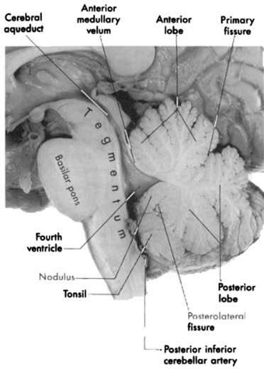
Function and Parts Spinal Cord
5
New cards
Second order neuron arises from the dorsal horn decussates and then travels to the thalamus (which projects third order neuron to somatosensory cortex) These columns consist of two bundles
fasciculus gracilis (slender bundle) and fasciculus cuneatus (wedge-shaped bundle)
6
New cards
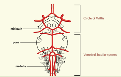
Vascularization
What are the major players in the vascularization
7
New cards
Processed in frontal, parietal and temporal lobe Optic
SA nerve, located at cerebrum not brainstem area, nerve deals with vision
8
New cards
SA nerves, General pathway of cochlear fibers
axons of neurons (bipolar) in cochlea exit as fibers of nerve going to medulla, pass through pons, make synapses in superior olivary complex, and midbrain inferior colliculus, go to thalamus (MGB medial geniculate body) to auditory cortex (temporal lobe)
9
New cards
Gag reflex
IX and X What two go through jugular foramen
10
New cards
Not
P,b, m,n,ng, f,v motor fibers to all muscles of tongue except palatoglossus muscle (Vagus nerve) what is the limbic system known for
11
New cards
Amygdala
what is it
12
New cards
Major landmarks (label and identify motor/sensory fibers that run through there): dorsal ramus, ventral ramus, spinal nerve, dorsal root, ventral root, dorsal horn, ventral horn, anterior median fissure.
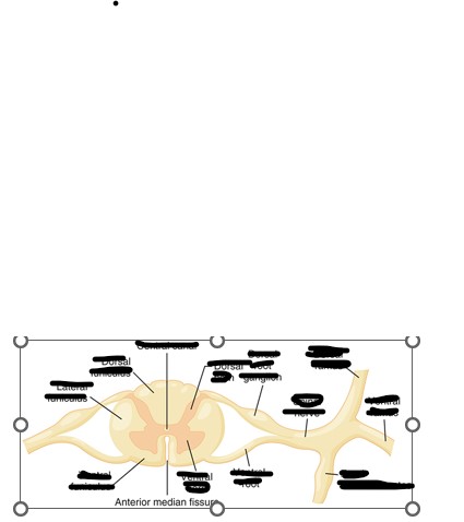
13
New cards
Identify the Four types of fibers
Identify the Four types of fibers Four fiber types:
• GSE fibers: to skeletal muscles
• GVE fibers: to smooth muscle, heart, glands
• GSA fibers: from skin
• GVA fibers: from lungs and digestive tract
• GSE fibers: to skeletal muscles
• GVE fibers: to smooth muscle, heart, glands
• GSA fibers: from skin
• GVA fibers: from lungs and digestive tract
14
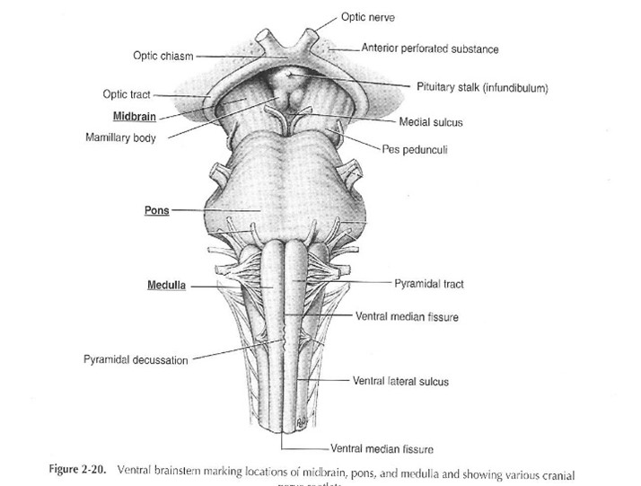
New cards
Where is the pons, medulla, midbrain?
• Midbrain – most superior portion
• Pons – middle portion
• Medulla – lowest portion
• Midbrain – most superior portion
• Pons – middle portion
• Medulla – lowest portion
Where is the pons, medulla, midbrain?
15
New cards
Label Pons Medulla Midbrain on different views
16
New cards
ventral/anterior
What View is this?
17
New cards
Label: superior and inferior colliculus, middle, inferior, superior peduncles, pineal body, dorsal median sulcus.
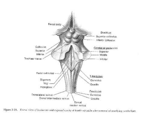
18
New cards
Functions of the medulla
• Hosting many cranial nerve nuclei
• Autonomic nervous system nuclei like respiratory and swallowing centers
• Place for crossing and decussating many motor tracts and sensory tracts
• reflexes like coughing, vomiting, gag (nucleus Salitarius) and swallowing (Nucleus ambiguous)
• Autonomic nervous system nuclei like respiratory and swallowing centers
• Place for crossing and decussating many motor tracts and sensory tracts
• reflexes like coughing, vomiting, gag (nucleus Salitarius) and swallowing (Nucleus ambiguous)
19
New cards
Functions of Pons:
• Bridge for tracts coming from cortex
• Hosting cranial nerve nuclei CN V, VI, VII, VIII.
• Hosting cranial nerve nuclei CN V, VI, VII, VIII.
20
New cards
Location of Cerebral Peduncles and Inferior Superior Colliculus
midbrain
21
New cards
The tectum contains what?
• Tectum – superior (vision) and inferior (hearing) colliculi
• Cerebral peduncles
• Cerebral peduncles
22
New cards
Which Cranial Nerves are in the midbrain
CN III, CN IV, CN VI
23
New cards
What is the substantia nigra? Where is it located? What is its function? What is it involved in?
• Substantia nigra: located in midbrain, function: produces dopamine which helps in motor movement and coordination. Involved in Parkinson’s disease.
24
New cards
What is the tegmentum?
• tegmentum is a general area within the brainstem. It is located between the ventricular system and basal / ventral structures at each level of the brainstem. It forms the floor of the midbrain whereas the tectum forms the ceiling. It is a multisynaptic network of neurons that is involved in many unconscious homeostatic and reflexive pathways
25
New cards
Label parts of the brain lateral view:
26
New cards
What are the major players in the vascularization
• Heart
• Aorta
• Carotid Artery System
• Vertebral/Basilar Artery System
• Aorta
• Carotid Artery System
• Vertebral/Basilar Artery System
27
New cards
There are two main arteries that come of the aortic arc what are they? What do they supply blood to?
External common carotid artery: the muscles of the face, eyes, oral cavity internal: goes toward the brain
Left subclavian artery: supplies oxygenated blood to the brainstem
Left subclavian artery: supplies oxygenated blood to the brainstem
28
New cards
What are the three main arteries that supply blood to the midbrain pons or medulla?
MIDBRAIN
(posterior cerebral artery)
PONS
(basilar artery)
MEDULLA
(vertebral artery)
(posterior cerebral artery)
PONS
(basilar artery)
MEDULLA
(vertebral artery)
29
New cards
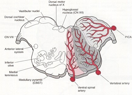
Label: vertebral, basilar, posterior spinal artery, superior cerebral artery, posterior inferior cerebellar artery (PICA), anterior inferior cerebellar artery (AICA), anterior spinal artery, middle cerebellar artery, posterior communicating artery, anterior communicating artery, anterior cerebellar artery.
30
New cards
List of arteries and where they supply blood
Basilar artery bifurcates and becomes posterior cerebral arteries. Feeds midbrain,
Internal carotid arteries have two main/major branches middle cerebral artery (provides blood for lateral surfaces of brain in frontal lobe and parietal lobe, temporal lobe and to language areas) and ACA anterior cerebral artery
Pica aica: cerebellum
• Internal Carotid (R/L)
– Anterior Cerebral Artery
• Medial zones of the frontal and parietal lobes
– Middle Cerebral Artery
– MCA: lateral aspects of frontal and parietal lobes
• Lateral zone of the hemisphere
Major artery supplying the midbrain is the Posterior Cerebral Artery
Posterior Cerebral Arteries (R/L) arise from the Basilar Artery
• Medial and lateral zone of the occipital lobe
• Medial and inferior lateral zone of temporal lobe
31
New cards
what two parts of the brain make up the diencephalon?
Thalamus and hypothalamus
32
New cards
Basal Ganglia parts (three of them)
caudate nucleus putamen and globus pallidus
33
New cards
The circle of willis arteries supply what?
the diencephalon and the basal ganglia
34
New cards
Label the areas of innervation in this image
35
New cards
which two arteries join the circle of willis?
• Internal carotid and basilar artery join the Circle of Willis
36
New cards
what is included in the circle of willis?
Posterior Communicating Artery, Internal carotid, anterior communicating artery, anterior cerebral artery, posterior cerebral artery,
37
New cards
what is not included in the circle of willis?
basilar artery, middle cerebral artery, superior cerebellar artery, vertebral artery
38
New cards
What is the venous drainage system?
• Consists of veins and dural sinuses
• Cerebral veins: deep and superficial empty deoxygenated blood into the sinuses
• Sinuses collect all the blood and direct it to the internal jugular veins which then take the blood back to the heart
• Cerebral veins: deep and superficial empty deoxygenated blood into the sinuses
• Sinuses collect all the blood and direct it to the internal jugular veins which then take the blood back to the heart
39
New cards
• They can be categorized or separated in terms of function, area of service, and type of fiber. Define each
o Function includes the distinction of general or specific
o area of service is divided into visceral and somatic
o Type of fiber indicates whether the nerve is a motor (efferent) and sensory (afferent).
o area of service is divided into visceral and somatic
o Type of fiber indicates whether the nerve is a motor (efferent) and sensory (afferent).
40
New cards
What are the twelve cranial nerves?
The twelve cranial nerves include olfactory (CN I), optic (CN II), oclomotor (CN III), trochlear (CN IV), trigeminal (CN V), abducens (CN VI), facial (CN VII), vestibulocochlear (CN VIII), glossopharyngeal (CN IX), vagus (CN X), accessory (CN XI), hypoglossal (CN XII).
41
New cards
What are the nerves involved in speech and hearing?
• CN- V (Trigeminal)
• CN- VI (Facial)
• CN- VIII (Vestibulocochlear)
• CN- IX (Glossopharyngeal)
• CN- X (Vagus)
• CN- X (Spinal Accessory)
• CN- XII (Hypoglossal
42
New cards
What are the sensory afferent nerves?
1 (olfactory),2 (optic),8 (vestibulocochlear)
43
New cards
Where is the first cranial nerve located? what type of fibers carry the information? what is the function?
Olfactory: CN I is at cerebrum, has sensory fibers that brings sensory information (smell) to the brain.
44
New cards
What is the second cranial nerve? where is it located? what does it do?
Optic: SA nerve, located at cerebrum not brainstem area, nerve deals with vision.
45
New cards
Describe the tract/pathway of the optic nerve
The pathway/tract for visual information starts with the first group of bipolar sensory neurons in the retina (back of eye) Optic nerve exits the posterior of the eye. Fibers decussate at the optic chiasm. then the fibers go to the superior colliculus (in midbrain) and some goes to the lateral geniculate body (nucleus) located in thalamus. Then they go to primary visual cortex area which is the occipital lobe.
46
New cards
What is the pathway for the olfactory nerve?
First group of neurons in nasal muscousa in nose and transfer to second group (located in olfactory bulbs) then to the cortex (not stopping by thalamus because that wouldn’t make sense because they would have to go back to the thalamus and back up to the cortex). Processed in frontal, parietal and temporal lobe
47
New cards
Where is the oculomotor nerve located?
Oculomotor: located between pons and midbrain
48
New cards
What are the functions of the 3rd cranial nerve?
All other muscles (superior rectus, medial rectus, inferior rectus, inferior oblique): innervated by oculomotor (CN III)
constriction of pupil - GVE
Holding eyelid open – GSE
constriction of pupil - GVE
Holding eyelid open – GSE
49
New cards
What are some problems involving the cranial nerve 3?
Ptosis: eyelid drooping
Strabismus: cross eyed:
tumor, stroke, brain injury can all impact eye movement
Diplopia: double vision
Strabismus: cross eyed:
tumor, stroke, brain injury can all impact eye movement
Diplopia: double vision
50
New cards
What is the 4th cranial nerve? where is it located? what is its functions?
Trochlear: Trochlear nerve in midbrain.
Superior oblique: moves eyes down innervated by (CN IV)
Smallest nerve (number of axons)
Greatest intracranial length
Moving the eyes downward
only cranial nerve that exits from dorsal aspect of brainstem
Superior oblique: moves eyes down innervated by (CN IV)
Smallest nerve (number of axons)
Greatest intracranial length
Moving the eyes downward
only cranial nerve that exits from dorsal aspect of brainstem
51
New cards
What is the 5th cranial nerve? where is it located? what are its branches? what type of fiber are each of these?
Trigeminal: At level of pons, opthalamic and maxillary branches are sensory, mandibular is sensory and motor. Biggest nerve among cranial nerves, general nerve, efferent and afferent functions (GVE< GVA< GSA< GSE)
B1: opthalamic: provides sensory information for forehead, eyelids, nose
B2: maxillary: sensory, provides for nose, lower eye, upper jaw/teeth
B3: mandibular sensory and motor, muscles of chewing (masseter, temporalis, anterior belly of digastricus, pterygoid branch (one of them)), controls tongue area, opening jaw.
Foramen that it passes through (middle fossa): superior (orbital fissure) foramen rodunume, foramen ovale (mandibular)
52
New cards
what are the three foramen that the branches pass through? which is for which branch?
Foramen that it passes through (middle fossa): superior orbital fissure (opthalamic), foramen rodunume(maxillary), foramen ovale (mandibular)
53
New cards
Function of the trigeminal nerve
GSA: touch, pain, temp. and vibration for face, mouth, ant. 2/3 of tongue
GVE: Chewing muscles
GVE: Chewing muscles
54
New cards
Problems with trigeminal nerve:
loss of above sensations, and difficulty chewing
55
New cards
which nerve controls the anterior 2/3 of the tongue in terms of sensory information?
Cranial Nerve V
56
New cards
The anterior 2/3 for taste is controlled by____
cranial nerve VII
57
New cards
Posterior 1/3 of tongue for sensory information AND taste is controlled by ______
the glossopharyngeal nerve
58
New cards
what type of nerve is the abducens? what does it control? problems that may occur?
motor nerve GSE nerve
Controls movement of single muscle
lateral rectus muscle of eye.
Problem(s) = eye rotates in (strabismus) and diplopia
Controls movement of single muscle
lateral rectus muscle of eye.
Problem(s) = eye rotates in (strabismus) and diplopia
59
New cards
what type of fibers make up the facial nerve?
Facial: Mostly motor but some sensory nerves for taste.
Parasympathetic proprioceptive sensors for tears.
GVE: parasympathetic innervation to lacrimal, submandibular and sublingual glands.
SVA: taste anterior 2/3 of tongue
Parasympathetic proprioceptive sensors for tears.
GVE: parasympathetic innervation to lacrimal, submandibular and sublingual glands.
SVA: taste anterior 2/3 of tongue
60
New cards
where is the nuclei for the facial nerve located?
Nuclei located in pons, fibers exit at pontomedullary junction (between pons and medulla)
Innervating facial muscles (speech production and swallowing)
Innervating facial muscles (speech production and swallowing)
61
New cards
What is the function of the facial nerve?
muscles of facial expression
Taste anterior two-thirds of tongue
Taste anterior two-thirds of tongue
62
New cards
Problems that occur with facial nerve
Bell’s Palsy
Problem(s) = Facial paralysis/paresis; taste loss
Consequences for speech: weak articulation
Consequences for swallowing: drooling
Problem(s) = Facial paralysis/paresis; taste loss
Consequences for speech: weak articulation
Consequences for swallowing: drooling
63
New cards
what is the 8th cranial nerve? location? function?
Sound and equilibrium
Vestibular (SSA)
Cochlear
Hearing and balance
Tinnitus or ringing
Located from pons and medulla
Vestibular (SSA)
Cochlear
Hearing and balance
Tinnitus or ringing
Located from pons and medulla
64
New cards
what are the two groups of fibers that the vestibulocochlear nerve is broken up into?
2 groups of fibers: cochlear and vestibular
65
New cards
What two nerves go through internal auditory meatus?
CN VII and CN VIII go through this foramen.
66
New cards
General pathways of cochlear fibers
General pathway of cochlear fibers: axons of neurons (bipolar) in cochlea exit as fibers of nerve going to medulla, pass through pons, make synapses in superior olivary complex, and midbrain inferior colliculus, go to thalamus (MGB medial geniculate body) to auditory cortex (temporal lobe).
67
New cards
Function of cranial nerve IX
receives sensory from (GSA)
posterior one-third of tongue
Tonsils
Pharynx
middle ear
supplies parasympathetic fibers to
Gag reflex (GVA)
stylopharyngeus muscle (Elevating larynx)
GSE and GVE nerve (sensory and motor fibers)
posterior one-third of tongue
Tonsils
Pharynx
middle ear
supplies parasympathetic fibers to
Gag reflex (GVA)
stylopharyngeus muscle (Elevating larynx)
GSE and GVE nerve (sensory and motor fibers)
68
New cards
what two nerves trigger the gag reflex?
IX and X
69
New cards
the jugular foramen houses what two nerves?
Jugular foramen: IX and X
70
New cards
Branches of the X cranial nerve
Pharyngeal branches:
• Superior, middle and inferior pharyngeal constrictors
• Levator veli palatini
Laryngeal branches
Muscles of the larynx
Sensory above and below larynx
Starts at sides of medulla, sensory and motor fibers,
If pharyngeal branch is affected they will have issues swallowing
levator veli palatini (elevates soft palate) to block VP port so that we don’t have nasal speech in sounds that don’t need to be. For nasal sounds soft palate needs to be lowered.
Larynx branch
Cricoarytenoid, thyrovocalis and thyromuscularis
Extrinsic muscles: up and down movement mylohyoid, diagstrics, geniohyoid
Intrinsic muscles: moving cartilages of larynx,
71
New cards
Functions of Cranial Nerve X and issues
Issues: paralysis of vocal folds: breathiness, cant breathe, cant change pitch (cricothyroid)
Sensory above and below vocal folds supraglottic and subglottic: if this doesn’t work properly you would have silent aspiration (can cause pneumonia)
VP closure, voice production, swallowing (pharyngeal phase)
Gag reflex
pain from the pharynx, larynx, trachea, esophagus, abdominal muscles
Sensory above and below vocal folds supraglottic and subglottic: if this doesn’t work properly you would have silent aspiration (can cause pneumonia)
VP closure, voice production, swallowing (pharyngeal phase)
Gag reflex
pain from the pharynx, larynx, trachea, esophagus, abdominal muscles
72
New cards
Accessory Nerve Function, Location, Type of Fiber
ACCESSORY
provides motor innervation
sternocleidomastoid muscle
upper part of trapezius muscle
GSE: motor
Nuclei located in medulla, innervation for sternocleidomastoid and trapezius muscle (accessory in respiration especially expiration).
Jugular foramen
provides motor innervation
sternocleidomastoid muscle
upper part of trapezius muscle
GSE: motor
Nuclei located in medulla, innervation for sternocleidomastoid and trapezius muscle (accessory in respiration especially expiration).
Jugular foramen
73
New cards
Hypoglossal Nerve, fiber type, issues, what is the exception of tongue muscles that this innervates?
Hypoglossal:
Motor nerve GSE intrinsic and extrinsic of tongue
Issues: articulation of lingual sounds, formation of bolus, transition of bolus during swallowing
bilabial, labial not affected
Hypoglossal canal
Idea about someone who has tumor on hypoglossal nerve, what speech sounds are involved? What are not?
Not: P,b, m,n,ng, f,v
motor fibers to all muscles of tongue except palatoglossus muscle (Vagus nerve)
74
New cards
what is the limbic system known for?
Limbic system = the brain’s emotional system
75
New cards
What is the acronym that helps you remember the functions of the limbic system
• H = Homeostasis
• O = Olfaction: (not involved is the thalamus)
• M = Memory
• E = Emotion
• O = Olfaction: (not involved is the thalamus)
• M = Memory
• E = Emotion
76
New cards
Cingulate Cortex: also known as, what is it? where is it?
• Also known as cingulate gyrus
• Arch-shaped band of cortical tissue between the corpus callosum and the lobes of the brain
• Functionally, helps identify negative emotions
• Arch-shaped band of cortical tissue between the corpus callosum and the lobes of the brain
• Functionally, helps identify negative emotions
77
New cards
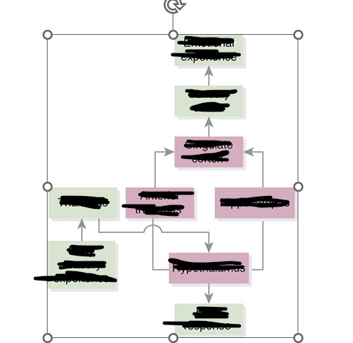
James Papez (1883–1958) proposed the Papez circuit, made up of the following structures:
• Limbic Lobe (Cingulate cortex +Uncus+ parahippocampal gyrus)
• Hippocampus (memory)
• Hypothalamus
• Amygdale
• Anterior Thalamic Nuclei
• Brainstem (reticular formation)
• Hippocampus (memory)
• Hypothalamus
• Amygdale
• Anterior Thalamic Nuclei
• Brainstem (reticular formation)
78
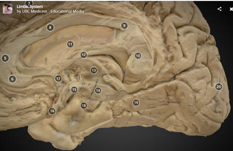
New cards
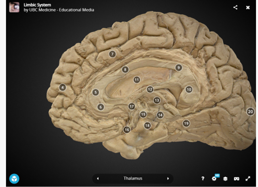
Label the above on this diagram:
Limbic Lobe (Cingulate cortex +Uncus+ parahippocampal gyrus)
• Hippocampus (memory)
• Hypothalamus
• Amygdale
• Anterior Thalamic Nuclei
• Brainstem (reticular formation)
Limbic Lobe (Cingulate cortex +Uncus+ parahippocampal gyrus)
• Hippocampus (memory)
• Hypothalamus
• Amygdale
• Anterior Thalamic Nuclei
• Brainstem (reticular formation)
79
New cards
Limbic Lobe (Cingulate cortex +Uncus+ parahippocampal gyrus)
• Hippocampus (memory)
• Hypothalamus
• Amygdale
• Anterior Thalamic Nuclei
• Brainstem (reticular formation)
• Hippocampus (memory)
• Hypothalamus
• Amygdale
• Anterior Thalamic Nuclei
• Brainstem (reticular formation)
80
New cards
Limbic Lobe (Cingulate cortex +Uncus+ parahippocampal gyrus)
• Hippocampus (memory)
• Hypothalamus
• Amygdale
• Anterior Thalamic Nuclei
• Brainstem (reticular formation)
• Hippocampus (memory)
• Hypothalamus
• Amygdale
• Anterior Thalamic Nuclei
• Brainstem (reticular formation)
81
New cards
Label the Papez Circuit
82
New cards
Amygdala: what is it? Where is it?
• What is it?
– Nuclear mass
• Where is it?
– Buried in the white matter of the temporal lobe, in front of the hippocampus
– Nuclear mass
• Where is it?
– Buried in the white matter of the temporal lobe, in front of the hippocampus
83
New cards
what part of the limbic system in our ability to preserve ourselves and respond to danger
amygdala
84
New cards
• Specific behaviors the amygdala regulates:
Feeding and drinking
– Fighting
– Mating
– Providing maternal care
– Responding to physical/environmental stress
– The brain’s shortcut for fear and self-preservation
– Fighting
– Mating
– Providing maternal care
– Responding to physical/environmental stress
– The brain’s shortcut for fear and self-preservation
85
New cards
Damage to the amygdala can cause the following:
– Flattened emotions
– Abnormal fear
– Aggression
– Anxiety
• Dysfunction may be connected to depression, anxiety, and posttraumatic stress disorder (PTSD)
– Abnormal fear
– Aggression
– Anxiety
• Dysfunction may be connected to depression, anxiety, and posttraumatic stress disorder (PTSD)
86
New cards
Damage to the cingulate cortex results in:
– Decreased social behavior
– Reduced time spent with others
– Decreased vocalizations
– Reduced time spent with others
– Decreased vocalizations
87
New cards
Limbic connection
• There are numerous connections between the prefrontal cortex and the limbic system.
• Cingulate cortex does appear to play a role in cognition.
– Attention
– Theory of Mind (ToM): the ability to understand that I have a mind, you have a mind, and that our minds are different from one another
• Cingulate cortex does appear to play a role in cognition.
– Attention
– Theory of Mind (ToM): the ability to understand that I have a mind, you have a mind, and that our minds are different from one another
88
New cards
Hypothalamus: what is it? where is it? what does it do?
Hypothalamus:
• What is it?
– A deep brain structure made up of a number of nuclei
• Where is it?
– Base of the fore brain
– Just above the brainstem
– Forms part of the walls of the 3rd ventricle
• What does it do?
– Linker : nervous system to endocrine system
– Regulator : Circadian cycles
* Sexual response
* Temp, hunger, thirst, anger, and fatigue
89
New cards
What shape is the thalamus?
almond shape
90
New cards
WHAT GOES INTO THE THALAMUS?***
• Processing station in the center of the brain
– All sensory systems (except olfaction)
– Motor inputs from cerebellum and basal ganglia
– Limbic inputs
– Blood supply
– Strokes lead to (Thalamic syndrome)
– Damage leads to coma, excessive daytime sleepiness
– Thalamic aphasia (Fluent, intact repetiotin)
– Sensory processing difficulties
– All sensory systems (except olfaction)
– Motor inputs from cerebellum and basal ganglia
– Limbic inputs
– Blood supply
– Strokes lead to (Thalamic syndrome)
– Damage leads to coma, excessive daytime sleepiness
– Thalamic aphasia (Fluent, intact repetiotin)
– Sensory processing difficulties
91
New cards
Function of Brainstem
Brainstem: recticular formation
• Receives hypothalamic and cortical output
– separate descending projections that run parallel to volitional motor system
• Output to somatic and autonomic effector systems
– cardiac, respiratory, bowels, bladder
– Coordinates brain-body response
• Receives hypothalamic and cortical output
– separate descending projections that run parallel to volitional motor system
• Output to somatic and autonomic effector systems
– cardiac, respiratory, bowels, bladder
– Coordinates brain-body response
92
New cards
Match the Cranial Nerve and its Function
A. innervating muscles of chewing
B. levator veli palatini
C. transverse muscles of the tongue
A. innervating muscles of chewing
B. levator veli palatini
C. transverse muscles of the tongue
a. trigeminal
b. vagus
c. hypoglossal
b. vagus
c. hypoglossal
93
New cards
Cranial Nerves ____ and ____ pass through the internal foramen
VII and VIII
94
New cards
which cranial nerve is not SA
a. II
b. I
c. VIII
d. V
a. II
b. I
c. VIII
d. V
Cranial Nerve V
95
New cards
What are the two branches of CN VIII
Vestibular and Cochlear
96
New cards
Which cranial nerve is responsible for mediating taste sensation to the posterior 1/3 of the tongue?
hypoglossal (CN IX)
97
New cards
. Which cranial nerve gives rise to the recurrent laryngeal nerve that innervates the larynx?
Vagus Nerve
98
New cards
What two (2) cranial nerves are involved with the “gag-reflex”?
X and XI
99
New cards
Which cranial nerve innervates motor functions to the intrinsic muscles of the tongue?
Hypoglossal (CN XII)
100
New cards
A lesion of CN-________ could result in deviation of the jaw to the same side of the lesion.
Trigeminal