Canine Ear Anatomy and Physiology: External, Middle, and Inner Ear Structure
1/167
There's no tags or description
Looks like no tags are added yet.
Name | Mastery | Learn | Test | Matching | Spaced |
|---|
No study sessions yet.
168 Terms
What is the primary structure of the canine ear responsible for sound localization?
The pinna
What are the two main surfaces of the pinna?
Concave (rostrolaterally facing) and convex (caudomedially facing)
What is the cutaneous marginal pouch?
A pouch located on the caudal margin of the pinna with no obvious function
What is the scapha in the context of the canine ear?
The thin, flat, pendulous portion of the pinna
What is the apex of the pinna?
The tip of the scapha
What type of cartilage forms the pinna?
Elastic auricular cartilage
What glands are present in the skin of the pinna?
Apocrine sweat glands, sebaceous glands, and hair follicles
Which surface of the pinna has more hair follicles?
The convex surface
What are the two main groups of muscles that move the pinna?
Rostroauricular muscles and caudoauricular muscles
What is the function of the auditory ossicles?
To transmit and amplify air vibrations from the tympanic membrane to the inner ear
What are the components of the middle ear?
Tympanic cavity, three auditory ossicles, and tympanic membrane
What is the tympanic membrane divided into?
Pars flaccida and pars tensa
What is the role of the auditory tube?
To connect the nasopharynx to the tympanic cavity
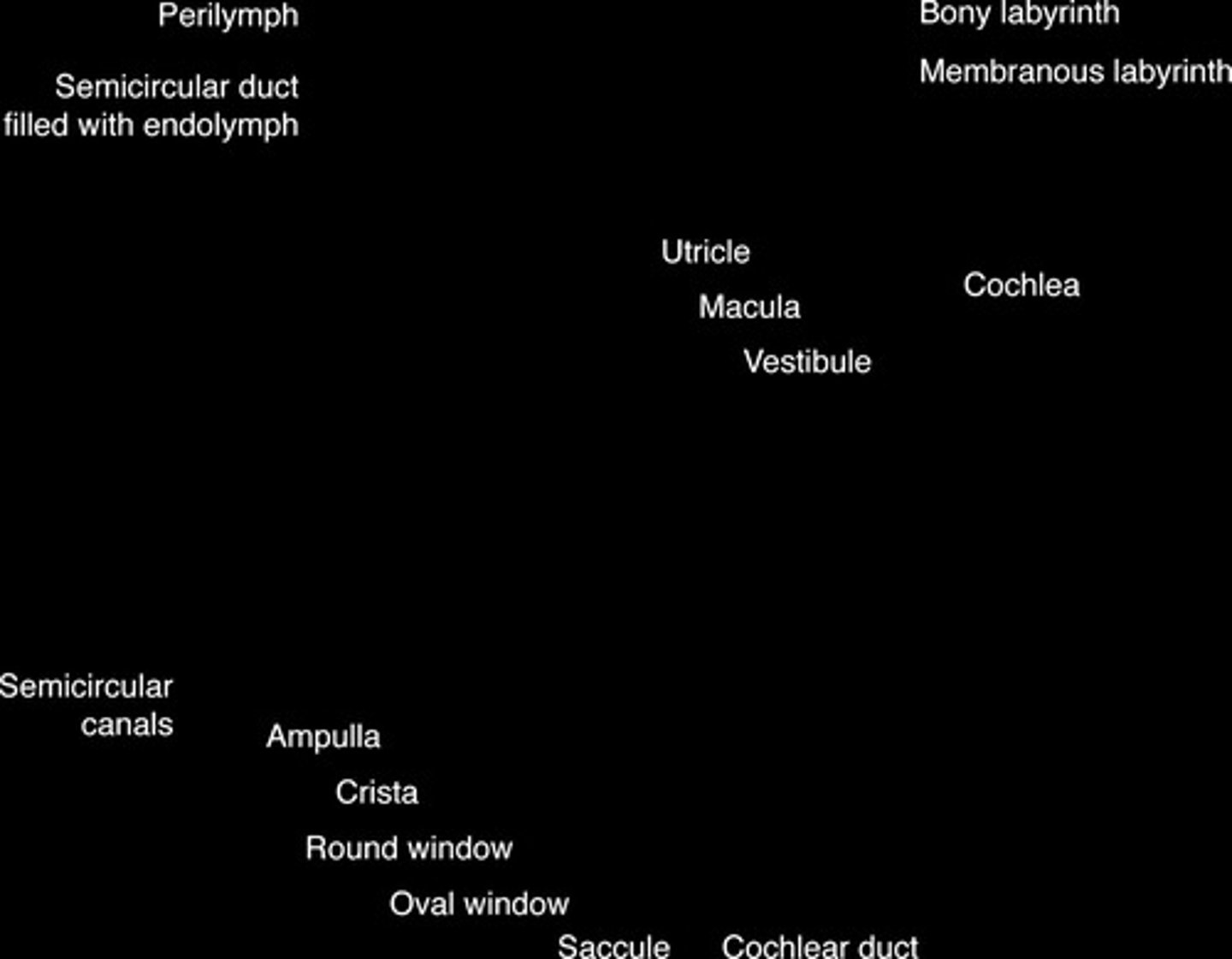
What is the bony labyrinth?
The structure in the inner ear that houses the membranous labyrinth and sensory organs for hearing and balance
What is the significance of the cochlear (round) window?
It is covered by a thin membrane and located in the promontory of the tympanic cavity
What factors may influence the occurrence of otitis externa in dogs?
Pinnal conformation, hair in the ear canal, temperature, and humidity
Which dog breeds are more likely to have otic infections?
Pure-bred dogs with pendulous pinnae and hirsute ear canals, such as cocker spaniels and poodles
What is the cavum conchae?
The groove bounded by the anthelix, tragus, and antitragus that continues into the external auditory meatus
What is the role of hairs in the external ear canal?
To decrease in number from distal to proximal and serve as landmarks for locating the tympanic membrane
What is the typical flora of the external ear canal?
Bacterial organisms and yeast
What is the difference in hair follicle types between cocker spaniels and greyhounds?
Cocker spaniels have excessive compound hair follicles, while greyhounds have sparsely distributed simple hair follicles
What is the function of the pinna in sound transmission?
To localize and collect sound waves and transmit them to the tympanic membrane
What anatomical feature separates the vertical and horizontal ear canals?
A prominent cartilaginous ridge
What is the function of the scutiform cartilage?
It is an L-shaped structure located in the rostroauricular muscles, but does not contribute to the external ear or its canal
What is the significance of the tympanic bulla?
It is part of the tympanic cavity and plays a role in sound transmission
What is the relationship between ear canal temperature and otitis externa?
Dogs with and without otitis externa show no differences in ear canal temperature
What are common organisms cultured from the external ears of healthy dogs?
Staphylococcus, Bacillus sp., yeast, Escherichia coli, Corynebacterium sp., and Micrococcus sp.
Which organisms are most commonly isolated from dogs with chronic otitis externa?
Staphylococcus (pseud)intermedius and Pseudomonas aeruginosa.
What is cerumen and what does it consist of?
Cerumen is an emulsion that coats the ear canal, composed of desquamated keratinized squamous epithelial cells and secretions from sebaceous and ceruminous glands.
How does the lipid content of cerumen differ in otitic ears compared to healthy ears?
The lipid content of cerumen from otitic ears is lower than that of healthy ears.
What is the function of sebaceous glands in the external ear canal?
Sebaceous glands secrete lipids that contribute to the composition of cerumen.
What are the two types of glands found in the external ear canal?
Sebaceous glands and ceruminous glands.
What anatomical structures form the external ear canal?
The external ear canal is composed of auricular and annular cartilage.
What is the role of ceruminous glands?
Ceruminous glands secrete an acidic material that lowers the pH of cerumen, creating an unfavorable medium for bacterial growth.
What is the pH range of the ear canal epithelium in dogs with normal ears?
The pH ranges from 4.6 to 7.2.
What anatomical feature separates the external ear canal from the middle ear?
The tympanic membrane.
What are the two sections of the tympanic membrane?
The pars flaccida and the pars tensa.
What histological changes occur in the external ear canal of dogs with otitis externa?
Epidermal and follicular hyperplasia, dermal inflammation, less active sebaceous glands, and dilated ceruminous glands.
What breeds of dogs are prone to otitis externa?
Cocker spaniels, springer spaniels, and Labrador retrievers.
What is the significance of the anthelix in the ear canal anatomy?
The anthelix is a low transverse ridge present on the medial wall of the ear canal.
What is the function of epithelial migration in the ear?
Epithelial migration helps in the self-cleaning function of the external ear.
What is the mean pH of the ear canal in male and female dogs with normal ears?
The mean pH is 6.1 in male dogs and 6.2 in female dogs.
How does the presence of ceruminous glands change in the ear canal of dogs with otitis?
Ceruminous gland hyperplasia and ectasia may occur, particularly in certain breeds.
What is the structure of the tympanic membrane?
It is a semitransparent three-layer membrane that separates the external ear canal from the middle ear.
What anatomical feature is located opposite the anthelix in the ear canal?
The tragus.
What is the role of the intertragic incisure?
It separates the tragus from the antitragus.
What are the auditory ossicles?
They are three small bones in the middle ear that transmit sound vibrations.
What is the primary component of cerumen in healthy ears?
Neutral lipids secreted by sebaceous glands.
What is the effect of instilling acetic acid in the ear canal?
It lowers the pH of the ear canal temporarily.
What is the anatomical position of the external ear canal opening?
The opening faces dorsolaterally.
What is the histological composition of the pars tensa?
It is thin, dense, contains more collagen than elastin fibers, and is keratinized.
What types of animals have been studied for epithelial migration?
Human beings, guinea pigs, and gerbils.
How are epithelial migratory patterns in the tympanic membrane studied?
By placing ink drops on the tympanic membrane and recording their movement.
What are the three types of epithelial migratory patterns identified in humans?
Two radial patterns migrating away from the umbo and one straight pattern migrating away from the manubrium of the malleus.
What is the effect of poor blood supply on epithelial migration in the tympanic membrane?
It results in a slowing of the epithelial migratory rate and abnormal migratory patterns.
What does a hyperinflated tympanic membrane indicate in humans?
Increased pressure in the middle ear.
What condition is indicated by a bulging pars flaccida in Cavalier King Charles spaniels?
Primary secretory otitis media (PSOM).
What is the pars tensa?
The thin, tough, gray structure of the tympanic membrane that contains the manubrium of the malleus.
What is the umbo?
The point of greatest depression in the tympanic membrane, opposite the distal end of the manubrium.
How long does it take for a normal tympanic membrane to regenerate after being ruptured?
Complete healing occurs between 21 and 35 days.
What is the germination center in humans believed to be?
The umbo.
What are the three parts of the tympanic cavity?
The epitympanic recess, the tympanic cavity proper, and the ventral cavity.
What is the average volume of the middle ear cavity in mesaticephalic dogs?
1.5 mL.
What types of epithelium line the tympanic cavity?
Simple squamous epithelium or simple cuboidal epithelium.
What gases are typically found in the middle ear?
Nitrogen (83.2%), oxygen (12.1%), and carbon dioxide (4.7%).
What are the three auditory ossicles?
The malleus, incus, and stapes.
What is the vestibular (oval) window?
An opening covered by a thin diaphragm where the footplate of the stapes attaches.
What is the role of the tensor tympani muscle?
To contract and make the tympanic membrane more taut, protecting the middle ear.
What is the primary function of the stapedius muscle?
To reduce the movement of the stapes and protect the middle ear from loud noises.
What is the bulla septum?
An elliptical opening in the dorsal wall of the ventral cavity that communicates with the tympanic cavity proper.
What is the significance of the promontory in the tympanic cavity?
It houses the cochlea and lies opposite the tympanic membrane.
What is the function of the cochlear (round) window?
To dissipate vibratory energy of the perilymph in the scala tympani of the cochlea.
What is the role of the Eustachian tube?
To equalize pressure in the middle ear and is opened by the contraction of specific muscles.
What types of microorganisms are part of the normal flora in the middle ear?
Low numbers of yeast and bacteria including E. coli, Staphylococcus sp., and Streptococcus sp.
What are the most common organisms isolated from dogs with infectious otitis media?
Yeast, Staphylococcus (pseud)intermedius, Pseudomonas aeruginosa, Proteus sp., beta-Streptococcus, Corynebacterium sp., and Enterococcus sp.
What is impedance audiometry used for?
It is an objective means of assessing the integrity of the middle ear, consisting of tympanometry and the acoustic reflex.
What does tympanometry measure?
It provides an indirect measurement of the pressure in the middle ear and compliance of the tympanic membrane, and can also measure external ear canal volume.
What might an abnormal tympanogram indicate?
It may indicate tympanic membrane rupture or middle ear effusion.
What is the purpose of the acoustic reflex?
To protect the middle ear from damaging levels of noise by contracting the stapedius muscle in response to loud sounds.
What does an absent acoustic reflex suggest?
An absent reflex in the presence of normal hearing and a normal tympanogram suggests retrocochlear disease.
What is the auditory tube and its function?
A canal extending from the nasopharynx to the tympanic cavity, it functions to equalize pressure on both sides of the tympanic membrane.
What are the three portions of the auditory tube?
Cartilaginous (proximal), junctional (connecting part), and osseous (distal).
What structures are housed in the inner ear?
The bony labyrinth, which contains the vestibule, semicircular canals, and cochlea.
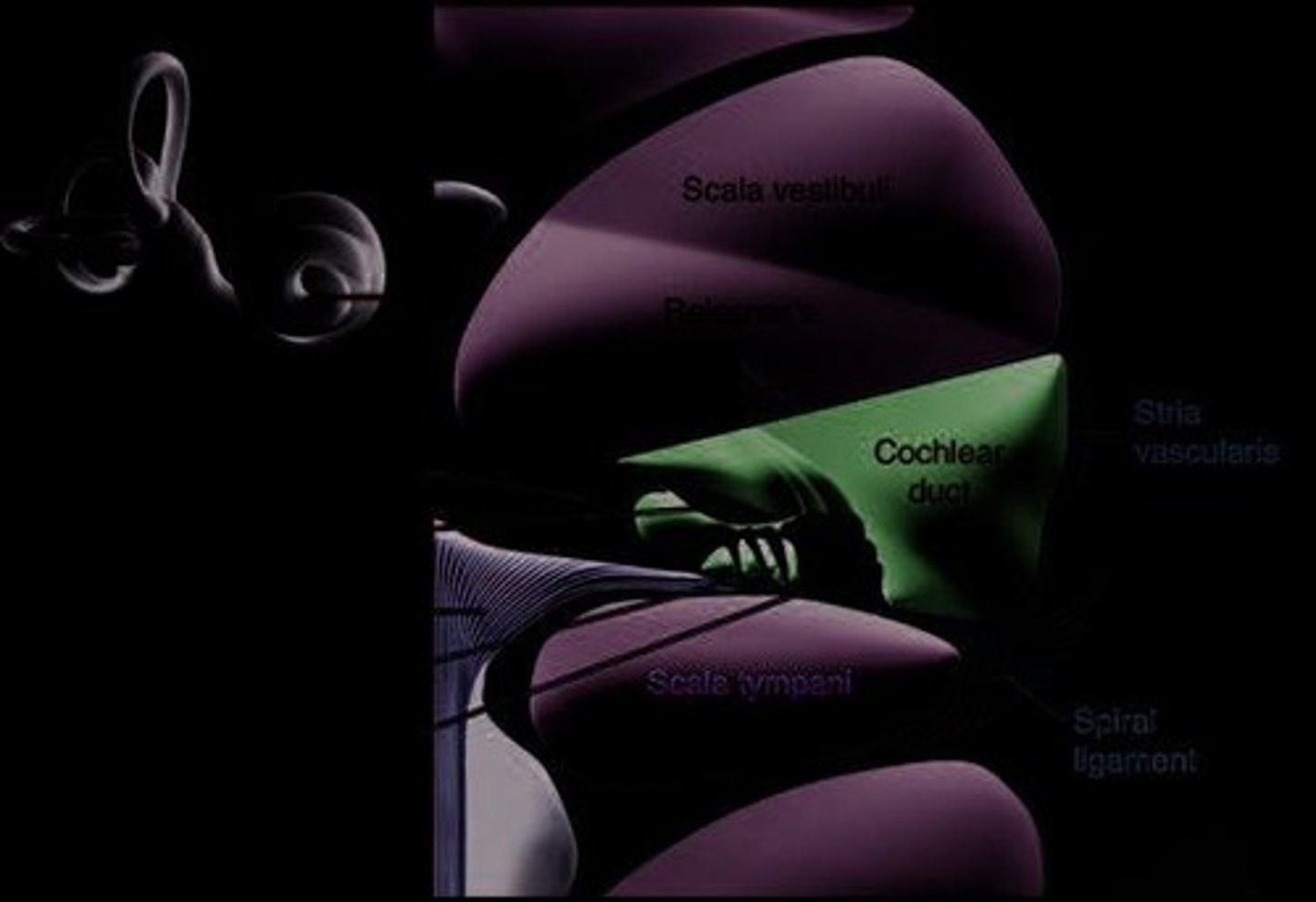
What is the role of the cochlear duct?
It is the auditory portion of the labyrinth, containing the organ of Corti, which is involved in hearing.
What are the three semicircular ducts responsible for?
They contain hair cells that detect acceleration of the endolymph caused by head rotation.
What are maculae and where are they located?
Receptors present in the utricle and saccule that contain hair cells responding to linear acceleration.
What is the function of the bony labyrinth?
It surrounds the membranous labyrinth and protects the sensory organs that control hearing and balance.
What is the organ of Corti?
A structure within the cochlear duct composed of supporting cells and hair cells, crucial for sound transduction.
How do sound waves travel from the external ear to the cochlea?
Sound waves collected by the pinna and external ear canal cause vibrations of the tympanic membrane, which are transmitted through the ossicles to the vestibular window.
What is the role of the tympanic membrane?
It vibrates in response to sound waves, transmitting these vibrations to the ossicles.
What arteries supply blood to the tympanic membrane?
The deep auricular and anterior tympanic branches of the maxillary artery (intrinsic) and the stylomastoid branch of the posterior auricular artery (extrinsic).
What is the vestibule in the inner ear?
An irregular oval space that communicates with the cochlea and semicircular canals, containing structures that help with balance.
What are the two windows located in the lateral wall of the vestibule?
The vestibular (oval) window and the cochlear (round) window.
What is the function of the hair cells in the organ of Corti?
They detect sound vibrations and convert them into electrical signals for transmission to the brain.
What is the role of the endolymph in the inner ear?
It surrounds the sensory cells in the membranous labyrinth and is involved in the detection of sound and balance.
What anatomical feature protects the structures within the osseous labyrinth?
The bony labyrinth itself, which encases the membranous labyrinth.
What is the significance of the tympanic membrane's blood supply?
It ensures the health and function of the membrane, which is crucial for hearing.
What arteries supply blood to the ear?
The external carotid artery and vertebral arteries.
What is the role of the deep auricular artery?
It forms a network in the periosteum of the manubrium supplying blood centrally and extends into the fibrous stratum of the tympanic membrane.
What is a potential complication during a myringotomy in dogs?
Significant bleeding due to venous variations in the tympanic membrane.