Unit 6 - Early Demise & Other Structural Anomalies
1/55
There's no tags or description
Looks like no tags are added yet.
Name | Mastery | Learn | Test | Matching | Spaced |
|---|
No study sessions yet.
56 Terms
What is the sonographic appearance of fetal early demise?
No fetal heart motion
Irregular shaped sac
Degenerative trophoblastic tissue
Subchorionic hemorrhage
What are the indications of fetal early demise?
Discrepancies in dates and size
Distortion of sac shape or contour
Abnormal decidual reaction
Abnormal growth in serial scans
Abnormal yolk sac
Early Demise indications: what is considered normal & abnormal growth in serial scans?
Normal sac grows 1.1 mm per day
<0.6 mm per day abnormal for sac growth
Normal embryo grows 1-2 mm per day
Early Demise indications: what is considered an abnormal yolk sac?
Yolk sac >5.6 mm between 5-10 weeks
Yolk sac <2 mm between 8-12 weeks
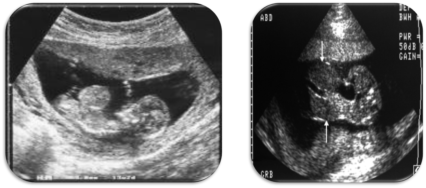
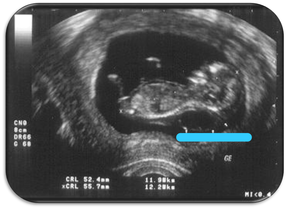
Characteristics that describe the Nuchal Translucency?
11-13w6d
CRL- 45-84mm
Midsagittal plane
Magnification
Neutral position
On-to-on measurement
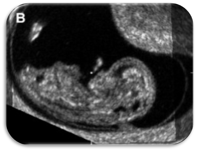
Characteristics that describe the Cardiac Activity in a fetus?
Late 1st Trimester
4 chamber heart
Ectopia cordis
Limb-body-wall complex
What is Ectopia Cordis?
Heart is found outside of the chest
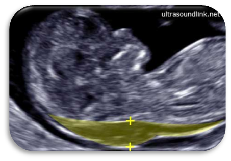
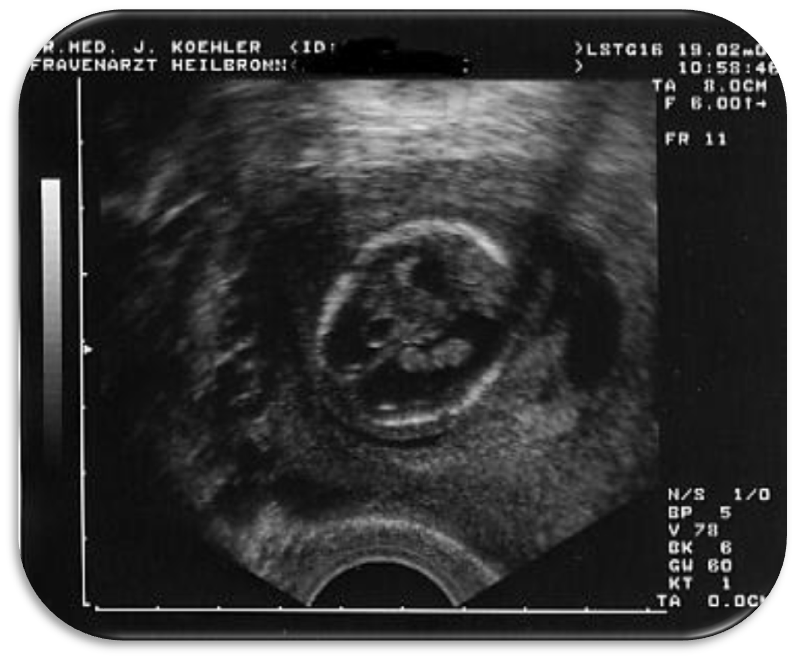
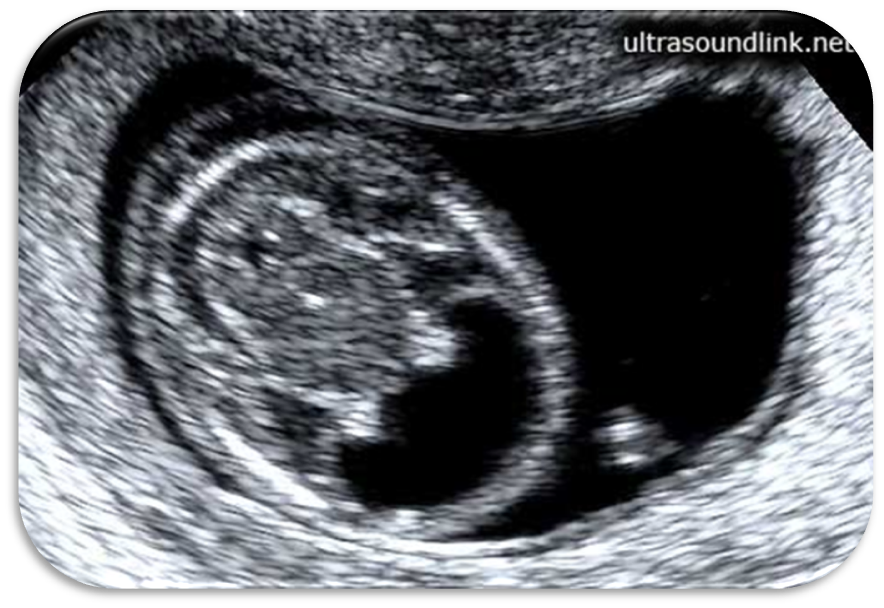
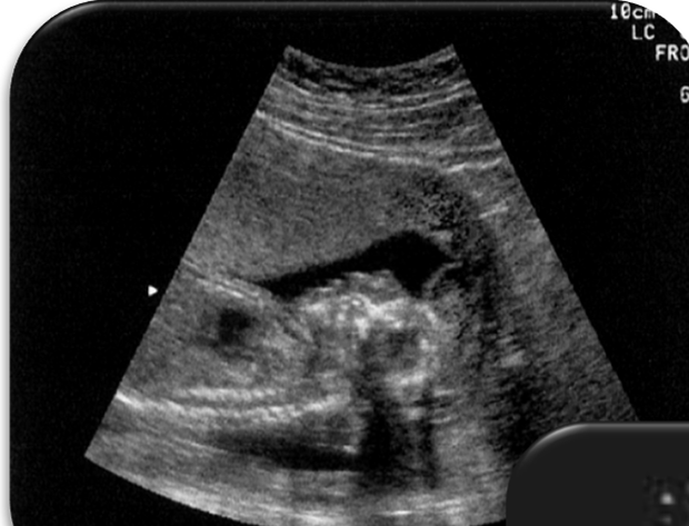
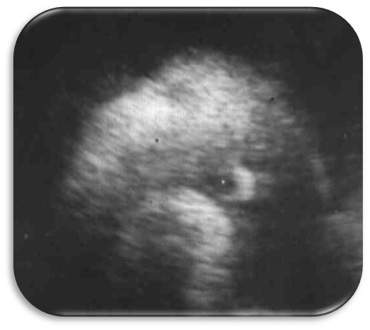
What is this US image?
Nuchal Translucency
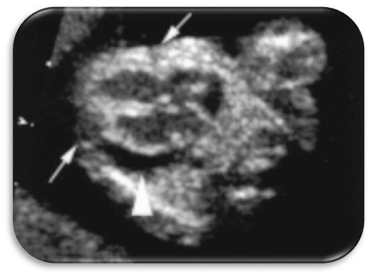
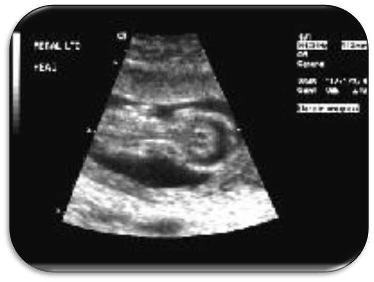
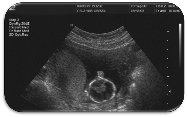
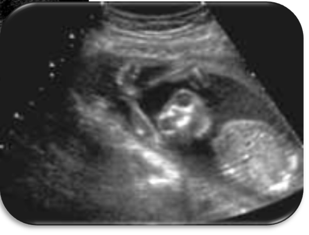
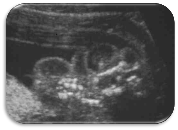
What is this US image?
Fetal Cardiac Heart
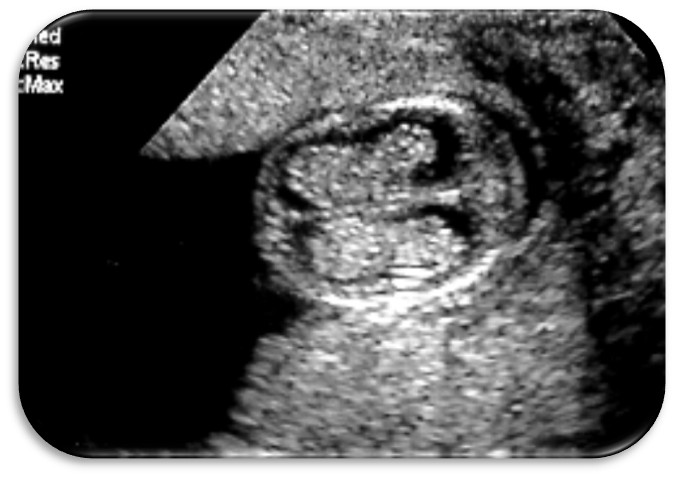
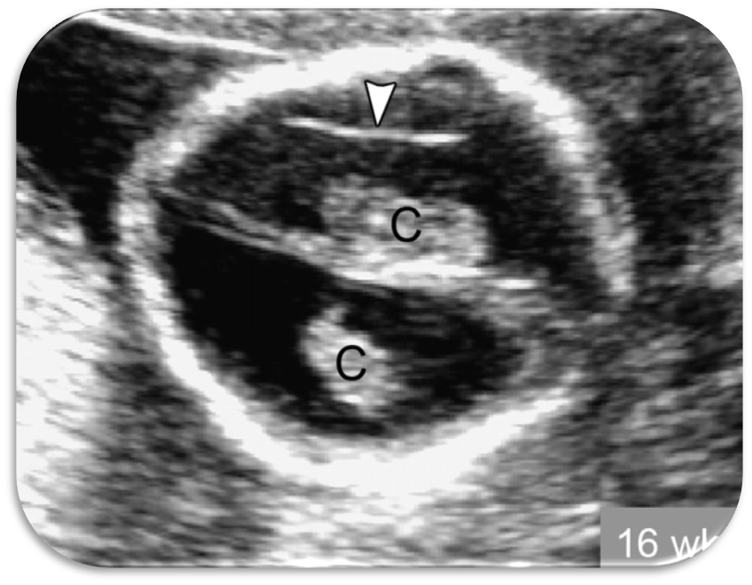
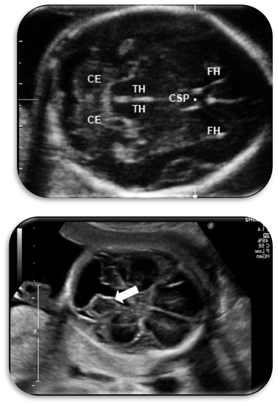
What is this US image?
Choroid Plexus
These structures fill the lateral ventricles.
Choroid Plexus
What is this US image?
Hydrocephalus
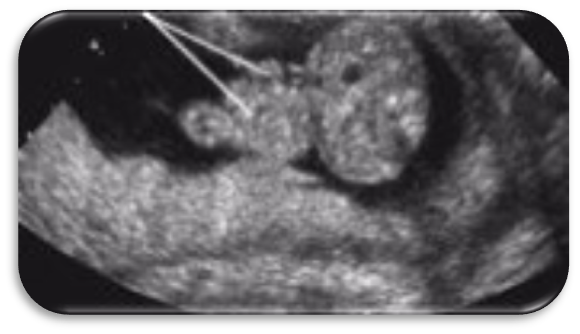
What is this US image?
Hydrocephalus
What is Hydrocephalus?
Fluid in the brain, blockage in the ventricular system
What is Hydranencephaly?
Brain necrosis from occlusion of internal carotid arteries
Characteristics of Hydranencephaly?
Blockage
Tissue shrinks & stops growing
No blood flow in the brain
What is the sonographic appearance of Hydranencephaly?
Loss of all intracranial anatomy
What is this US image?
Hydranencephaly
What is this US image?
Hydranencephaly
What is this US image?
Hydranencephaly
Imaged later in pregnancy
What is Anencephaly?
Absence of the brain tissue, cranium did not ossify
Cranial ossification is not complete yet
Repeat US at 12-14 wks
Amniotic fluid can _______ the remaining brain tissue that is affected by Anencephaly.
Degenerate
What is this US image?
Anencephaly
What is this US image?
Anencephaly
What is the difference between Anencephaly & Acrania?
Acrania ~ Still have brain tissue, just no cranium
Anencephaly ~ Skull and brain tissue are absence
What are these US images showing?
Acrania
What is this US image?
Acrania
What is Iniencephaly?
Defect of occipital bone at foramen magnum
Retroflexion of the head {posterior}
Open spinal defects
What is this US image?
Iniencephaly
What is this US image?
Iniencephaly
What is Dandy Walker Malformation?
4th ventricle enlargement
Agenesis or dysgenesis of cerebellar vermis
Often accompanied with enlarged lateral ventricles
What are these US images showing?
Dandy Walker Malformation
Bowel Herniation characteristics?
Echogenic mass at base of umbilical cord
Normal in 1st trimester
Out and back into abdomen 8-12 weeks
What is a Gastroschisis?
Bowel and other organs protruding outside the abdomen through an open [has a NO skin covering]
Always found to the right side of the umbilical cord
Bowel extends separately from umbilical cord
What is this US image?
Gastroschisis
Gastroschisis & Bowel-Only Omphaloceles are both associated with _________ _______.
Chromosomal disorders
What is an Omphaloceles?
Congenital hernia of the umbilicus, covered by a membrane
What are these US image?
Omphaloceles
Characteristics describing Obstructive uropathy?
Enlarged bladder into abdomen
Sono @ 10-12 weeks
What is this US images?
Normal fetal bladder
What is this US image?
Enlarged fetal bladder
What is Exstrophy?
Blood is located outside of the abdomen cavity
What is Bladder Outlet Obstruction [BOO]?
Blockage at the level of the urethra
What is Megacystitis?
Enlarged Bladder
Cloacal anomalies are associated with the…
Urinary System
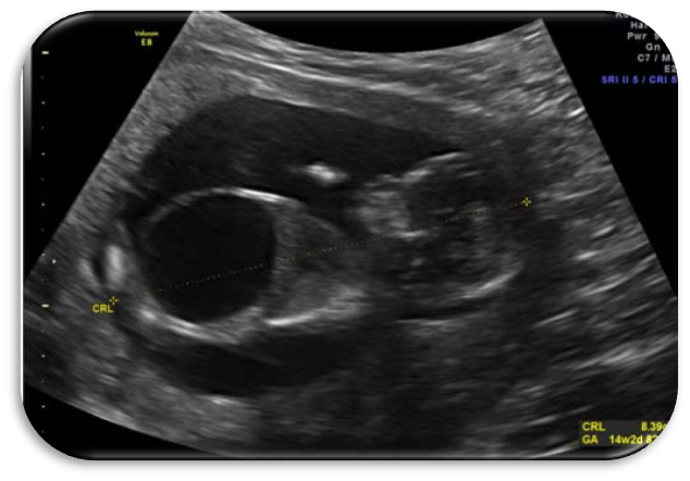
Cystic Hygroma is associated with…
Chromosome abnormalities, esp. in the 1st trimester [13, 18, 21]
What is Cystic Hygroma?
Cystic mass on the baby’s neck
If not resolved by 18 weeks, bad
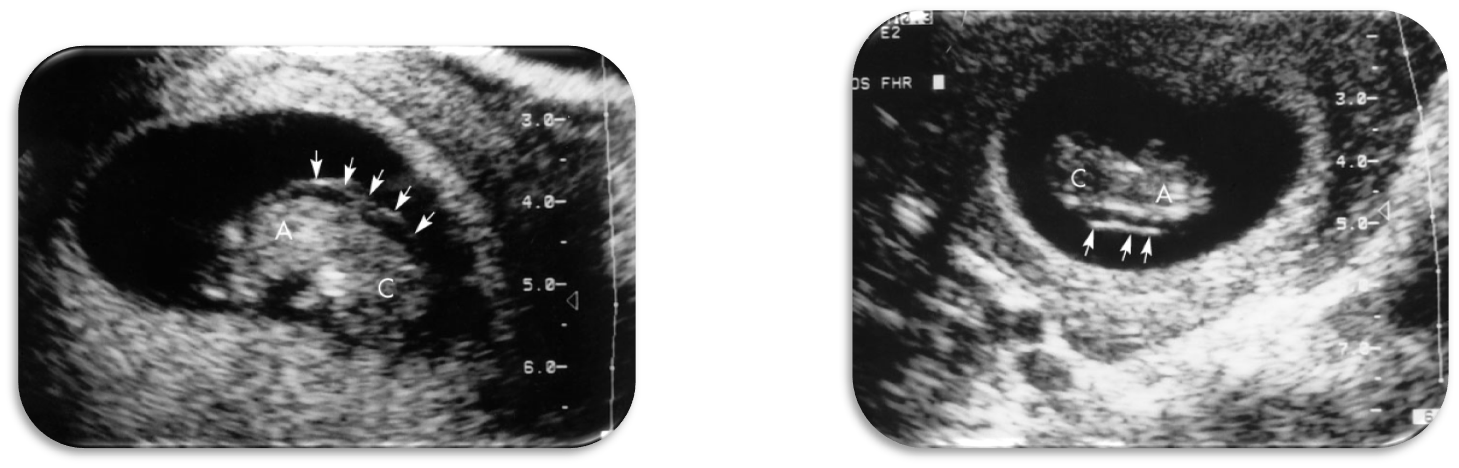
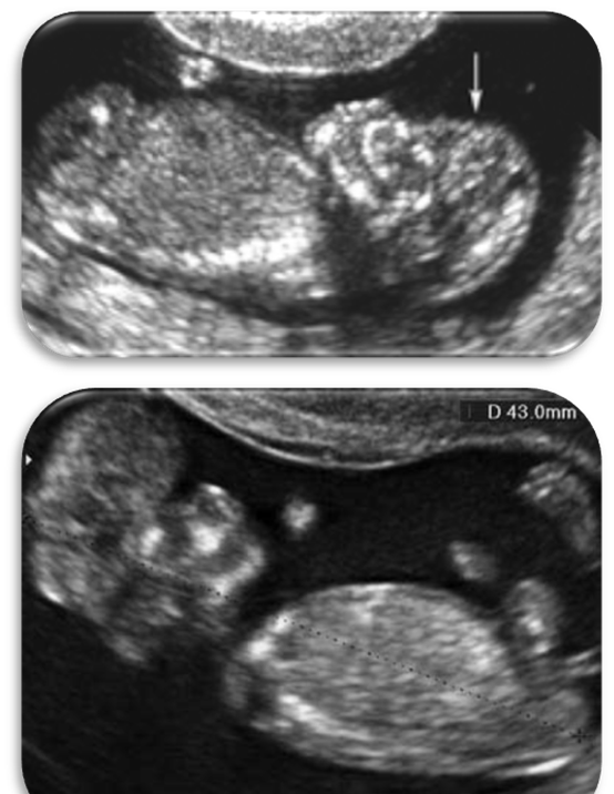
What are these US images?
Cystic Hygroma
What is the sonographic appearance of Cystic Hygroma?
Nuchal thickening
“Hammocking” effect
Embryo lying on amniotic membrane
Pseudomembrane gives false impression
What is this US image?
Cystic Hygroma
What are 1st Trimester Umbilical Cord Cysts?
Cysts within the cord
2mm- 7.5mm
What are the differential diagnoses for Umbilical Cord Cysts?
Amniotic inclusion cysts
Omphalomesenteric duct cysts
Allantoic cysts
Vascular anomalies
Neoplasms
Wharton’s jelly abnormalities
What is the prognosis for Umbilical Cord Cysts?
If resolve by 2nd trimester = normal delivery
IF PERSISTENT in 2nd trimester + associated with other abnormalities = further studies and genetic evaluation
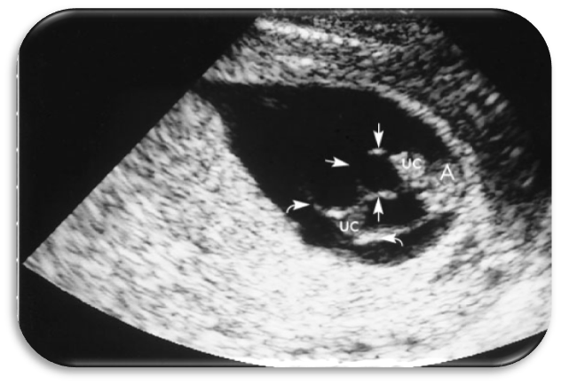
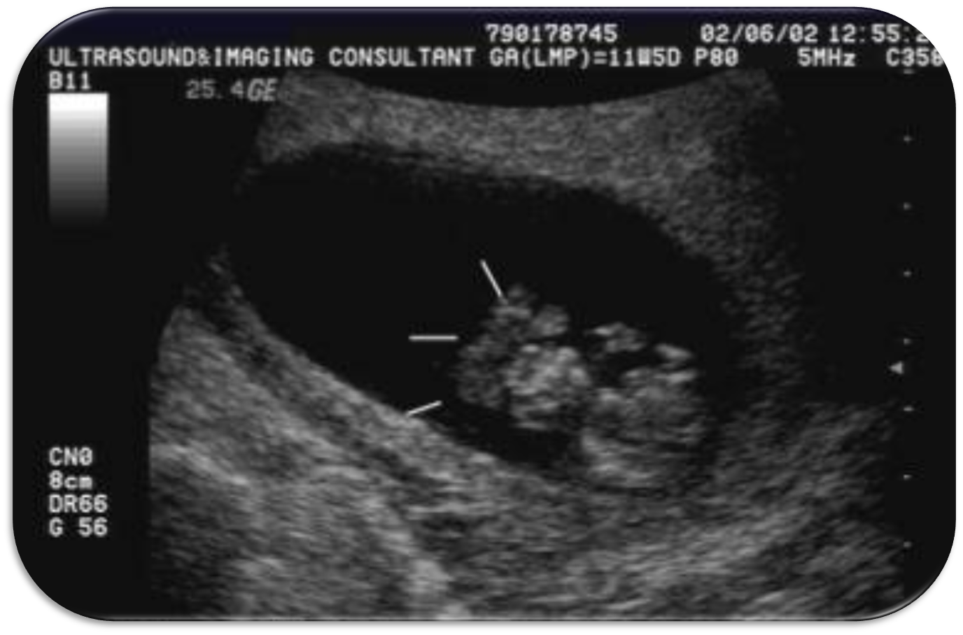
What is this US image?
Umbilical Cord Cysts
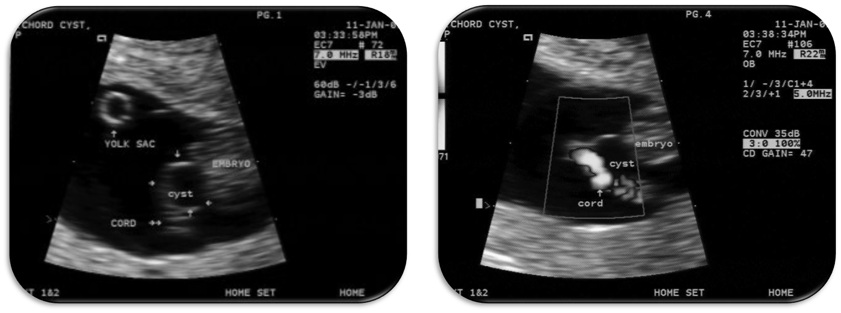
What are these US images?
Umbilical Cord Cysts
Abnormalities identified before __ ______ represented only about 22% of all sonographically detected abnormalities in the study population.
14 weeks