Post-Partum & Infertility
 Call Kai
 Learn
Learn Practice Test
 Spaced Repetition
 Match
Match Flashcards
 Knowt Play
1/55

There's no tags or description
Looks like no tags are added yet.
Name | Mastery | Learn | Test | Matching | Spaced | Call with Kai | Chat |
|---|
No analytics yet
Send a link to your students to track their progress
56 Terms
Puerperium
period of time from expulsion of placenta to return of uterus to pre-pregnancy size and function
Can last 6-8 weeks
Post-Partum presentation to U/S:
C-section scars can develop hematomas and abscesses
Retained parts of conception (placenta)
Infection (fever, abd pain)
Persistent bleeding
Post-Partum Anatomy
involution of uterus
Reversal of the growth changes that take place during pregnancy
50% reduction in size during first week postpartum
Lactation begins at this time
when discontinued, the normal reproductive organ functions (ovulation and menstruation) resume
Post Partum U/S appearance:
involution of uterus
Reversal of the growth changes that occur during pregnancy
50% reduction in size during week 1 postpartum
Lactation begins at this time
When discontinued the normal reproductive organ functions (ovulation + menstruation) occur
Post Partum U/S appearance:
uterus may appear heterogenous due to vascular changes and resolution of tissue edema and fluid
Endometrial cavity may measure up to 2 cm in AP
Common finding to see free fluid in the endometrial cavity
Represents blood and other fluids/substances as endo sloughs after post-birth
Post Partum U/S appearance:
uterine length can measure from 14.5 - 25 cm
TRV / Coronal width of the uterus can range from 7-14 cm
Size decreases w/ time
Ligaments are flaccid after delivery
Assume normal pre-pregnancy firmness within a few weeks to about a month
What might or might not you see in a Post-Partum U/S?
Broad Ligaments
Contains uterine vessels and fallopian tubes
Potential site for hematoma or abscess formation
Ovaries seen 50% of the time
Usually in true pelvis in non-pregnant state
Extra pelvic space during much of postpartum
Post-Partum Bleeding
Postpartum hemorrhage - blood loss >500 ml during the third stage of labor or immediately after a vaginal delivery
Uterine muscles must contract rapidly and firmly after delivery to occlude the uterine vessels at the point of placental separation
The C-section delivery requires >1,000 mL loss to classify as having postpartum hemorrhage
Occurs after 5% of all deliveries but is potentially the most lethal puerperium complication
35% of all maternal deaths
Post-Partum bleeding risks:
abnormal placental attachment increases risk of hemorrhage
Placenta accreta, increta, and percreta
Antepartum uterine rupture → emergency delivery & hysterectomy
Rare
Primary (Acute) Hemorrhage
acute hemorrhage is severe bleeding immediately following delivery
Occurs within first 24 hours post delivery
Associated w/ acute clinical problems
Difficult labor (dystocia)
Chorioamnionitis
Placenta previa & accreta, increta or percreta
Incomplete manual placental removal
Uterine antony - not contracting
Secondary (Delayed) Hemorrhage
occurs more than 24 hours post delivery
Several days to weeks later
Usually due to retained parts of conception and/or infection
Puerperal Infection
uterine tenderness usually the first sign of infection
Fever of over 100.4 on any two successive days after the first 24 hours postpartum
Most puerperal infections are urinary tract infections, but they can also be breast infections, thrombophlebitis, or endometritis
Other Signs / Symptoms of Puerperal Infection:
leukocytosis
Chills, headache, malaise, anorexia
Uterus is typically soft, tender, and large
Lochia, or vaginal discharge, may be diminished or profuse and odorous
Endometritis
endometrium infection
Presents within 3-4 days post-delivery
Usually caused by migration of normal vaginal flora, which may result in postpartum bleeding
Endometritis U/S findings:
dilated / irregular endometrial cavity
Shadowing echoes d/t gas
Cul-de-sac fluid
Retained Products of Conception (RPOC)
incomplete expulsion of miscellaneous products of conception during labor and delivery
Can cause infection
If left untreated, the following may result:
Infection
Bleeding
Intrauterine adhesions
RPOC U/S appearance:
Hyperechoic mass in the endometrial canal
remnants of placental tissue (called a placental polyp), are b/c of incomplete placental expulsion from the uterus
A heterogenous mass may contain necrotic tissue, clot, or infection
may appear similar to endometritis
Postpartum Ovarian Vein Thrombophlebitis (POVT)
rare
more likely to occur w/ c-sections
more common in Rt ovarian vein (80-90%)
inflammation of a vein d/t a thrombus found in the lumen of the vessel
POVT relates to Virchow’s triad:
hypercoagulability of blood during pregnancy and the postpartum period
venous stasis
venous wall damage as a result of uterus expansion and contraction
POVT symptoms:
can be mistaken for other processes
pelvic pain
fever
right-sided pelvic mass
What is the modality of choice evaluating POVT?
CT
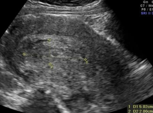
POVT U/S:
dilated anechoic to hypoechoic tubular structure extending superiorly from the adnexa
lack of a color or spectral Doppler signal
echogenic thrombus in the lumen confirms the presence of the thrombus
may extend into the IVC
the thrombus also may image as a mass between the uterus and the psoas muscle
most common site is where the IVC and the rt ovarian vein meet
Postpartum Ovarian Vein Thrombophlebitis Treatment:
potentially life threatening
IV anticoagulants such as:
heparin
blood thinners (warfarin)
IVC filters
helps keep the embolus from traveling into the heart /lungs
prevents pulmonary emboli or death
follow-up imaging studies help in monitoring thrombus resolution
Post-Partum C-section Incision
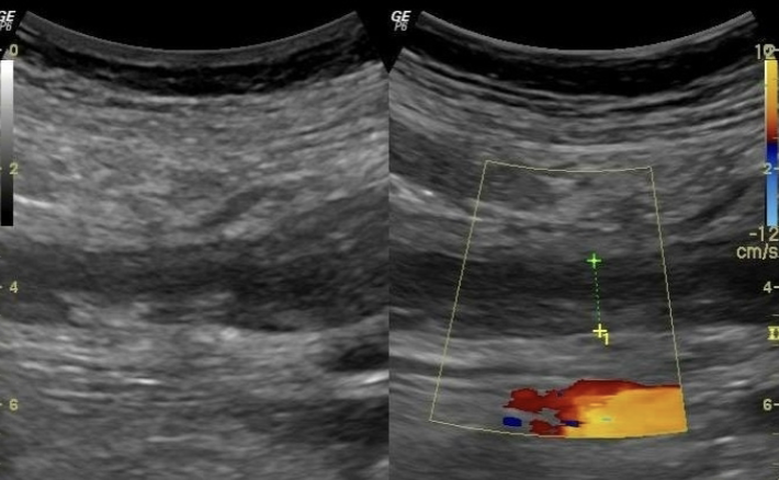
watch for hematoma formation
will present as anechoic areas along incision line of >2cm
may have decreased hematocrit
VBAC - Vaginal birth after cesarean
increased risk of uterine rupture
Bladder Flap Hematoma
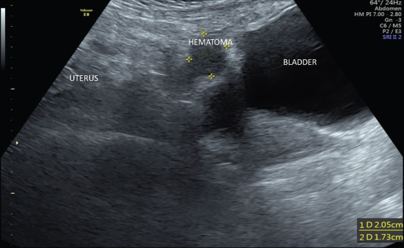
During a C-section, the surgeon incises the peritoneum btwn the bladder and the uterus to access the lower uterine segment
this potential space is called the bladder flap
where most hematomas are found post c-section
Bladder Flap Hematoma U/S appearance:
anechoic w/ ill-defined borders
very small (<1cm) to very large (>15cm)
complex masses b/c of the blood components clotting, internal septations, or debris within the hematoma
Infertility
80% of couples attempting pregnancy conceive naturally within their first 6 months
when naturally conceiving doesn’t occur after 12 consecutive months, this is called infertility
women >35 y/o w/ a failure to conceive after 6 months of attempts
Causes of infertility include:
tubal & pelvic pathology of the female partner (35%)
male problems (35%)
ovulatory dysfunction in the female (15%)
unexplained fertility (10%)
other unusual problems (5%)
Other causes of infertility can include:
cigarette smoking
body weight disorder
sexually transmitted disease
the delay of childbearing
Female factors of causing infertility:
ovulatory dysfunction (40%)
Tubal dysfunction (adhesions, endometriosis) (30-50%)
uterine dysfunction (5-10%)
cervical (5-10%)
immunologic (5%)
unknown (5%)
Male factors of causing infertility:
varicocele (40%)
idiopathic (25%)
testicular failure (10%)
tubular obstruction (5-10%)
cyptorchidism (5-10%)
other semen disorders (10%)
With Infertility, U/S evaluates the:
structural anatomy
uterine shape, check for masses
endometrium
thickness + echogenicity
ovaries
presence and number of follicles
presence of cysts for other pathology
fallopian tubes
Patency - can confirm w/:
hyterosalpingogram (xray)
sonohysterogram / saline infused sonogram (SIS)
Congenital Uterine Anomalies causing infertility:
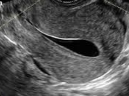
bicornuate uteus has small association w/ fetility complications
septate uterus has high association w/ intertility
2 uterine cavities w/ single fundus
problems w/ implantation
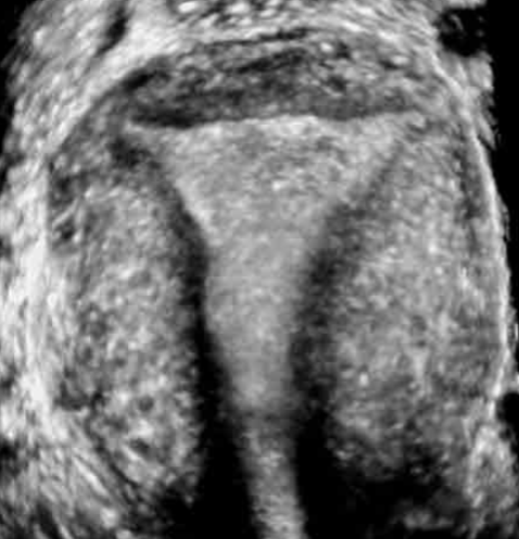
T-shaped Uterus
associated w/ DES exposure
Diethylstillbestrol exposure in utero occurred btwn 1950-70 to help threatened AB
high risk for cervical incompetence
What can a progesterone deficiency lead to?
early pregnancy loss + infertility
can supplement w/ vaginal gel, shorts, or oral tablets
The ________ can be evaluated for changes throughout the menstrual cycle.
endometrium
Irregular endometrial shape may be caused by:
submucosal fibroids + polyps
evaluated by Saline Infusion Sonography (SIS)
Polyps have narrpw base and fibroids have a more broad base
Main problem w/ fibroids + polyps is impeded implantation
internal uterine scars are called synechiae
When evaluating fallopian tubes for infertility, check for:
hydrosalpinx ( fluid-filled fallopian tube)
tubal patency - inject fluid into tube and check for spillage in the posterior cul-de-sac
no spillage and pain = blocked tube
Ovaries during normal ovulation period:
follicular stage → many follicles <5mm
FSH levels encourage growth and one follicle becomes dominant
Dominant follicle grows 2-3mm per dat until average diameter is reached → 22mm
then ruptures (ovulation)
corpus luteum is left behind
Polycystic Ovarian Syndrome (PCOS)
follicles grow but do not develop properly
immature follicles continue to produce estrogen and androgens while inhibiting the pituitary function (more LA than FSH)
prevents normal ovulation
Causes of PCOS:
obesity
diabetes
thyroid, pituitary, or adrenal gland dysfunction
PCOS U/S findings:
round ovaries w/ multiple cysts in the periphery → String of Pearls sign
ovary may appear normal
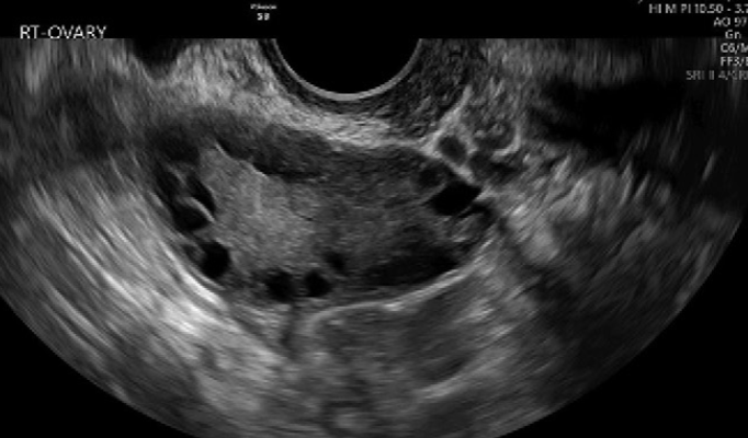
Endometriosis
may be diffuse or local
ectopic placement of the endometrium outside the uterine cavity
most common site is the ovary
but can spread throughout the pelvic + sometimes throughout the body
focal form is endometrioma
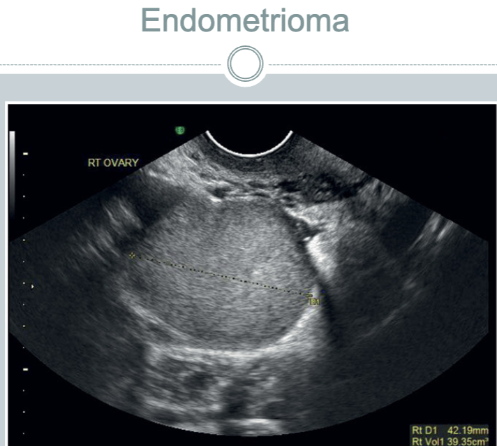
Adhesions
Scar tissue tha obstructs the Fallopian tube or fimbriated ends of the tube
Infertility Treatments
controlled follicle stimulation w/ medication and timed intercourse
IUI (intrauterine insemination)
IVF (invitro fetilization)
surrogate / gestational carrier
Ovarian Induction Therapy
ovarian stimulation achieved in a controlled setting
baseline U/S perfoemed to assess anatomy + check for cysts that could interfere with medication
Mediciations such as Clomiphene Citrate (Clomid) or letrozole are administered on days 3-5 of a normal cycle
Muliple U/S’s to measure follicle size + number
measure any follicles >1cm
may need hCG injection to trigger ovulation
Ovarian Induction Therapy & the Endometrium
Assess thickness & echogenicity
thickness changes from 2-3mm to 12-14mm
an endometrium that measures <8mm in thickness and has an irregular echogenicity has increased risk of infertility
Intrauterine Insemination (IUI)
used to treat male factor infertility in most cases
may use sperm donor or partner’s sperm
sperm sample is obtained, other substances and proteins from the semen are “washed” and then the sperm are introduced to the uterine cavity with a catheter at the time of natural or induced ovulation
donor / surrogate sperm may also be used
In Vitro Fertilization (IVF)
fertilization of ova outside the body
mature ova are collected through an egg retrieval procedure and mixed in a dish w/ spermatozoa
resulting embryos are placed back into uterus
IVF treatment usually consists of:
ovarian monitoring, needle aspiration of oocytes, incubation of oocytes, fertilization, and transferring the embryos into the uterus
aspiration of the oocytes may be done laparoscopically, the transabdominal or transvaginal ultrasound
Intracytoplasmic Sperm Injection (ICSI)
sometimes d/t poor sperm motility or quality, another procedure is needed to help fertilization occur
ICSO involves the injection of one sperm into the cytoplasm of an egg, thereby improving the likelihood of fertilization and subsequent embryo development
Surrogate / Gestational Carrier
If a couple’s infertility prevents them from carrying a pregnancy, the couple may still have a child using their own sperm + ovum, using a GA carrier
alternatively, IVF-ET of donor sperm and/or ova also addresses a couple’s treatment needs
in some situations, crypto-preserved embryos are obtained from a donor couple
“snowflake” babies handled like an adoption, but of an embryo
Complications associated w/ assisted reproduction:
Ovarian Hyperstimulation Syndrome (OHSS)
Multiples
Ectopic Pregnancy
Heterotopic Pregnancy
Ovarian Hyperstimulation Syndrome (OHSS)
more common in pt’s w/ PCOS
may be mild to severe
enlarged ovaries
multiple cysts
abdominal ascites
pleural effusion
Multiples
number of cases of multiples has decreased w/ decreased numbers of embryos transferred
used to be about 25-30%
now overall IVF birth rates have fallen to 8.4% for IVF procedures specifically
fetal reduction is rarely done today
Ectopic & Heterotopic
increased incidence in ectopic and heterotopic pregnancies w/ ART
risk is about 2-5%, which is higher than the 1-2% in normal pregnancies
likely d/t underlying tubal infertility issues