A&P II Exam 2, The Peripheral Nervous System
1/101
There's no tags or description
Looks like no tags are added yet.
Name | Mastery | Learn | Test | Matching | Spaced | Call with Kai |
|---|
No analytics yet
Send a link to your students to track their progress
102 Terms
peripheral nervous systems (PNS)
links the CNS to the body and external environment
PNS detects sensory stimuli and delivers info to CNS as sensory input
CNS processes input and transmits impulse through PNS to muscle cells and glands as motor output
what are the divisions of the PNS
sensory division, somatic sensory division, visceral sensory division, motor division, somatic motor division, visceral motor division (autonomic motor nervous system, ANS)
sensory division of the PNS
consists of sensory (afferent) neurons; sends sensory stimuli to CNS
somatic sensory division of the PNS
detects both internal and external stimuli
general sense detect stimuli from skin
special sensory detect stimuli from special sense organs (eyes, skin, skeletal muscles, etc.)
visceral sensory division of the PNS
relays internal info (blood pressure) from organs (abdominopelvic and thoracic cavities); ex urinary bladder
motor division of the PNS
motor (efferent) neurons; carry out motor functions of nervous system
somatic motor division of the PNS
voluntary motor functions; triggers skeletal muscle contractions
visceral motor division (autonomic motor nervous system, ANS) of the PNS
maintains many aspects of homeostasis (involuntary motor functions of the body); divided into sympathetic and parasympathetic
what events occur when sensory neurons detect stimuli
detected stimuli transmitted along sensory neurons (spinal or cranial) to cerebral cortex → in cortex, sensory info is interpreted, integrated, and appropriate motor response is selected and initiated (such as fight or flight, or rest and digest)
the motor division of the PNS is divided in the parasympathetic and sympathetic divisions. What does the sympathetic nervous system do?
also known as the fight or flight division
does homeostasis activities surrounding physical work and visceral response of emotion
the motor division of the PNS is divided in the parasympathetic and sympathetic divisions. What does the parasympathetic nervous system do?
also known as the rest and digest division
does digestion and maintains the body’s homeostasis at rest
why is the sympathetic nervous system referred to as the fight or flight division of the ANS in the PNS
the sympathetic nervous system carries out the “fight or flight” response in the body by prepping the body for emergency situations. plays a vital role in maintaining homeostasis when the body is engaged in physical work. mediates the body’s response to motion.
how is the body effected when going into “fight or flight” via the sympathetic nervous system
heart rate increase, increases diameter of airways and increases diameter of pupils
blood flow diverted from the GI tract and kidneys to organs involved in activity, such as skeletal muscles
liver increases conversion of glycogen to glucose for ATP production
why is the parasympathetic division of the ANS in the PNS referred to as “rest and digest”
the parasympathetic nervous system carries out the rest and digest function in the body; plays a role in body maintenance functions, such as digestion and urine formation
also known as the craniosacral division based on association with cranial nerves and pelvic nerves from sacral plexus
how is the body effected when going into “rest and digest” via the parasympathetic nervous system
heart rate decrease, decreased diameter of airways, and decreased diameter of pupils
promotes salivation, lacrimation, urination, digestion, and defecation
liver converts absorbed glucose to glycogen stores
peripheral nerves
main organ of the PNS; axons of many neurons bound together by connective tissue; nerves of PNS contact (innervate) majority of the structures in the body (which they control)
mixed nerves
contain both sensory and motor neurons
sensory nerves
contain only sensory neurons; also some involved in muscle stretch and tension
spinal nerves
originate from the spinal cord and innervate structures below the head and the neck; all 31 pairs of spinal nerves are mixed nerves
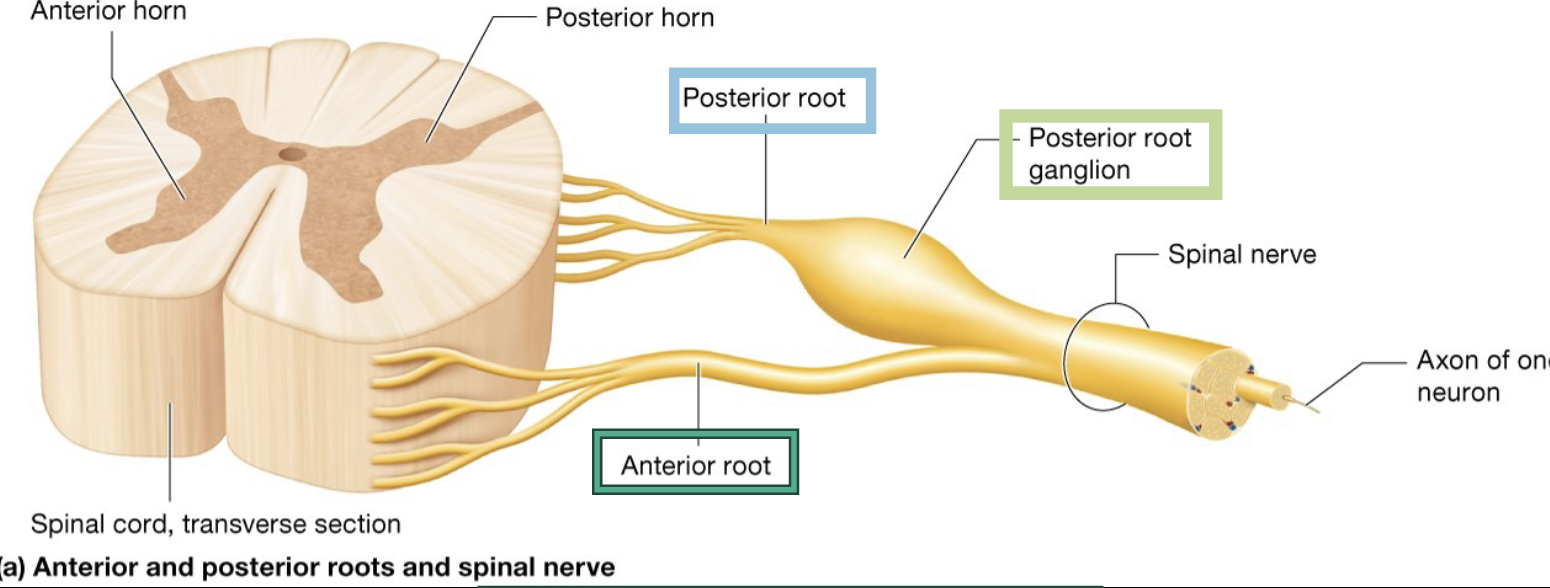
what are the functions of the parts of the spinal nerves
posterior root - carry sensory neurons from posterior horn
posterior root ganglion (dorsal root ganglion) - swollen area in posterior root; houses cell bodies of sensory neurons
anterior root - motor neurons from anterior horn
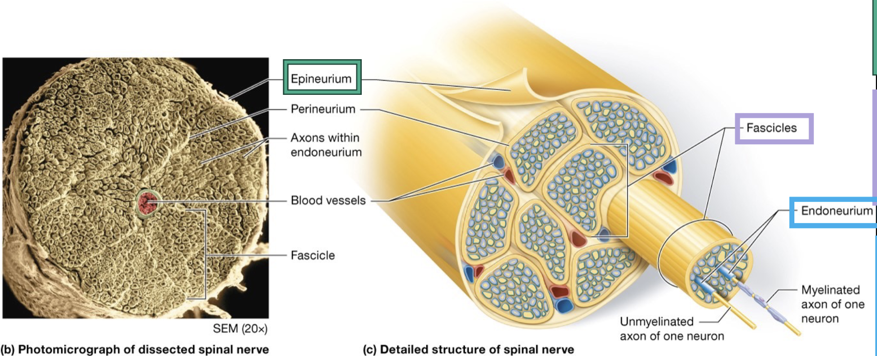
what makes up the epineurium, fascicles, and endoneurium of the peripheral nerves and what is their function
epineurium - outermost layer of connective tissue; hold motor and sensory axons together
fascicles - small groups of bundled axons surrounded by connective tissue perineurium
endoneurium - connective tissue surrounding each individual axon within fascicless
cranial nerves
attach to brain; mostly innervate structures in the head and neck
not formed by the fusion of sensory and motor roots (like spinal nerves)
allow for purely sensory, mixed, and mostly motor nerve
spinal nerve
short; divides into two mixed nerves; both carry somatic motor and sensory info
posterior ramus - travels of the posterior side of the body
anterior ramus - travels to anterior side of the body and/or to an upper or lower limb
how many pairs of spinal nerves are there
31
of the 31 spinal nerves, how many are cervical, thoracic, lumbar and sacral, and coccygeal
cervical = 8
thoracic = 12
lumbar and sacral = 5
coccygeal = 1
nerve plexus
anterior rami of cervical, lumbar, and sacral spinal nerves merged together; complicated networks of nerves
right and left cervical plexuses
anterior rami of C1-C5, with a small contribution from hypoglossal nerve (CN XII)
each nerve has cutaneous branches that innervate skin of neck and sections of head, chest, and shoulders
motor branches innervate specific muscle in neck
phrenic nerve
major motor branch of C4 with contribution from C3 and C5 (3-5 to stay alive); innervates diaphragm
part of the cervical plexus
hiccups
spasms of the diaphragm that cause of forceful inhalation of air; one way to end hiccups is to apply firm pressure to the muscles of the neck that overlie the phrenic nerve for about 5-10 seconds, the pressure will interrupt aberrant impulses causing the diaphragm to contract inappropriately, pressure is not adequate to stop the nerve from firing completely or interfering with breathing
right and left brachial plexuses
lateral to 5th cervical through 1st thoracic vertebrae; provides motor and sensory innervation to upper limbs; includes nerve roots from c1-T1
brachial plexus begins with formation of large nerve trunks
C5 and C6 typically unite to form superior trunk
C7 usually forms middle trunk
C8 and T1 unite to form inferior trunk
medial cord
the anterior division of the inferior trunk of the brachial plexus that descends down the medial arm
lateral cord
the anterior division of superior and middle trunks of the brachial plexus combined; descends down the lateral arm
posterior cord
posterior division of each trunk of brachial plexus combined; lies in posterior arm
what are the 5 major nerves of the brachial plexus
axillary nerve
radial nerve
musculocutaneous nerve
median nerve
ulnar nerve
axillary nerve
structures near axilla, including deltoid and teres minor muscle and skin over deltoid region
radial nerve
innervates triceps barchii muscle and most of extensor muscles of forearm; also skin over posterior thumb, 2nd digit, 3rd digit, and lateral half of 4th digit
musculocutaneous digit
innervates bicets brachii and skin covering lateral arm
median nerve
innervates wrist and digital flexors, some intrinsic muscles of hand and skin over anterior thumb, 2nd, 3rd digits, and lateral half of 4th digit
ulnar nerve
innervates flexor muscles in forearm (not innervated by median nerve), most of intrinsic hand muscles, and skin of 5th digit and medial side of 4th digit
thoracic spinal nerves ——- form plexuses
do not (except T1)
the posterior ramus of thoracic spinal nerves innervates…
deep back muscles
the anterior ramus of the thoracic spinal nerves travels between two ribs as an ——-
intercoastal nerve
left and right lumbar plexus
derived from anterior rami of L1-L; innervate pelvic structures and lower extremity after splitting into two divisions
what are the lumbar plexus divisions
obturator nerve and femoral nerve
obturator nerve
anterior division’s largest member
enters thigh from pelvis via obturator foramen
branches of nerves innervate adductor muscles in thigh, hip joint, and skin over medial aspect of thigh
femoral nerve
posterior divisions largest member; largest branch of lumbar plexus
travels from psoas, through pelvis and under inguinal ligament to enter thigh to innervate; anterior thigh muscles and skin over anterior and medial thigh and leg, as well as knee joint
right and left sacral plexuses
formed from anterior rami of spinal nerves L4-S4; nerve branches innervate structures of pelvis, gluteal region, and much of lower extremity; each plexus is divided into anterior and posterior divisions
what are the sacral plexuses
sciatic nerve, common fibular nerve (common peroneal(, and tibial nerve
sciatic nerve
longest and largest nerve in the body
travels through the greater sciatic notch in the pelvis into the thigh
innervates hip joint in posterior thigh before it divides into tibial and common fibular nerves
common fibular nerve (common peroneal)
made up of axons from posterior division of sacral plexus
descends along lateral leg to supply part of knee joint and skin of anterior and distal leg
divides into superficial and deep branches; superficial branch serves lateral leg and dorsum of foot; deep branch supplies ankle dorsiflexors and two muscles of dorsum
tibial nerve
larger branch of sciatic nerve branches innervates most of hamstring muscles as nerve descends distally
innervates parts of knee and ankle joint
smaller nerve branches serves posterior and lateral skin of leg as well as skin and muscles of foot
what is the role o the RNS in sensation
stimuli are first detected by sensory neurons’ stimulus is transmitted by sensory neurons to CNS, where stimulus is integrated and interpreted by CNS neurons
sensory transduction
stimulus is converted into electrical signal
what are the steps of sensory transduction
ion channels in axolemma are closed
stimulus is detected by sensory receptor → sodium ion channels open → sodium ions flow into axoplasm → temp. depolarization (less -) (receptor potential)
if enough sodium ions enter, membrane potential may reach threshold → voltage-gated sodium ion channels open → actions potential is propagated along axon towards CNS
what are the types of sensory receptors in sensory transduction
rapidly adjusting receptors and slowly adapting receptors
rapidly adjusting receptors
receptors that respond rapidly with high intensity to stimuli; stop sending signals after certain time period (called adaptation); receptors detect initiation of stimuli but ignoer ongoing stimuli
slowly adapting receptors
respond to stimuli with constant action potentials that dont diminish over time
how are sensory receptors classified when concerning surrounding cells
encapsulated nerve endings (surrounded by specialized supportive cells) and free nerve endings (lack supportive cells)
how are sensory receptors classified when concerning location and origin of stimulus
exteroceptors - at or near external surface
interceptors - in vessels or tissues of organs (blood vessels, tissues, organs, think visceral)
proprioceptors - in muscles, joint, and inner ear
how are sensory receptors classified when concerning the type of stimulus detected
mechanoreceptors - mechanical energy
thermoreceptors - temperature energy
nociceptors - damage to tissue (pain)
photoreceptors - light energy
chemoreceptors - chemical energy
osmoreceptors - osmotic pressure of body fluids
what are the classes of mechanoreceptors (on the exterior surface of the skin)
merkel cell fibers, tactile corpuscles, ruffini endings, lamellated corpuscles, hair follicle receptors, proprioceptors
merkel cell fibers
slow adapting; detect discriminative touch stimuli; tell the difference between the objects touching you
tactile corpuscles (meissner corpuscles)
rapidly adapting; transmit discriminative touch stimuli; tell the difference between objects
ruffini endings (bulbous corpuscles)
slowly adapting receptors that respond to stretch and movement
lamellated corpuscles (pacinian corpuscle)
rapidly adapting receptors detect vibration and deep pressure stimuli (pain)
hair follicle receptors
free nerve endings surrounding the base of hair follicles in thin skin; not on palms and soles; respond to stimuli that cause hair to bend
proprioceptors
in musculoskeletal system; detect movement and position of joints and body parts
types of thermoreceptors
cold
in superficial dermis
temp range: 10-40 C
warm
in dermis
temp range: 32-48 C
how do thermoreceptors work
activated with temp detection; free nerve endings that adapt rapidly but generate impulses at lower frequency for prolonged stimulus; temps above or below thermoreceptors range stimulate nociceptors instead, resulting in painful sensations
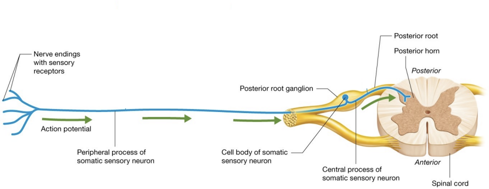
what are first order somatic sensory neurons and what are the three components
pseudounipolar neurons; composed on cell body, peripheral process, and central process
cell body
cell bodies of spinal nerves are in posterior (dorsal root ganglion), just lateral to spinal cord; cell boies of cranial nerves are in cranial nerve ganglia in head and neck
peripheral process
long axon that transmits action potentials from source of stimulus (receptor) to neuron’s central process
central process
exits cell body and travels through posterior root; enters spinal cord at posterior horn (or brainstem for cranial nerves) where they deliver their action potentials
how does an action potential travel down somatic sensory nerve
peripheral process transmits action poetnial from sensory receptors to neuron’s central process
central process transmits actions potential from peripheral process to posterior horn, eventually synapsing on second-neuron in spinal cord or brain stem
action potential propagated down peripheral process does not generally reach cell body ; instead, usually transmitted to central process in area when peripheral and central processes come into contact near cell body
receptive fields
areas served by particular neurons; neurons with more branches innervates larger receptive fields
body regions whose primary function is sensing environment (fingertips) contain — neurons with — receptive fields
many, smaller
body regions that are not as involved in sensing environment (skin of forearm) have —- neurons with — receptive fields
fewer, larger
two point discrimination threshold
mthod for measuring the relative size of receptive fields
referred pain
phenomenon whereby pain that originates in an organ is perceived as cutaneous pain
occurs because many spinal nerves carry both somatic and visceral neurons; so visceral sensations travel along the same pathways as somatic sensations
rgenerally located along dermatome for particular neurons
upper motor neurons
neurons of primary motor cortex makes decision to move and initiate that movement; not in contact with muscle fibers itself
lower motor neurons
recieve messages from upper motor neurons; in contact with skeletal muscle fibers; release acetylcholine onto muscle fibers to initiate contraction
lower motor neurons
multipolar neurons whose cell bodies are in either anterior horn of the spinal cord or brainstem; axons are in the PNS
motor neuron pools
groups of lower motor neurons that innervate same muscles; clustered in anterior horn of spinal cord
large motor neurons
majority of neurons within pools; stimulate skeletal muscle fibers to contract by excitation-contraction mechanism
smaller motor neurons
found within neuron pools; innervate intrafusal fibers; part of specialized stretch receptors
diagram of brain and motor response
reflexes
programmed, autonomic responses to stimuli; occur in reflex arc (three-step sequence of events); usually protective negative feedback loops
how do reflexes work
reflexes begin with sensory stimulus and finish with rapid motor response
neural integration between sensory stimulus and motor response occurs in CNS, at spinal cord or brainstem
mechanoreceptors in muscles and tendons monitor muscle length and force of contraction; communicate information to spinal cord, cerebellum, and cerebral cortex
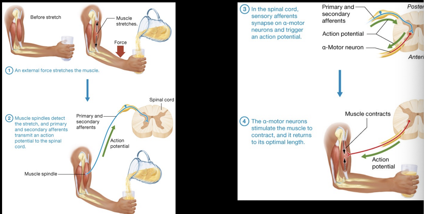
muscle spindles
tapered structures found scattered among regular contractile muscle fibers (extrafusal muscle fibers)
intrafusal fibers have contractile filaments of actin and myosin at their poles; innervated by motor neurons
contractile filaments are absent in central area of intrafusal fibers
what are the two structural and function classes of sensory neurons that innervate intrafusal fibers
primary afferents - respond to stretch when first initiated
secondary afferents - respond to both static length of muscle and position of limb
golgi tendon organs
mechanoreceptors located within tendons near muscle-tendon junction
monitor tension generated by muscle contraction
contain single somatic sensory axon that fires more rapidly as greater tension is generated with each contraction; info is sent to the CNS
how are reflexes classified
number of synapses between neurons involved in arc
type of organ in which the reflex takes place, either visceral or somatic
simplest reflex (monosynaptic reflexes)
involve only a single synapse within spinal cord between sensory and motor neuron
polysynaptic reflex
complicated reflex arc involving multiple synapses
patellar (knee-jerk). reflex and jaw-jerk reflex
examples of simple stretch reflexes
steps of a simple reflex stretch
golgi tendon reflexes
polysynaptic reflexes; protects muscles and tendons from damaging forces
causes muscle relaxation; opposite of simple stretch reflex action
when tension in muscle and tendon increases dramatically, golgi tendon organs signal spinal cord and cerebellum
motor neuron innervating muscle are inhibited while antagonist muscles are simultaneously activated
flexion or withdrawal reflex
involves rapidly conducting nociceptive afferents and multiple synapses in spinal cord; act to withdraw limb from painful stimuli
crosses-extension reflex
occurs simultaneously on opposite sides of the body for balance and postural support while other limb is withdrawn from painful stimulus
cranial nerve reflexes
polysnaptic reflex arcs that involve cranial nerves; gag reflex and corneal blink reflex