Osteology Written Exam

0.0(0)
Studied by 0 people
call kaiCall Kai
Locked
learnLearn
examPractice Test
spaced repetitionSpaced Repetition
heart puzzleMatch
flashcardsFlashcards
GameKnowt Play
Card Sorting

1/206

encourage image

There's no tags or description

Looks like no tags are added yet.

Last updated 12:35 PM on 8/31/26
Name
Mastery
Learn
Test
Matching
Spaced
Call with Kai
Chat

No analytics yet

Send a link to your students to track their progress

207 Terms
1
New cards
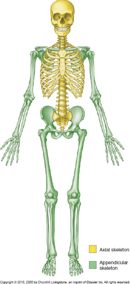
Axial skeleton and appendicular skeleton
What are the two major divisions of the skeleton, and what does each include? | Axial skeleton: head, neck, trunk. | Appendicular skeleton: limbs.
2
New cards
Axial skeleton
Is the skull part of the axial or appendicular skeleton?
3
New cards
<p>Axial - torso and head</p><p>Appendicular - limbs </p>

Axial - torso and head

Appendicular - limbs

Distinguish the axial skeleton from the appendicular skeleton in the image.

<p>Distinguish the axial skeleton from the appendicular skeleton in the image.</p>
4
New cards
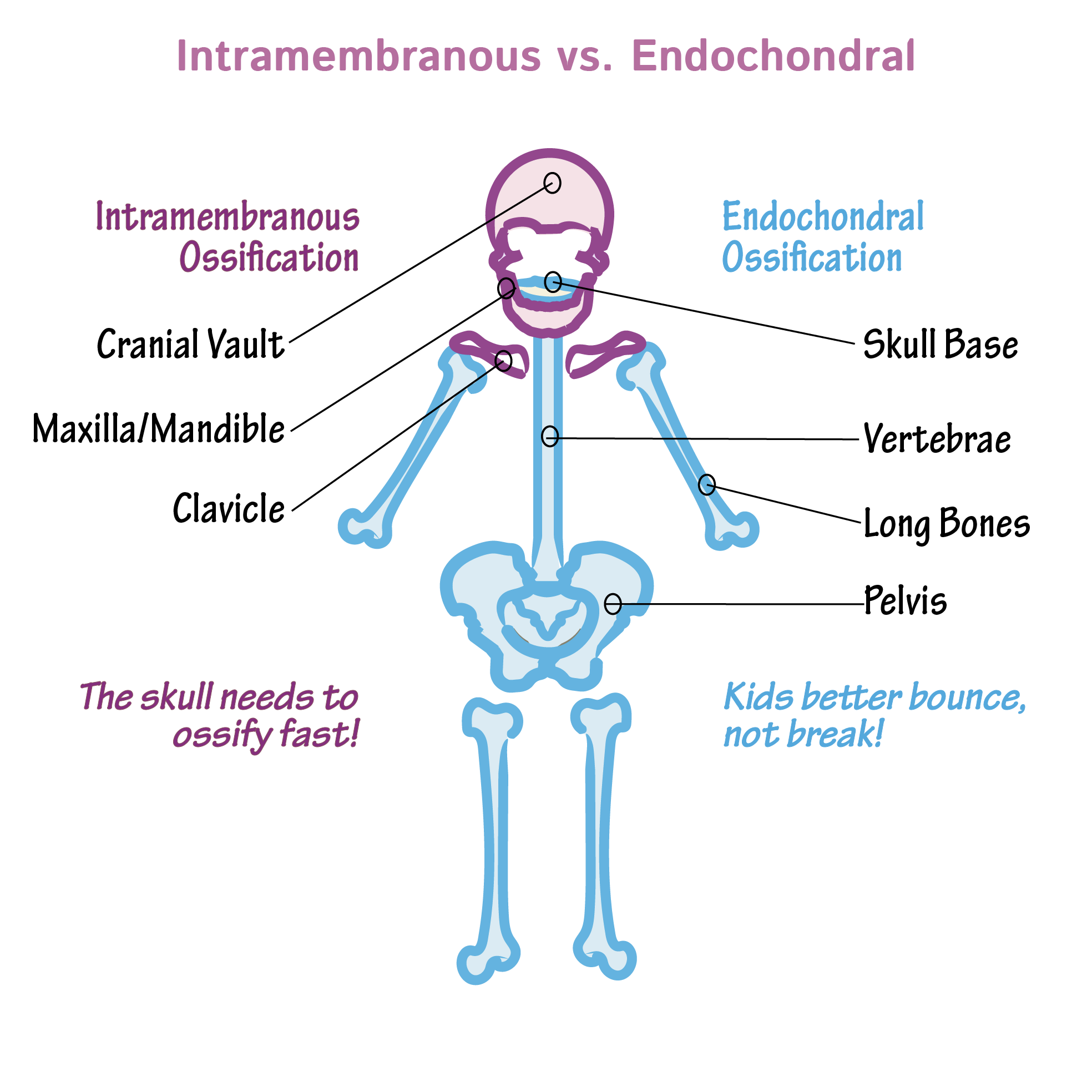
<p>Intramembranous ossification</p>

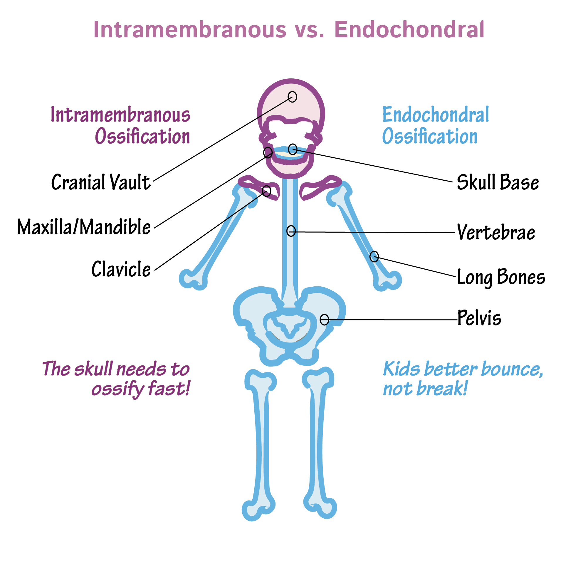
Intramembranous ossification

What type of ossification is associated with the cranial vault?
5
New cards
<p>Intramembranous ossification</p>

Intramembranous ossification

What type of ossification are the maxilla and mandible grouped under?

6
New cards
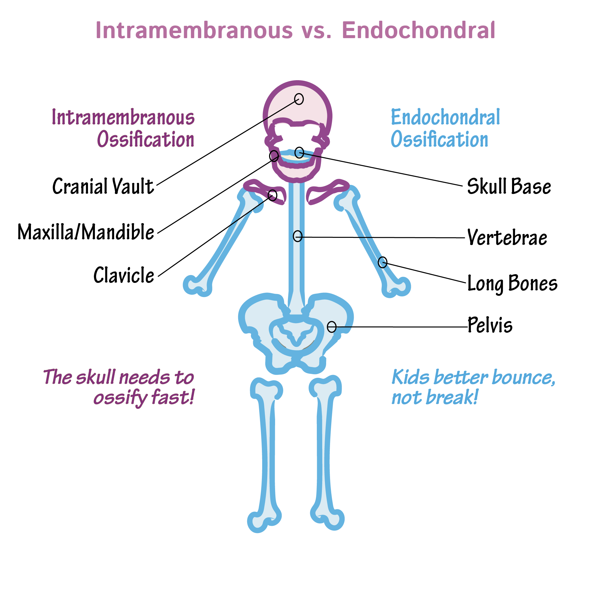
<p>Endochondral ossification</p>

Endochondral ossification

What type of ossification is associated with the skull base?

7
New cards
<p>Endochondral ossification</p>

Endochondral ossification

What type of ossification are vertebrae, long bones, and the pelvis grouped under?
8
New cards
<p>“The skull needs to ossify fast!” / “Kids better bounce, not break!”</p>

“The skull needs to ossify fast!” / “Kids better bounce, not break!”

What memory phrases are paired with intramembranous and endochondral ossification, respectively?
9
New cards
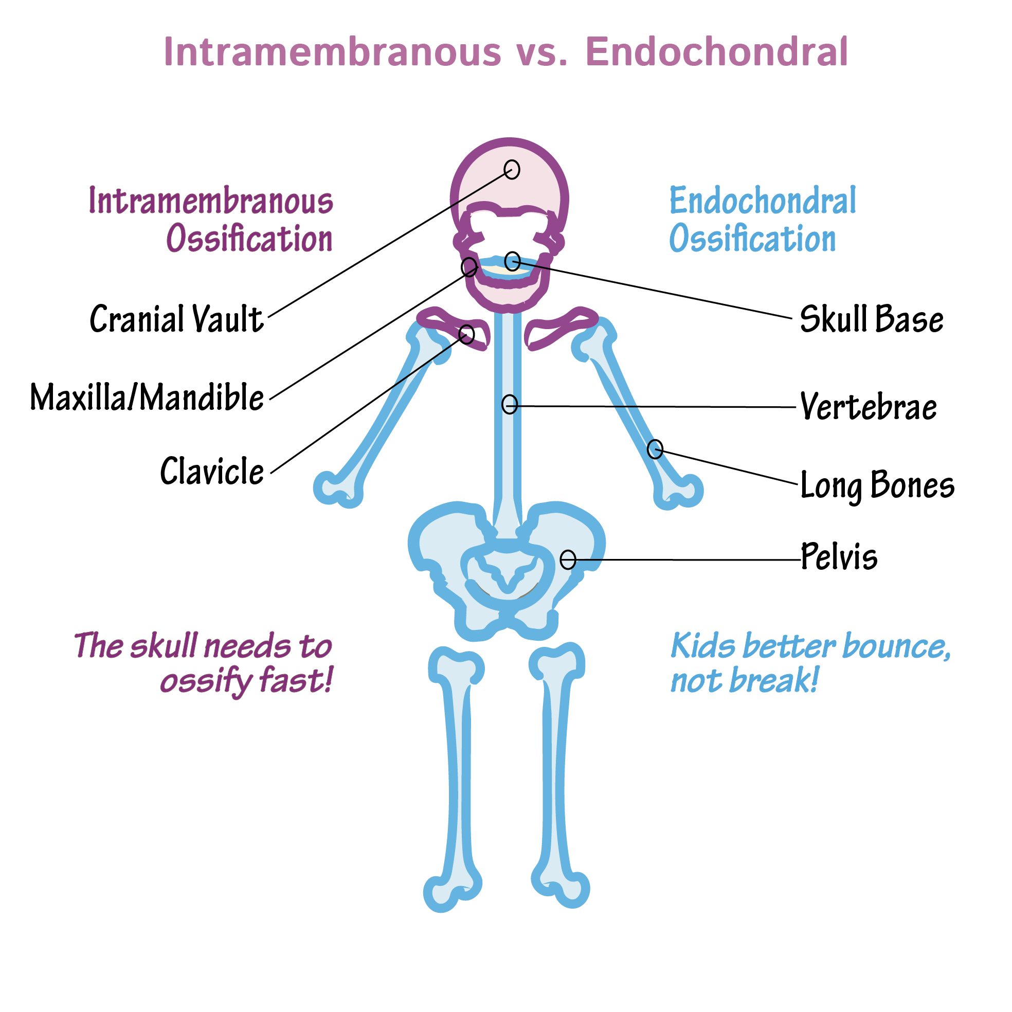
<ul><li><p>Cranial Vault</p></li><li><p>Maxilla/Mandible</p></li><li><p>Clavicle</p></li></ul><p></p>
  • Cranial Vault

  • Maxilla/Mandible

  • Clavicle


Which structures are formed by intramembranous ossification?

10
New cards
Long, sesamoid, flat, short, irregular
What five categories are used to classify bones by shape?
11
New cards
  • Cranial/skull vault = flat bone

  • facial region = irregular bone


What bone-shape classifications are demonstrated in the skull?

12
New cards
<p>Bone-shape categories</p>

Bone-shape categories

Identify the five bone-shape categories shown in the image.
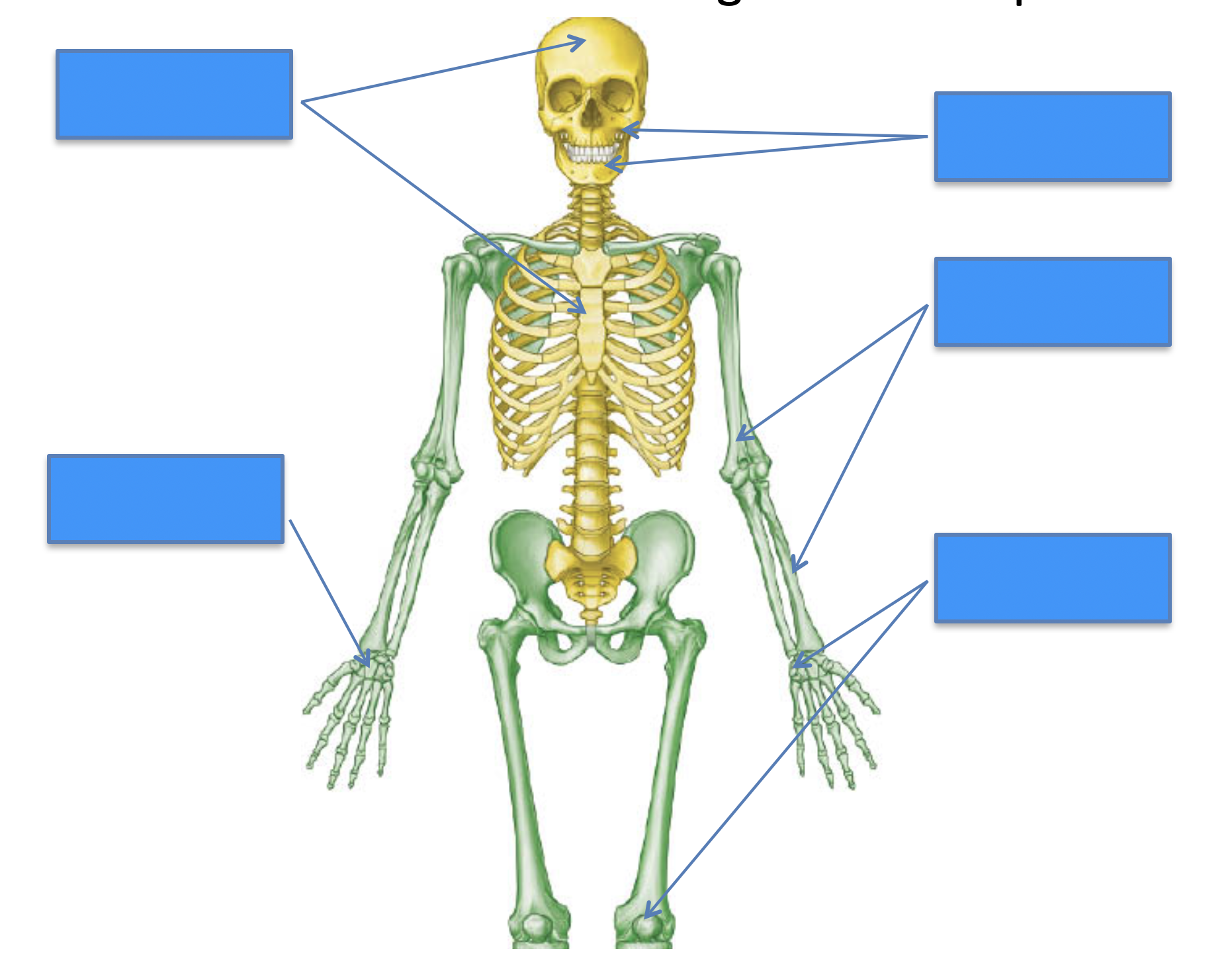
<p>Identify the five bone-shape categories shown in the image.</p>
13
New cards

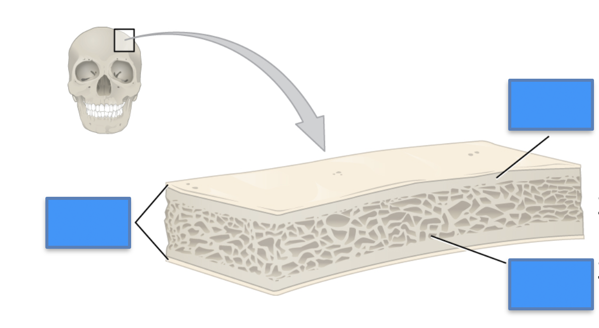
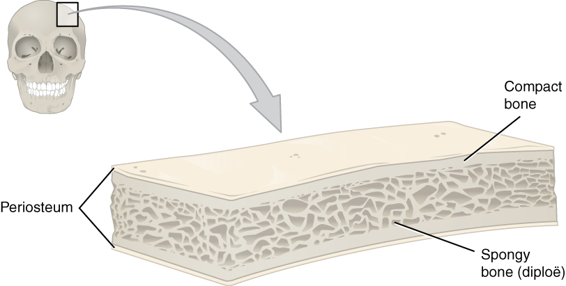
Outer plate → diploë → inner plate

  • Outer plate: compact bone

  • Diploë: spongy/trabecular bone

  • Inner plate: compact bone


What are the three layers of skull bone in the diploë arrangement, from outside to inside? What type of bone are they made of?

14
New cards

Outer plate → periosteum

Inner plate → endosteum

What covers the outer and inner plates of skull bone?

15
New cards
Diploë
What is the central spongy/trabecular bone between the outer and inner compact plates called?
16
New cards
  1. Brain protection

  2. Reduced weight

  3. Bone marrow reservoir

  4. Shock absorption


What four functions are listed for the diploë arrangement?
17
New cards
<p></p>


Label this image

<p>Label this image</p>
18
New cards
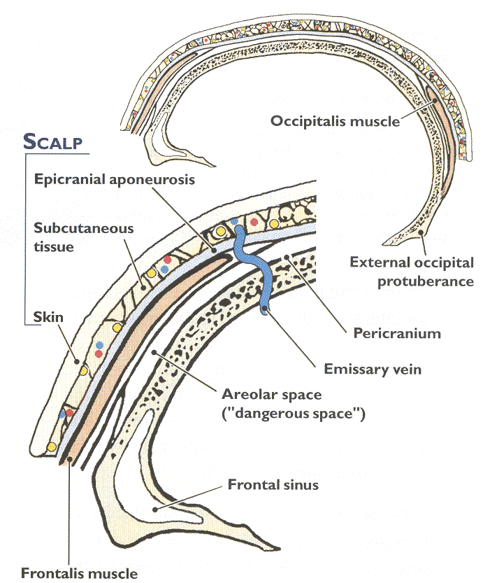
<p>Emissary veins pass through small holes in the skull; diploic veins lie within diploic/spongy bone</p>

Emissary veins pass through small holes in the skull; diploic veins lie within diploic/spongy bone

How are emissary veins and diploic veins positioned relative to the skull?
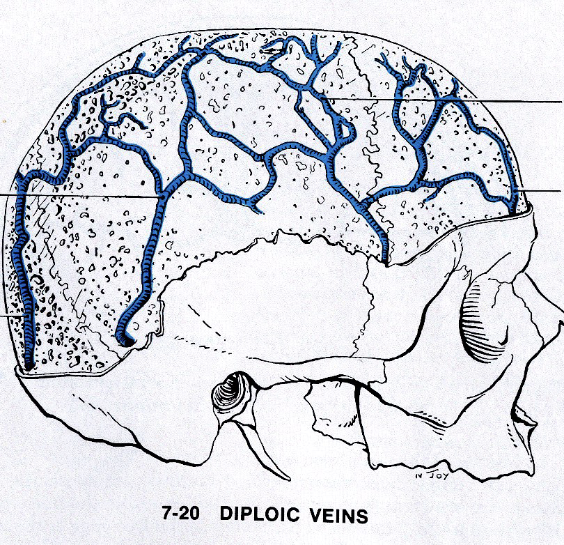
<p>How are emissary veins and diploic veins positioned relative to the skull?</p>
19
New cards
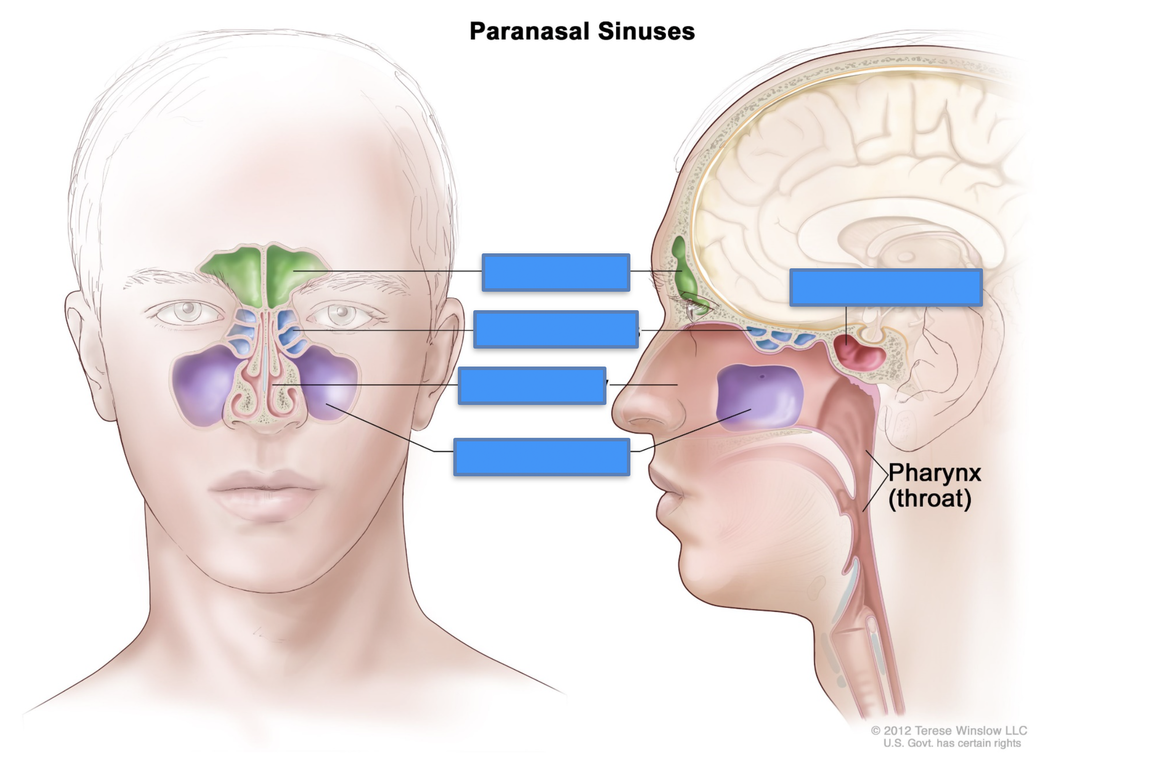
Frontal, ethmoid, maxillary, sphenoid sinuses
What four paranasal sinuses are shown?
20
New cards
  • Frontal = forehead/frontal region

  • ethmoid = upper nasal region between the orbits

  • maxillary = cheek/maxillary region

  • sphenoid = posterior to the nasal cavity


Match each paranasal sinus with its general location.

21
New cards
<p>Paranasal sinuses </p>

Paranasal sinuses

Label this image

<p>Label this image</p>
22
New cards
Neurocranium and viscerocranium
What are the two major parts of the skull?
23
New cards

Neurocranium = cranial/brain-containing portion

viscerocranium = facial skeleton

Which major skull division corresponds to the cranial portion, and which corresponds to the facial skeleton?

24
New cards
<p></p>


How do you divide the neurocranium from the viscerocranium

25
New cards
8 bones: frontal, occipital, ethmoid, sphenoid, 2 parietal, 2 temporal
How many bones make up the neurocranium, and which bones are included?
26
New cards

Single: frontal, occipital, ethmoid, sphenoid

Paired: parietal, temporal

Which neurocranial bones are single vs paired?

27
New cards

15 bones: mandible, vomer, hyoid, nasal (paired), maxilla (paired), zygomatic (paired), palatine (paired), lacrimal (paired), and inferior nasal concha (paired)

How many bones are listed in the viscerocranium group, and which bones are included?

28
New cards

Single: mandible, vomer, hyoid

Paired: nasal, maxilla, zygomatic, palatine, lacrimal, inferior nasal concha

Which bones listed in the viscerocranium group are single vs paired?
29
New cards
Paired skull bones: parietal, temporal, nasal, maxilla, zygomatic, palatine, lacrimal, inferior nasal concha
Which skull bones in these groups are paired?
30
New cards
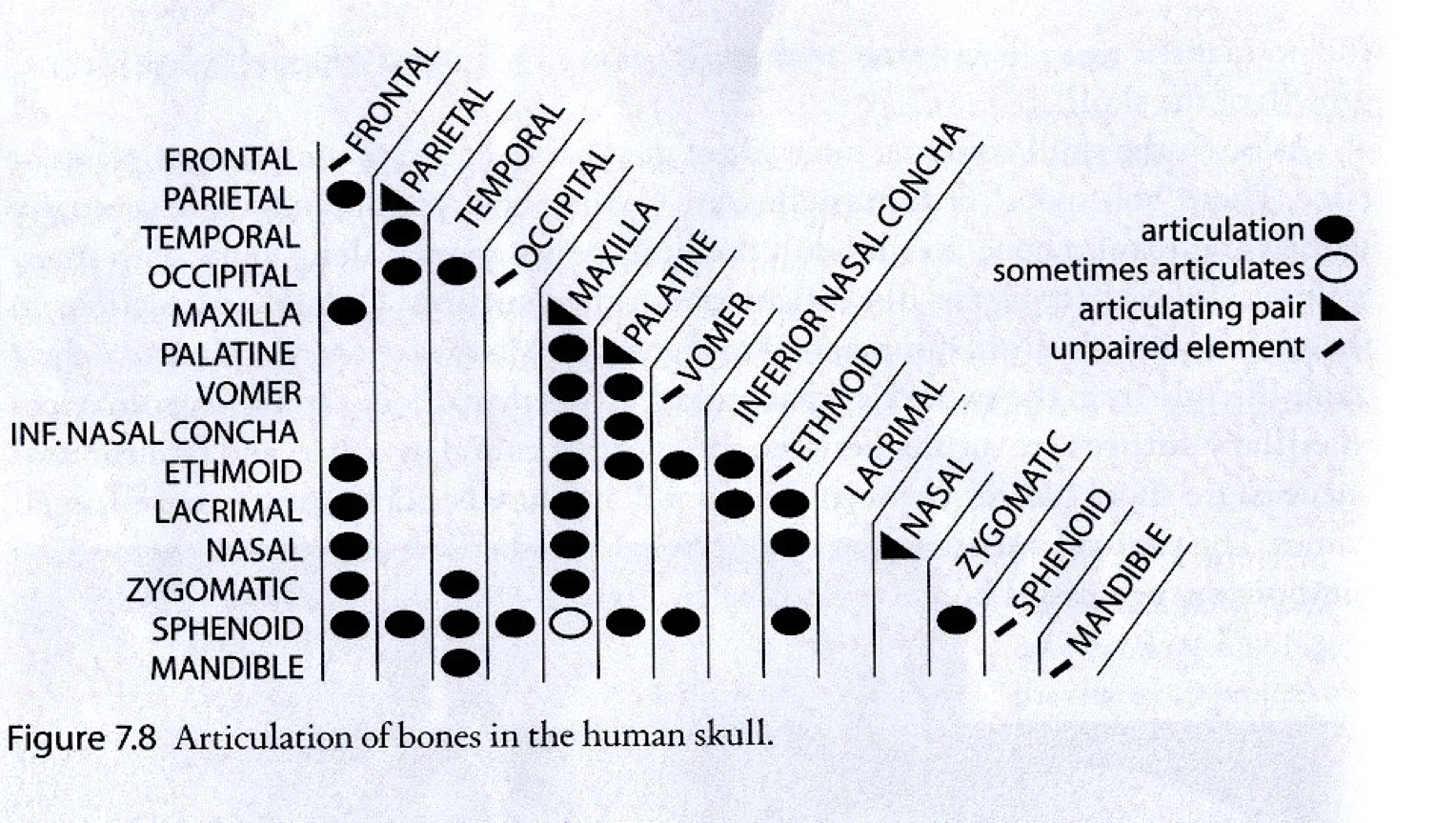
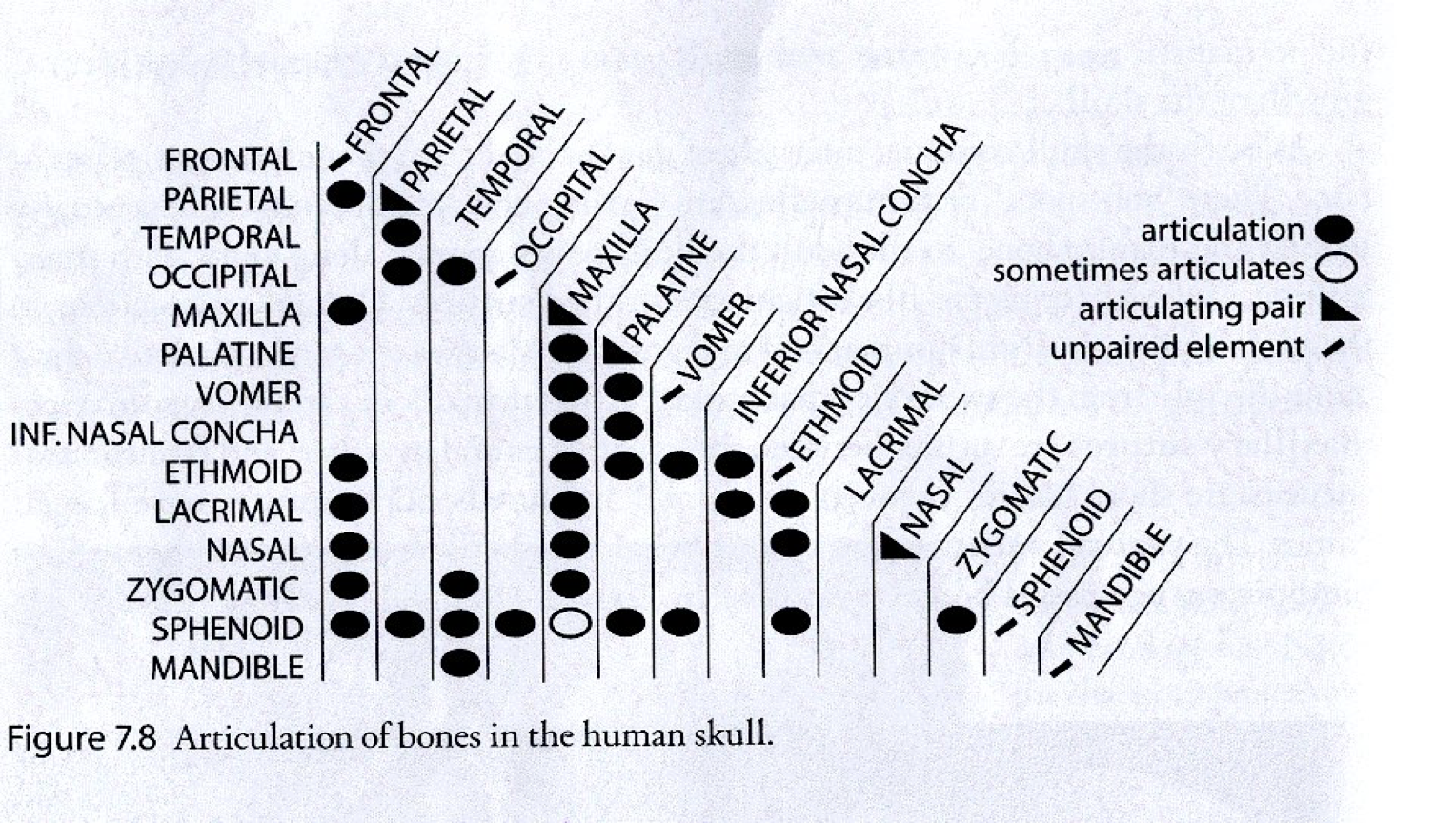
<p>Solid circle = articulation; open circle = sometimes articulates; triangle = articulating pair; slash = unpaired element</p>

Solid circle = articulation; open circle = sometimes articulates; triangle = articulating pair; slash = unpaired element

What do the symbols on the skull bone articulation chart mean?
31
New cards
<p>Solid circle = articulation; open circle = sometimes articulates; triangle = articulating pair; slash = unpaired element</p>

Solid circle = articulation; open circle = sometimes articulates; triangle = articulating pair; slash = unpaired element

Use this chart to study which skull bones articulate with one another.
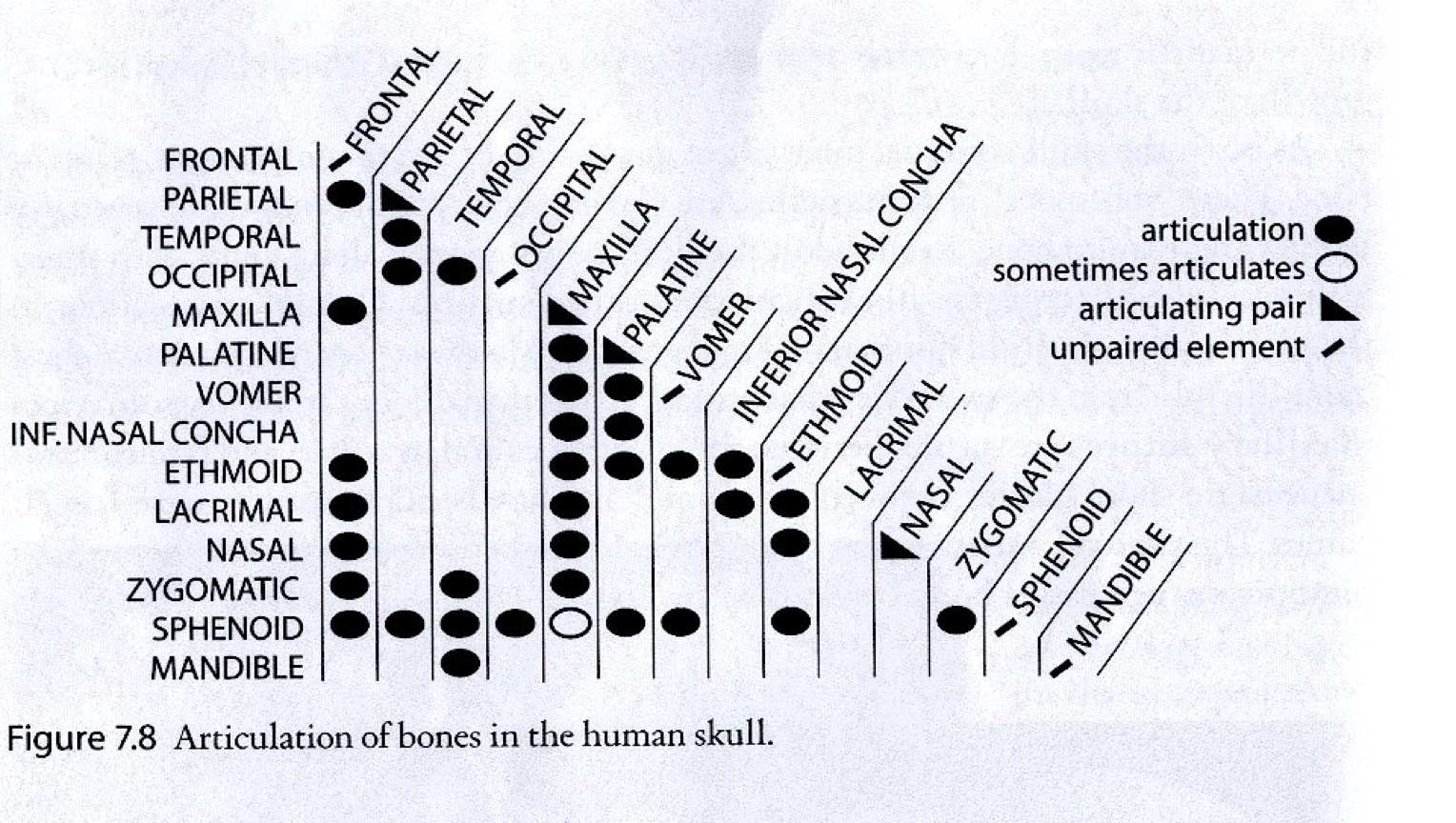
<p>Use this chart to study which skull bones articulate with one another.</p>
32
New cards
Gomphosis
What is a tooth in its socket, described as a “peg in a hole”?
33
New cards
Suture (synarthrosis)
What skull articulation consists of bones joined by connective tissue?
34
New cards
Synchondrosis
What articulation consists of bones joined by cartilage?
35
New cards

Gomphosis and suture = fibrous; synchondrosis = cartilaginous; TMJ = synovial

Classify the major skull articulation types shown. (IMAGE?)

36
New cards
Skull articulation types (IMAGE NEEDED — SLIDE 14)
Identify the articulation types shown and classify them as fibrous, cartilaginous, or synovial.
37
New cards
Coronal suture
Which suture is formed by the frontal and parietal bones?
38
New cards
Sagittal suture
Which suture is formed by the right and left parietal bones?
39
New cards
Lambdoid suture
Which suture is formed by the occipital and parietal bones?
40
New cards
Squamosal suture
Which suture is formed by the temporal and parietal bones?
41
New cards
Coronal, sagittal, lambdoid, squamosal sutures (IMAGE NEEDED — SLIDE 15)
Identify the major sutures shown in the image.
42
New cards
Median palatine suture
Which suture is formed by the right and left maxillae?
43
New cards
Transverse palatine suture
Which suture is formed by the maxilla and palatine bone?
44
New cards
Internasal suture
Which suture is formed by the right and left nasal bones?
45
New cards
Metopic/interfrontal suture — formed by right and left frontal bones; usually not present in the adult
Which suture is formed by the right and left frontal bones, and what is notable about it in adults?
46
New cards
Median palatine, transverse palatine, metopic, internasal sutures (IMAGE NEEDED — SLIDE 16)
Identify the sutures shown in the image.
47
New cards
Wormian (intrasutural) bones
What are bones located within skull sutures called?
48
New cards
Lambdoid suture
What is the most common location for Wormian/intrasutural bones?
49
New cards
Wormian/intrasutural bones (IMAGE NEEDED — SLIDE 17)
Identify the Wormian/intrasutural bones in the image.
50
New cards
Closure of the suture so that it becomes bone
What is stenosis of a skull suture as defined here?
51
New cards
Pterion
What landmark is the junction of the greater wing of the sphenoid, squamous portion of the temporal bone, frontal bone, and parietal bone?
52
New cards
Lambda
What landmark is the junction of the lambdoid and sagittal sutures between the occipital and parietal bones?
53
New cards
Bregma
What landmark is the junction of the coronal and sagittal sutures between the frontal and parietal bones?
54
New cards
Vertex
What is the superior point of the cranium in the midline with the skull in anatomical position?
55
New cards
Asterion
What landmark is the junction of the parietomastoid, occipitomastoid, and lambdoid sutures?
56
New cards
Glabella
What is the elevation superior to the root of the nose?
57
New cards
Nasion
What landmark is the junction of the frontonasal and internasal sutures?
58
New cards
Inion
What is the most prominent point of the external occipital protuberance?
59
New cards
Major skull landmarks (IMAGE NEEDED — SLIDE 19)
Identify pterion, lambda, bregma, vertex, asterion, glabella, nasion, and inion in the image.
60
New cards
Major skull landmarks — alternate views (IMAGE NEEDED — SLIDE 20)
Identify the major skull landmarks from these alternate views.
61
New cards
Anterior, middle, and posterior cranial fossae
What are the three cranial fossae?
62
New cards
Posterior border of the lesser wing of the sphenoid + anterior border of the chiasmatic sulcus
What marks the boundary between the anterior and middle cranial fossae?
63
New cards
Ridge of the petrous portion of the temporal bone + posterior border of the sella turcica (dorsum sellae)
What marks the boundary between the middle and posterior cranial fossae?
64
New cards
Cranial fossae and boundaries (IMAGE NEEDED — SLIDE 21)
Identify the anterior, middle, and posterior cranial fossae and their boundaries.
65
New cards
Crista galli; frontal crest; foramen cecum; foramina of cribriform plate; cribriform plate; orbital part of frontal bone; lesser wing of sphenoid; anterior clinoid process; body of sphenoid
What major structures are labeled in the anterior cranial fossa?
66
New cards
Anterior cranial fossa structures (IMAGE NEEDED — SLIDE 22)
Identify the labeled structures of the anterior cranial fossa.
67
New cards
Tuberculum sellae; chiasmatic sulcus; hypophysial fossa; dorsum sellae; posterior clinoid process; middle clinoid process
What central sphenoid/sella structures are labeled in the middle cranial fossa?
68
New cards
Optic canal; superior orbital fissure; foramen rotundum; foramen ovale; foramen spinosum; foramen lacerum; opening of carotid canal
What major openings are labeled in the middle cranial fossa?
69
New cards
Trigeminal impression; grooves/hiatuses for greater and lesser petrosal nerves; tegmen tympani; arcuate eminence; groove for middle meningeal artery
What temporal/petrous-region features are labeled in the middle cranial fossa?
70
New cards
Greater wing of sphenoid
Which sphenoid region forms much of the lateral middle cranial fossa in this diagram?
71
New cards
Middle cranial fossa structures (IMAGE NEEDED — SLIDE 23)
Identify the labeled structures of the middle cranial fossa.
72
New cards
Clivus; foramen magnum; internal occipital protuberance; internal occipital crest
What major midline structures are labeled in the posterior cranial fossa?
73
New cards
Internal acoustic meatus; jugular foramen; hypoglossal canal; jugular tubercle; grooves for sigmoid, transverse, and inferior petrosal sinuses
What openings and grooves are labeled in the posterior cranial fossa?
74
New cards
Superior border/ridge of the petrous portion of the temporal bone
What temporal-bone landmark helps form the anterior boundary of the posterior cranial fossa?
75
New cards
Posterior cranial fossa structures (IMAGE NEEDED — SLIDE 24)
Identify the labeled structures of the posterior cranial fossa.
76
New cards
Head, neck, condyle, trochlea, facet
What surface markings are associated with articulation with other bones?
77
New cards
Tuberosity, tubercle, crest, line, spine
What surface markings are associated with attachment of ligaments, tendons, or fascia?
78
New cards
Depressions: fossa, sulcus. Openings: foramen, canal, fissure.
What depressions and openings are associated with proximity or entry of vessels, nerves, or tendons?
79
New cards
Sinus, antrum, ramus, body, process, notch
What other external/internal bone features are listed?
80
New cards
Supraorbital → infraorbital → mental foramina
Which three facial foramina line up vertically?
81
New cards
Supraorbital, infraorbital, mental foramina (IMAGE NEEDED — SLIDE 26)
Identify the three vertically aligned foramina.
82
New cards
Venous sinuses
What structures create grooves on the internal surface of the skull?
83
New cards
Superior sagittal, inferior sagittal, straight, transverse, sigmoid, occipital, cavernous, intercavernous, sphenoparietal, superior petrosal, inferior petrosal, basilar sinus, confluence of sinuses
What major venous sinuses are labeled in the diagrams?
84
New cards
Venous sinus grooves and locations (IMAGE NEEDED — SLIDE 27)
Identify the named venous sinuses in the image.
85
New cards
Parietal bone (IMAGE NEEDED — SLIDE 29)
Identify the parietal bone in the different skull views shown.
86
New cards
Superior temporal line and inferior temporal line
What two temporal lines are found on the lateral skull/parietal region?
87
New cards
Superior vs inferior temporal lines (IMAGE NEEDED — SLIDE 30)
Identify the superior and inferior temporal lines.
88
New cards
Groove for superior sagittal sinus and depressions for arachnoid granulations
Which internal parietal/skull-vault features are emphasized here?
89
New cards
Internal parietal features (IMAGE NEEDED — SLIDE 31)
Identify the groove for the superior sagittal sinus and the depressions associated with arachnoid granulations.
90
New cards
Emissary veins
What veins are associated with the parietal foramina?
91
New cards
Parietal foramina (IMAGE NEEDED — SLIDE 32)
Identify the parietal foramina.
92
New cards
Frontal bone (IMAGE NEEDED — SLIDE 33)
Identify the frontal bone in the different skull views shown.
93
New cards
Frontal bone external landmarks (IMAGE NEEDED — SLIDE 34)
Identify the labeled external features of the frontal bone, including the squamous portion, metopic/interfrontal suture, supraorbital margin/notch/foramen, and superciliary region.
94
New cards
Trochlear fossa (IMAGE NEEDED — SLIDE 35)
Identify the indicated frontal-bone feature.
95
New cards
Supraorbital notch
The supraorbital foramen may also appear as what?
96
New cards
Supraorbital foramen/notch, nasion, metopic suture (IMAGE NEEDED — SLIDE 36)
Identify the labeled frontal-region structures in the image.
97
New cards
Occipital bone (IMAGE NEEDED — SLIDE 37)
Identify the occipital bone from posterior, inferior, and lateral views.
98
New cards
Basilar part; external occipital crest; external occipital protuberance; superior and inferior nuchal lines; foramen magnum; occipital condyle; pharyngeal tubercle
What major external/inferior features of the occipital bone are emphasized in this view?
99
New cards
External occipital features (IMAGE NEEDED — SLIDE 38)
Identify the major external/inferior occipital structures shown.
100
New cards
OC = occipital condyle → occipital bone; JF = jugular foramen → occipital + temporal bones; CC = carotid canal → temporal bone; arrows = tympanic canaliculus → temporal bone
Match the abbreviations/features with their associated bones.