neurology exam two
1/124
There's no tags or description
Looks like no tags are added yet.
Name | Mastery | Learn | Test | Matching | Spaced |
|---|
No study sessions yet.
125 Terms
frontal, parietal, temporal, occipital, insular
five lobes of the brain
gyrus
a convoluted ridge between anatomical grooves
sulcus
narrow groove
central sulcus
divides parietal and frontal lobes
lateral sulcus (sylvian fissure)
separates temporal lobe from frontal and parietal lobes
superior longitudinal fissure
divides left and right hemispheres
broadmann's areas
numbered brain regions based on types of neurons and the function of that region
main divisions of the frontal lobe
prefrontal cortex, premotor area, supplemental area, primary motor area
motor areas of the frontal lobe
primary motor cortex, premotor cortex, supplementary motor area
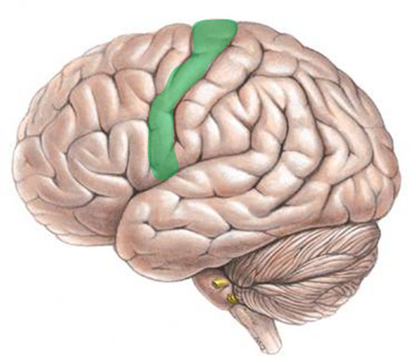
precentral gyrus
- also known as the primary motor cortex; is responsible for the control of simple motor movement
- brodmann area 4
voluntary motor activation

the origin of the corticospinal and corticobulbar tracts
the pre-central gyrus is responsible for
primary motor strip (aka the precentral gyrus)
narrow strip that goes from near the top of the head right down along where your ear is located
responsible for producing the contractions of all the muscles needed for the chosen movement with the brainstem and the spinal cord
determines how much force each muscle group must exert and then sends this information to the spinal motor neurons and interneurons that generate the movement itself, as well as the postural adjustments that accompany it
premotor region
motor planning
involved in sensory-guided movement (rather than internally generated movement)
for complex movements
mirror neurons in premotor regions
helps us internalize the actions of others
the premotor region is strongly connected with
the parietal lobe, motor strip/precentral gyrus, SMA, and prefrontal cortex
the pathology of the premotor region is
ideomotor apraxia
ideomotor apraxia
inability to imitate gestures or mimic using tools (i.e., "use a hammer")
from disorder in left parietal lobe and premotor cortex
great example:
https://www.youtube.com/watch?v=EvOYeqM-6CE
supplementary motor area (SMA)
portion of area six and eight, including medial surface
involved in internally-generated movement (i.e., making a decision to move, rather than responding to an external stimulus)
planning and rehearsal of the motor act
decision-making about movement
what area controls sequential movements
the supplementary motor areas
cerebellum
the supplementary motor area works with the _____________ to specify the precise sequence of contractions of the various muscles that will be required to carry out the selected motor action
the tissue makeup of an association cortex determines
what kind of neural information can be processed by cells in different areas of the brain
motor association areas are sites where
motor plans, programs, and commands are formulated with input from auditory and somatosensory processing, particularly touch, as well as other modalities
motor associations are very important for
the articulation of clear speech
the front of the frontal lobe
prefrontal area
prefrontal area
responsible for the ability to concentrate and attend, elaboration of thought
executive functioning
personality and emotional traits
planning
reigns in the impulsive aspects of our behavior
highly connected to the limbic system, which is primarily responsible for our emotional life
damage to the prefrontal area results in
-impairment of recent memory, inattentiveness, inability to concentrate, behavior disorders, difficulty in learning new information
-inappropriate social and/or sexual behavior
-emotional lability (especially when both hemispheres are damaged)
the prefrontal cortex is
the single largest brain region in human beings
estimated to constitute 29% of the total cortex
the prefrontal cortex is the only area of the brain that is
connect with all of the senses
cognitive processes
attention, memory, visuospatial processes, and linguistic processes
executive functions used for cognition to "execute" or complete tasks
inhibition of responses
accessing memory
organization of stimuli and memories
planning and sequencing: motor planning of a response
judgement
etc.
middle frontal gyrus
aka dorsolateral cortex
9, 10, 46, and 8 cognitive association areas
- implementing memory strategies
- working memory
- organization of material for encoding
- memory verification and evaluation
highest-level executive functions of the middle frontal gyrus
decision making
controlling executive functions
inferior frontal gyrus
consists of 44, 45, and 47 areas
responsible for risk aversion (area 47)
- addiction resistance (people with addiction tend to have lower activation in this area)
-implications in autism
-implications in tourette syndrome
inferior frontal gyrus: pars opercularis
area 44
-overlies insula
-people with autism may have smaller bilaterally
damage to the prefrontal cortex results in
disorders of categorizing, a decrease in voluntary motor behavior, decreased will and energy, a tendency to engage in repetitive or preservative behavior, difficulty in shifting response set, and abnormalities of affect and emotion, particularly apathy, indifference, and shallowness also occur
lobotomy
a psychosurgical procedure once used to calm uncontrollably emotional or violent patients
-the procedure cut the nerves connecting the frontal lobes to the emotion-controlling centers of the inner brain
walter freeman
doctor who streamlined lobotomies and made them extremely common
he shocked patients into unconsciousness instead of anesthesia and used an ice pick through the eye to get to the brain
orbitofrontal cortex
a region of the brain in which impulses involving reward processing, body response gnosis and interpretation, fear knowledge, drive and emotional regulation, mood
superior frontal gyrus
responsible for memory and memory strategies, memory verification, conjugate eye movement
oculomotor apraxia is a result of damage in this area
broca's area
controls language expression - an area of the frontal lobe, usually in the left hemisphere, that directs the muscle movements involved in speech
inferior frontal gyrus: pars triangularis
area 45
expressive language function
pars opercularis (44) and pars triangularis (45) make up broca's area
critical for motor planning for speech
expressive language function In dominant hemisphere
broca's aphasia (nonfluent aphasia)
area 44 and 45 make up
broca's area
expressive language impairment
ability to produce language is impaired
brain lateralization
the two hemispheres of the brain - the left and the right - are specialized for different functions
even though they work together, each area has plays a bigger role in some functions
left hemisphere (for most right-handed people) is known for
language (speech, reading, and writing)
logic, math, and analytical thinking
broca's and wernicke's areas (speech production and comprehension)
the right hemisphere, for many individuals, includes the
inferior frontal gyrus (right homologue of broca's area)
-located in the frontal lobe of the right hemisphere
-while broca's area on the left controls speech production, the right inferior frontal gyrus helps control
prosody (emotional tone and intonation in speech)
understanding and expressing emotional content through voice
turn-taking and social aspects of speech
both broca's aphasia and acquired apraxia of speech (AOS) are linked to damage in
the left frontal lobe, especially in areas around broca's area
broca's area is located in
the left hemisphere, specifically the left inferior frontal gyrus (brodmann areas 44 and 45)
suppelementary motor areas (sma) and the premotor cortex are involved in
planning and programming the movements needed for speech
broca's aphasia
condition resulting from damage to broca's area, causing the affected person to be unable to speak fluently, to mispronounce words, and to speak haltingly
comprehension is often better than expression
apraxia of speech (AOS)
an impairment of motor programming and planning of speech movements
phineas gage
railroad worker who survived a severe brain injury to his prefrontal cortex that dramatically changed his personality and behavior; the case played a role in the development of the understanding of the localization of brain function
afferent neurons
otherwise known as sensory or receptor neurons--carry nerve impulses from receptors or sense organs toward the central nervous system
primary sensory cortex
occupies broadmans 1, 2, and 3
processes somatosensory information such as:
vibration
propioception
touch
astereognosis (stereognosis)
somatosensory association cortex
occupies broadman’s 5 and 7
interprets sensory experience during motor movements
sensory movements used to refine motor action
involved in the fine movements associated with speech
plays role in writing sensory and motor experience
angular gyrus
- occupies broadman's area 39
-primarily involved in semantic processing, particularly in the left hemisphere
-plays a key role in reading, especially when making semantic associations, and is important for understanding speech and written language
-involved in arithmetic fact retrieval, shifting attention toward salient stimuli related to motion, emotion, value, or meaning
-helps in the ability to discriminate left from right and is associated with verbal working memory
-may play a role in understanding metaphors and self-recognition, as well as in theory of mind
damage to the angular gyrus results in
angular gyrus syndrome:
agraphia, alexia, gerstmann's syndrome, poor memory, behavioral manifestations such as depression, frustration, and irritability
gerstmann's syndrome
a cognitive impairment caused by damage to the left parietal lobe, specifically the angular gyrus
-may result from a stroke or other damage to the parietal lobe
-characterized by four main symptoms: writing disability (agraphia or dysgraphia), difficulty with arithmetic (acalculia or dyscalculia), inability to distinguish right from left, and inability to identify fingers (finger agnosia)
-some adults with the syndrome may also experience aphasia
parietal lobe: supramarginal gyrus
-occupies BA 40
- closely rated to the angular gyrus (BA 39)
- involved in phonological system; stores auditory representations of phonemes (auditory images)
- helps us sound out words
- damage can result in phonological dyslexia, difficulty reading new and nonwords
supramarginal gyrus
spatiomotor tasks - identifying functional orientation of object
phonological processing of language - phonological processing of speech and written language
the right hemisphere of the supramarginal gyrus is
involved in learning phonological sequences
the left hemisphere of the supramarginal gyrus is
more active in well-learned sequences
Some Say Marry Money But My Brother Says Big Brains Matter More
sensory, motor, or both in cranial nerves
upper motor neurons originate in the
motor cortex
most cranial nerve nuclei have _________ innervation
bilateral
CN I - olfactory nerve
function: smell
location: telencephalon
clinical sign of damage: anosmia (loss of smell)
CN II - optic nerve
function: vision
location: diencephalon
clinical sign of damage: vision loss, visual field defects
CN III - oculomotor nerve
•eye movement, eyelid elevation, pupil constriction
•damage: ptosis, eye deviates down/out, diplopia
CN IV - trochlear nerve
•superior oblique muscle (eye down & in)
•damage: vertical diplopia
CN VI - abducens nerve
•lateral rectus muscle (eye out)
•damage: medial strabismus, diplopia
CN V - trigeminal nerve
sensory: Face, scalp, cornea, oral & nasal cavities
motor: muscles of mastication (chewing)
nuclei: pons (chief sensory, spinal trigeminal, mesencephalic)
corticobulbar tract: bilateral innervation
damage:
LMN lesion: ipsilateral jaw weakness, jaw deviates to weak side, loss of corneal reflex (afferent limb)
UMN lesion: minimal effect due to bilateral innervation
branches:
ophthalmic: sensory
maxillary: sensory
mandibular: mixed
functions:
sensory: tactile facial sensation
motor: mastication (masseter, temporalis, medial & lateral pterygoid), tensor veli palatini, mylohyoid, anterior digastric, tensor tympani
muscles of mastication:
masseter, temporalis, medial pterygoid, lateral pterygoid
CN VII - Facial Nerve
Motor: Muscles of facial expression (like smiling, frowning)
Sensory: Taste from the front 2/3 of the tongue
Parasympathetic: Controls lacrimal (tear) and salivary glands
Nuclei: Located in the pons
Corticobulbar tract:
Upper face: Bilateral control (both sides of brain)
Lower face: Contralateral control (opposite side of brain)
Damage:
LMN lesion: Complete facial paralysis (like Bell's palsy) on one side of the face
UMN lesion: Weakness in the lower part of the face (forehead stays unaffected)
Motor Functions:
Controls muscles of facial expression, posterior belly of digastric (raises hyoid), stylohyoid, and stapedius (ear muscle)
Sends impulses to tear and salivary glands
Sensory Functions:
Taste: Anterior 2/3 of the tongue
General sensation: Skin on the ear and small area behind the ear
Damage Effects:
Motor Damage: Paralysis of one side of the face, including emotional movements (like smiling). The face may look asymmetrical, with wrinkles smoothing out on the affected side.
CN VIII - Vestibulocochlear
Vestibular: Balance, equilibrium
•Cochlear: Hearing
•Nuclei: Pons/Medulla junction
Damage:
•Vestibular: Vertigo, imbalance
•Cochlear: Sensorineural hearing loss
Has been known as the Auditory Nerve
The nerve originates in the ear and carries sensory info.
There are 2 branches: the cochlear which is responsible for hearing and the vestibular branch which is responsible for balance
CN IX - Glossopharyngeal
Sensory: Taste posterior 1/3 tongue, carotid body
Motor: Stylopharyngeus (swallowing)
Parasympathetic: Parotid gland
Nuclei: Medulla
Damage:
Loss of gag reflex (afferent limb)
Dysphagia
The motor component of this nerve
supplies the stylopharyngeus muscle
Carries visceral sensory information from the carotid sinus (of the carotid artery) and body.
Provides general sensory information from the skin of the external ear, the internal surface of the tympanic membrane, the upper pharynx, and the posterior one-third of the tongue.
Provides taste sensation from the posterior one-third of the tongue
Damage to CNIX
Lesions to CNIX also typically involve CNX Reduced pharyngeal sensation
Decreased gag Reduced pharyngeal elevation Sometimes glossopharyngeal neuralgia is reported (radiating throat pain)
stylopharyngeus muscle
elevates and opens pharynx, elevates the larynx
neuralgia
a stabbing, burning, and often severe pain due to an irritated or damaged nerve
CN XII - Hypoglossal
Motor: Intrinsic & extrinsic tongue muscles
Nuclei: Medulla
Corticobulbar: Contralateral innervation (mostly)
Damage:
LMN lesion: Ipsilateral tongue weakness, atrophy, fasciculations, tongue deviates toward lesion
UMN lesion: Contralateral tongue weakness, deviates away from lesion
Oh Oh Oh To Touch And Feel Very Good Velvet AH
olfactory, optic, oculomotor, trochlear, trigeminal, abducens, facial, vestibulocochlear, glossopharyngeal, vagus, accessory, hypoglossal
trigeminal neuralgia
characterized by severe lightning-like pain due to an inflammation of the fifth cranial nerve
severe facial pain due to trigeminal nerve dysfunction
90% of trigeminal neuraligia is due to
compression by the Superior Cerebellar Artery (SCA)
treatment options for trigeminal neuraligia
Medical Management
Carbamazepine (anti-convulsant)
Gabapentin (mechanism unclear, but effective)
2. Surgical Interventions
Microvascular decompression
Radiofrequency ablation
Gamma Knife surgery
afferent
sensory
efferent
motor
somatic
body wall and skeletal muscle
controls voluntary muscles and relays conscious sensory input
visceral
organs and glands
controls involuntary functions & unconscious sensory input
general
widespread functions
special
unique functions of cranial nerves
CN X - Vagus
Motor Functions:
Controls muscles for phonation (voice), swallowing, and the soft palate
Supplies muscles of the pharynx, most of the larynx, and one muscle of the tongue
Parasympathetic: Controls heart, lungs, and GI tract
Sensory Functions:
Sensation from the thoracic & abdominal organs, larynx, esophagus, and trachea
Provides taste from the epiglottis and sensory from the ear and pharynx
Branches Important for Speech/Swallowing:
Pharyngeal, Superior Laryngeal, Recurrent Laryngeal
Unique Feature:
The only nerve that extends from the medulla and travels all the way to the abdomen
It's called "Vagus" because it "wanders" from the brainstem to the colon
Damage Effects:
LMN lesion: Affects voice, swallowing, and gag reflex
Unilateral: Weakness in the soft palate and voice
Bilateral: Significant issues with resonance, voice, phonation, and clarity of speech
motor association areas are extremely important for
highly coordinated and specific functions like, clearly articulated speech, chewing gum, and chewing gum and speaking clearly at the same time!
the prefrontal area doesn’t fully develop until
twenty-five
the frontal lobe is highly connected to the
limbic system
result of lobotomy on brain
more white matter damage than gray
some of the tracts could not recover over time
priming
repeating words
grammarian
leaving out connecting words like is, the, etc.
theory of mind
not everyone shares the same thoughts
all cranial nerve nuclei have bilateral innervation besides
the lower face (CN VII) and the tongue (CN XII)
CN I, II, III, and IV are located in the
midbrain
CN V, VI, VII, VIII, and are located in the
pons
the largest cranial nerve is
CN V: Trigeminal
Bell's Palsy
A condition that causes sudden, temporary weakness or paralysis of the muscles on one side of the face due to facial nerve inflammation - IPSILATERAL
IX, X, XI, and XII are located in the
medulla