PT21: Lower Limb Overview
 Call Kai
 Learn
Learn Practice Test
 Spaced Repetition
 Match
Match Flashcards
 Knowt Play
1/100

There's no tags or description
Looks like no tags are added yet.
Name | Mastery | Learn | Test | Matching | Spaced | Call with Kai | Chat |
|---|
No analytics yet
Send a link to your students to track their progress
101 Terms
Lower limbs primary function
weight bearing
Lower limbs secondary function
locomotion
Hip joint
structure favors stability; supported by strong ligaments of fibrous capsule; extensive muscular support
Lower limb region
extends from pelvis to phalanges
Gluteal region
buttocks and hips
Superior gluteal region
iliac crest
Lateral gluteal region
greater trochanter
Inferior gluteal region
gluteal fold
Medial gluteal region
intergluteal cleft
Thigh region
femoral region
Proximal-anterior thigh region
inguinal ligament
Proximal-posterior thigh region
gluteal fold
Distal-anterior thigh region
knee
Distal-posterior thigh region
popliteal fossa
Leg region
calf and shin
Anterior leg region
knee
Posterior knee region
popliteal fossa
Distal knee region
ankle
Foot region (pes/pedis)
dorsum; plantar surface
Proximal foot region
ankle
Distal foot region
toes
Lower limb osteology
pelvic bone, femur, patella, tibia, fibula, 7 tarsals, 5 metatarsals, 14 phalanges
Pelvic bones
paired; fused ilium, ischium, and pubis
Pelvic girdle
comprised of pelvic bones and sacrum; distributes body mass to lower limbs
Femur
long bone of thigh; articulates with pelvic bone and tibia and patella
Tibia and fibula
long bones of leg; both articulate with talus
Tibia
weight bearing; articulates with femur and patella
Patella
sesamoid bone; helps quadriceps muscles move tibia; protection for knee joint
Tarsal proximal row
talus and calcaneus
Tarsal distal row
navicular, cuboid, medial cuneiform, intermediate cuneiform, and lateral cuneiform
Foot bones
tarus, metatarsus, antetarsus
Hallux
first toe
Superficial fascia
deep to skin; composed of loose connective tissue, adipose tissue, cutaneous nerves, superficial veins, and lymphatics
Deep fascia types
fascia lata, crural fascia, intermuscular septa, retinacula
Fascia lata
deep fascia of thigh; consists of iliotibial tract and saphenous opening
Iliotibial tract/band
lateral thickening of deep fascia; stability for lateral thigh/knee
Saphenous opening
hiatus below inguinal ligament; great saphenous vein drains into femoral vein here
Intermuscular septa of thigh
from deep aspect of fascia lata; attaches to linea aspera
What are the three compartments that the intermuscular septa of the thigh separate?
anterior, posterior, medial
Crural fascia
deep fascia of leg; distal continuation of fascia lata from iliotibial tract to foot
Intermuscular septa of leg
from deep aspect of crural fascia; attach to fibula
What are the three compartments that the intermuscular septa of leg separates?
anterior, lateral, posterior
Posterior compartment of leg layers
triceps surae and deep flexors
Retinacula
extensions of deep fascia; tendon stabilization
Plantar aponeurosis
modified deep fascia; supports and protects structures on plantar aspect of foot; contributes to arch support
Anterior thigh general actions
knee extensors and hip flexors
Anterior general thigh innervation
femoral n.
Medial thigh general actions
hip adductors and hip flexors
Medial thigh innervation
obturator n.
Posterior thigh general actions
knee flexors and hip extensors
Posterior thigh innervation
sciatic n.
Anterior leg general actions
dorsiflexion and toe extension
Anterior leg innervation
deep fibular n.
Lateral leg general actions
eversion and plantar flexion
Lateral leg innervation
superficial fibular n.
Posterior leg general actions
plantar flexion and knee flexion
Posterior leg innervation
tibial n.
Lumbar plexus
nerve network formed from fibers in ventral rami of spinal nerves L1-L4; innervates anterior and medial thigh; supplies part of abdominal wall and external genitalia
Sacral plexus
nerve network formed from fibers in ventral rami of spinal nerves L4-S4; innervates gluteal region, posterior thigh and leg; supplies part of pelvis and perineum
Lumbar plexus nerves
femoral nerve and obturator nerve
Sacral plexus nerves
sciatic nerve and gluteal region nerves
Sciatic nerve branches
common fibular n. and tibial n.
Tibial nerve branches
medial and lateral plantar nerves
Common fibular nerve branches
superficial and deep fibular nerves
L2-L3 myotome action
hip flexion
L2-L3 myotome memory device
lift my knee
L2-L3 myotome nerve
femoral n.
L3-L4 myotome action
knee extension
L3-L4 myotome memory device
kick the door
L3-L4 myotome nerve
femoral nerve
L4-L5 myotome action
ankle dorsiflexion
L4-L5 myotome memory device
foot points to the sky
L4-L5 myotome nerve
deep fibular n.
L5-S1 myotome action
toe extension
L5-S1 myotome memory device
toes point to the sun
L5-S1 myotome nerve
deep fibular n.
S1-S2 myotome action
plantar flexion
S1-S2 myotome memory device
on the tips of my shoes
S1-S2 myotome nerve
tibial n.
S2-S3 myotome action
toe abduction
S2-S3 myotome memory device
toes spread free
S2-S3 myotome nerve
medial and lateral plantar nerves
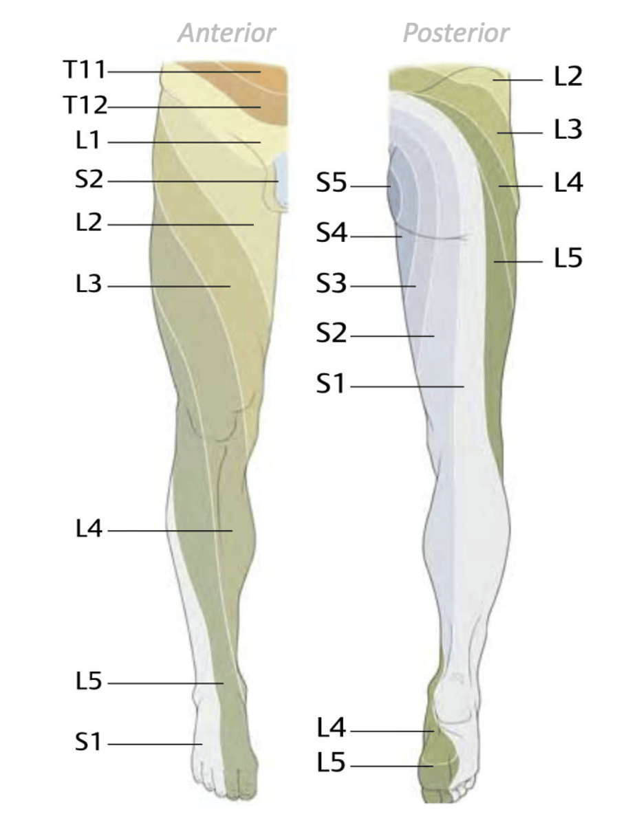
Cutaneous innervation
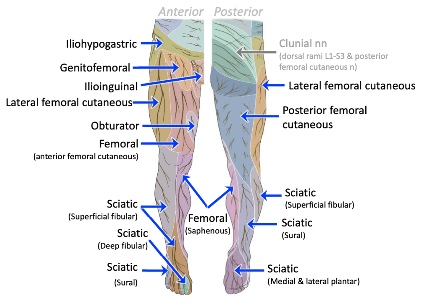
Lower limb dermatomes
spiral pattern due to limb rotation during embryonic development; most areas overlap
Lower limb arterial pathway
abdominal aorta -> common iliac a. -> external/internal iliac aa. -> femoral a.
Femoral artery branches
profundus femoris a. and popliteal a.
Profundus femoris artery branches
lateral and medial circumflex femoral arteries; 3-4 perforating arteries
Popliteal artery branches
anterior and posterior tibial arteries
Anterior tibial artery branch
dorsalis pedis artery
Posterior tibial artery branches
fibular a., medial and lateral plantar arteries
Superficial veins of lower limb
veins that run independent from arteries
Deep veins above the knee
true veins that accompany arteries
Deep veins below the knee
venae comitantes accompany arteries
Veins of lower limb
superficial and deep layers; all drain toward IVC; contain valves
Great saphenous vein drains into...
femoral vein
Great saphenous vein course
arises from medial side of foot and passes through saphenous opening
Small saphenous vein drains into...
popliteal vein
Small saphenous vein course
arises from lateral side of foot
Popliteal nodes
receives lymph from vessels traveling with small saphenous vein; in popliteal fossa
Superficial inguinal nodes
receives lymph from vessels traveling with great saphenous vein; in femoral triangle