Equine Forelimb Radiology
1/87
Earn XP
Description and Tags
Sourced from Quizlet
Name | Mastery | Learn | Test | Matching | Spaced | Call with Kai |
|---|
No study sessions yet.
88 Terms
- To confirm a special diagnosis
- Standard radiographic series if evaluation is inconclusive
- Pre-purchase exams
- Additional special views for surgery
What are the indications for equine limb rads?
- Sedation (adapt to swaying)
- Brush hair coat to remove debris
- Remove splint/bandage
What are steps in preparing patients before rads?
handle
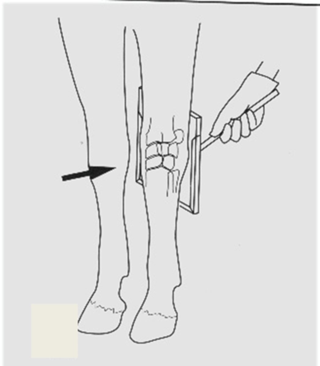
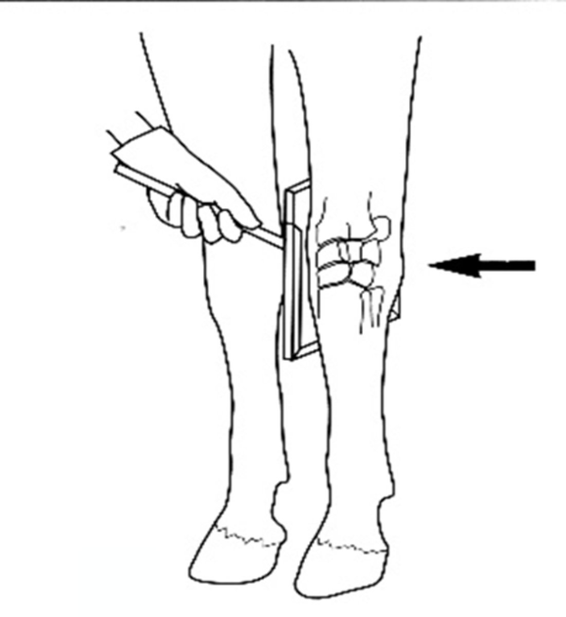
Cassette holders should have a long ____________ for the safety of the holder during exposure (follow ALARA!)
Area where there is a lot of muscle, and soft tissue may attenuate the radiographs
Why might you need a grid when radiographing stifles?
Describes path of x-ray beam as it enters and exits region of interest
What do the names of radiographic views tell you ?
Beam enters the lateral surface and exposes the cassette on the medial aspect of the limb
Describe the path of the beam in a lateromedial (LM) view
parallel
When thinking about radiographic views, it is assumed that the beam is _______________ to the ground
Carpus
When moving distally down the limb, when does naming terminology switch from cranial/caudal to dorsal/palmar/plantar?
Horses are standing for rads and you are trying to place the expensive X-ray tube as far away from the horse's hooves as possible
Why do you normally have more LM than ML views in horses?
- Dorsal/cranial
- Lateral
Where are markers always placed?
Left or Right (not lateral)
What do the actual letters of the markers indicate?
Accessory carpal bone
How can you tell the lateral side of a carpus if you do not have a marker?
Fetlock
At what joint is it nearly impossible to tell medial or lateral (or fore or hind) without a marker?
- Medial
- Lateral
- Proximal
- Distal
Ex. Dorsolateral-palmaromedial oblique (DLPMO)
What directional terms are used in naming oblique views?
before
When naming oblique views, the degrees angled should be placed _____________ the positional term it affects
- Enters 30 degrees proximal from the perpendicular plane (dorsal plane)
- Leaves 30 degrees distally from the palmar aspect of the limb
Describe the path of the beam in a Dorso-30o-proximal palmarodistal oblique view
To view the joint space without superimposition and cope with the natural angulation of the horse's joints (weight bearing during exposure)
Why do you take more oblique views in horses?
- Right/Left (limb)
- Dorsal/Palmar or Cranial/Caudal
- Proximal/Distal
- Medial/Lateral
What is the proper order for naming the directions in oblique views?
You get the same radiograph!
Why can you take a view in the opposite direction (ex. lateromedial or mediolateral) for convenience purposes (don't want to put the X-ray tube under the horses abdomen)?
Changes the angulation and alignment of the whole limb
Why is it important that the horse be weight bearing on the limb being radiographed?
- Technique
- Check that series is complete for region
- Soft tissue analysis (Swelling, if yes, where? (association with joint space); Subcutaneous gas; Mineralization)
- Axis of extremity (angular deformity, deviated due to fracture malalignment)
- Bone opacity: lucency or sclerosis
- Articular and bone margins
- Periosteal, endosteal new bone formation
- Normal anatomy vs. pathology vs. artefact
What are important considerations for actual image interpretation?
- Mediolateral
- Cranio 45 medial-caudolateral
+/- Cranioproximal-craniodistal (skyline)
What are the standard views for the shoulder (glenohumeral joint)?
- Shoulder
- Elbow
What are the forelimb joints that are radiographed non-weight bearing?
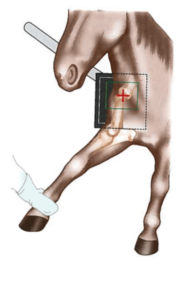
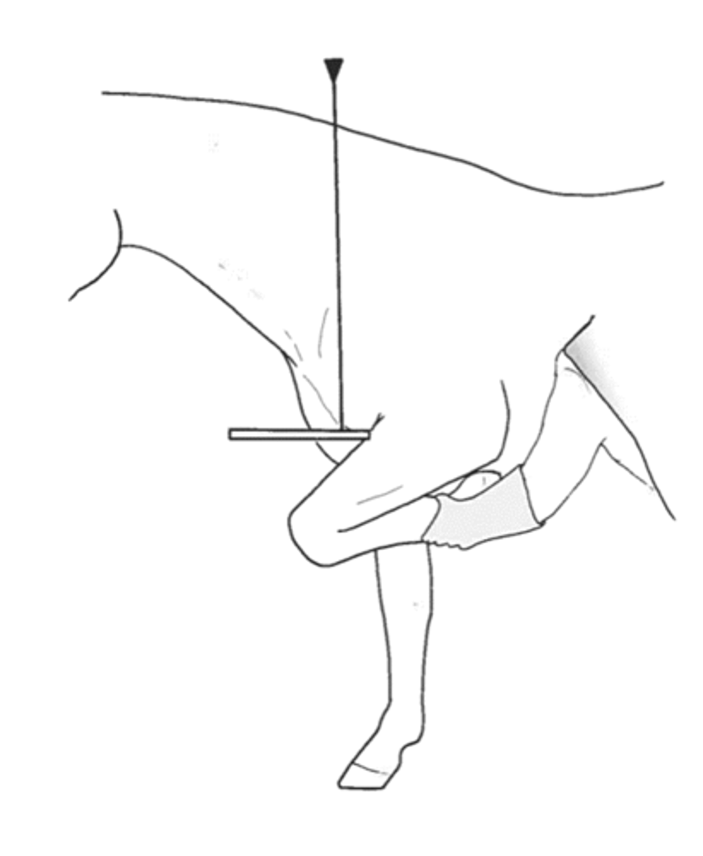
Pull the limb cranially (removes superimposition of spine and first rib)
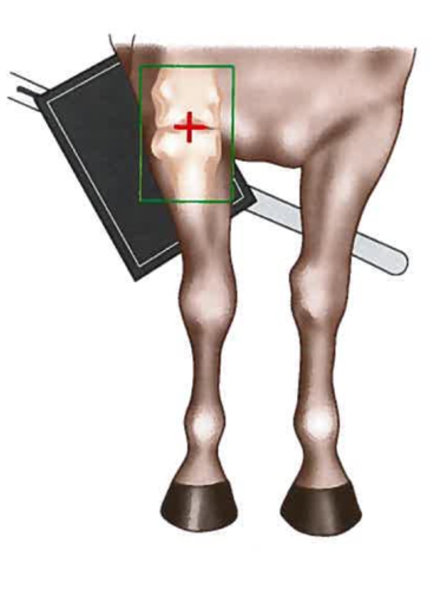
How is the horse positioned for shoulder radiographs?
- Tubercles of the humerus
- Articular surface of humeral head
- Glenoid cavity
- Supraglenoid tubercle
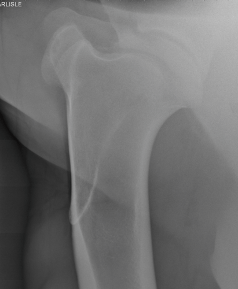
What structures are highlighted by a mediolateral shoulder rad?
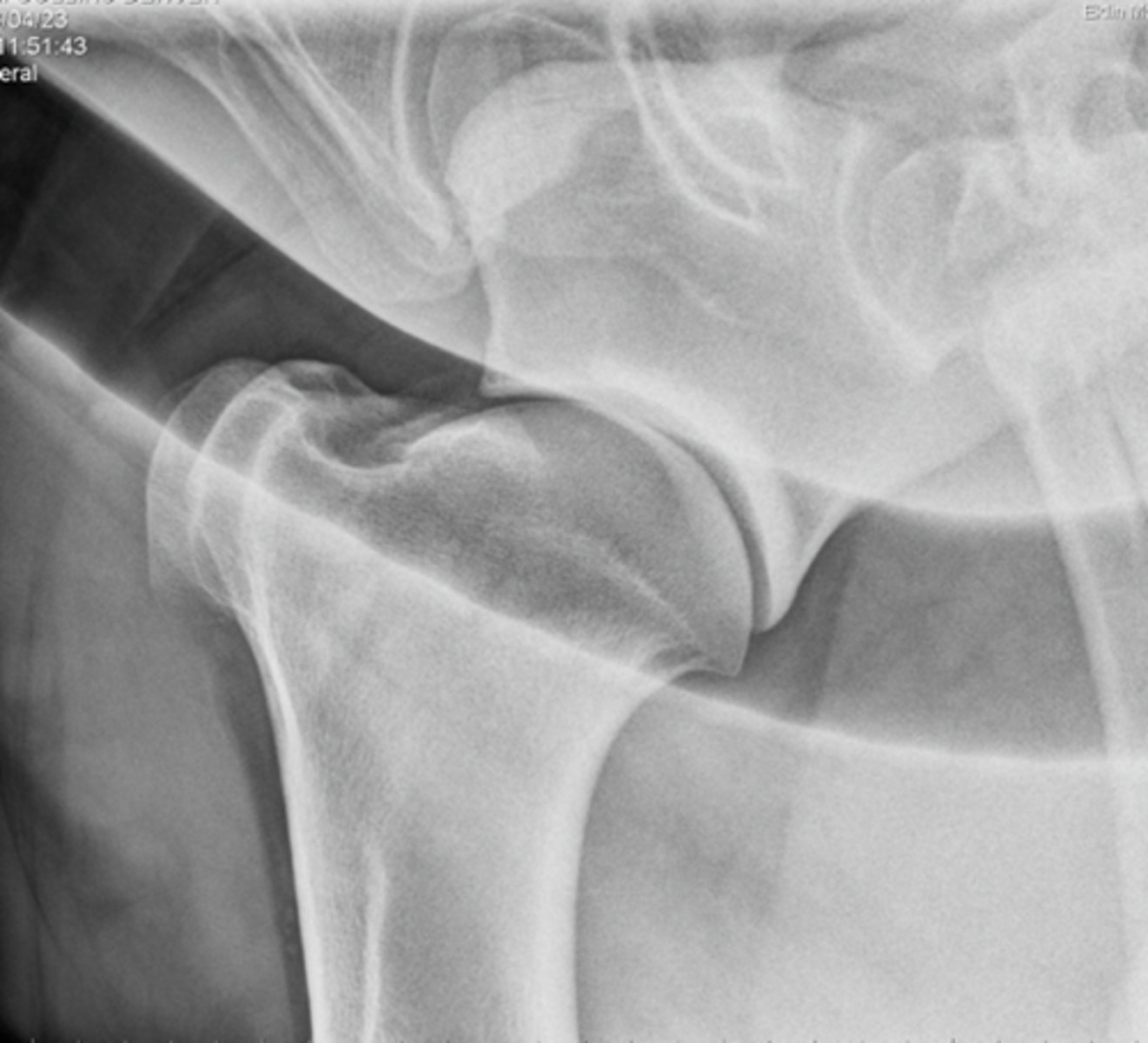
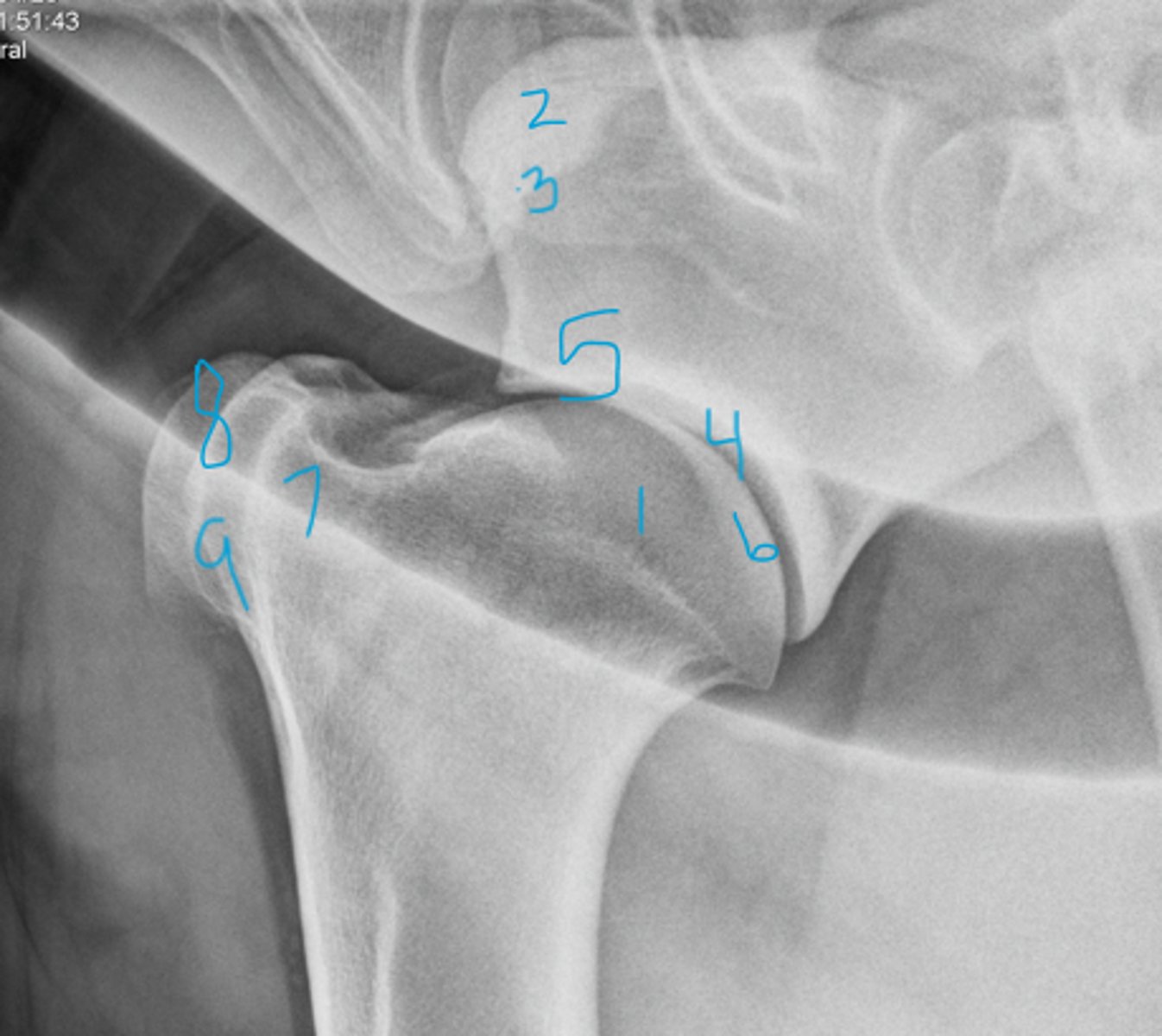
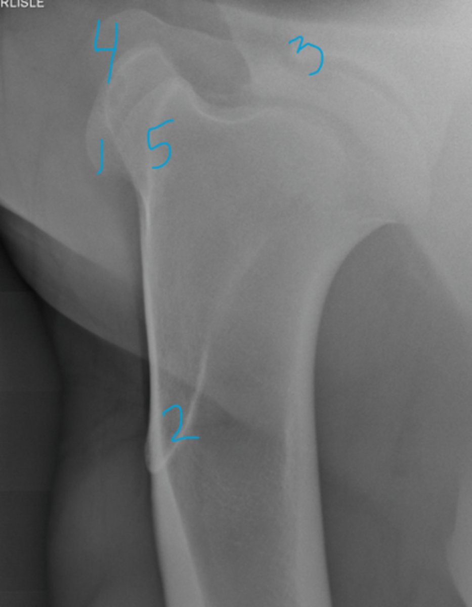
Mediolateral
What view is being taken?

1 - Head of humerus
2 - Supraglenoid tubercle
3 - Coracoid process
4 - Medial rim of glenoid
5 - Glenoid notch
6 - Lateral rim of glenoid
7 - Greater tubercle
8 - Intermediate tubercle
9 - Lesser tubercle
Name the structures
Cranial 45 Medial-Caudolateral view
What view is being taken?
- Craniodorsal aspect of greater tubercle
- Cranial aspect of intermediate tubercle
- Deltoid tuberosity
What structures are highlighted by a cranial 45 medial-caudolateral view?
1 - Intermediate tubercle
2 - Deltoid tuberosity
3 - Coracoid process
4 - Greater tubercle
5 - Lesser tubercle
Name the structures
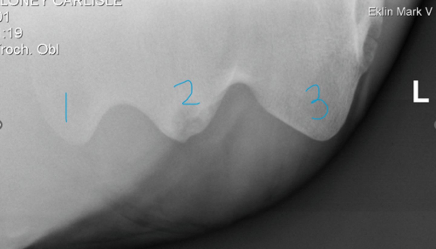
CranioProximal- Craniodistal (Skyline)
What view of the shoulder joint is taken to highlight the humeral tubercles?
1 - Lesser tubercle
2 - Intermediate tubercle
3 - Greater tubercle
Name structures
L marker indicates left humerus and the marker is always placed on the lateral side (greater tubercle is lateral)!
How do you know which limb this is and that 3 is the greater tubercle?

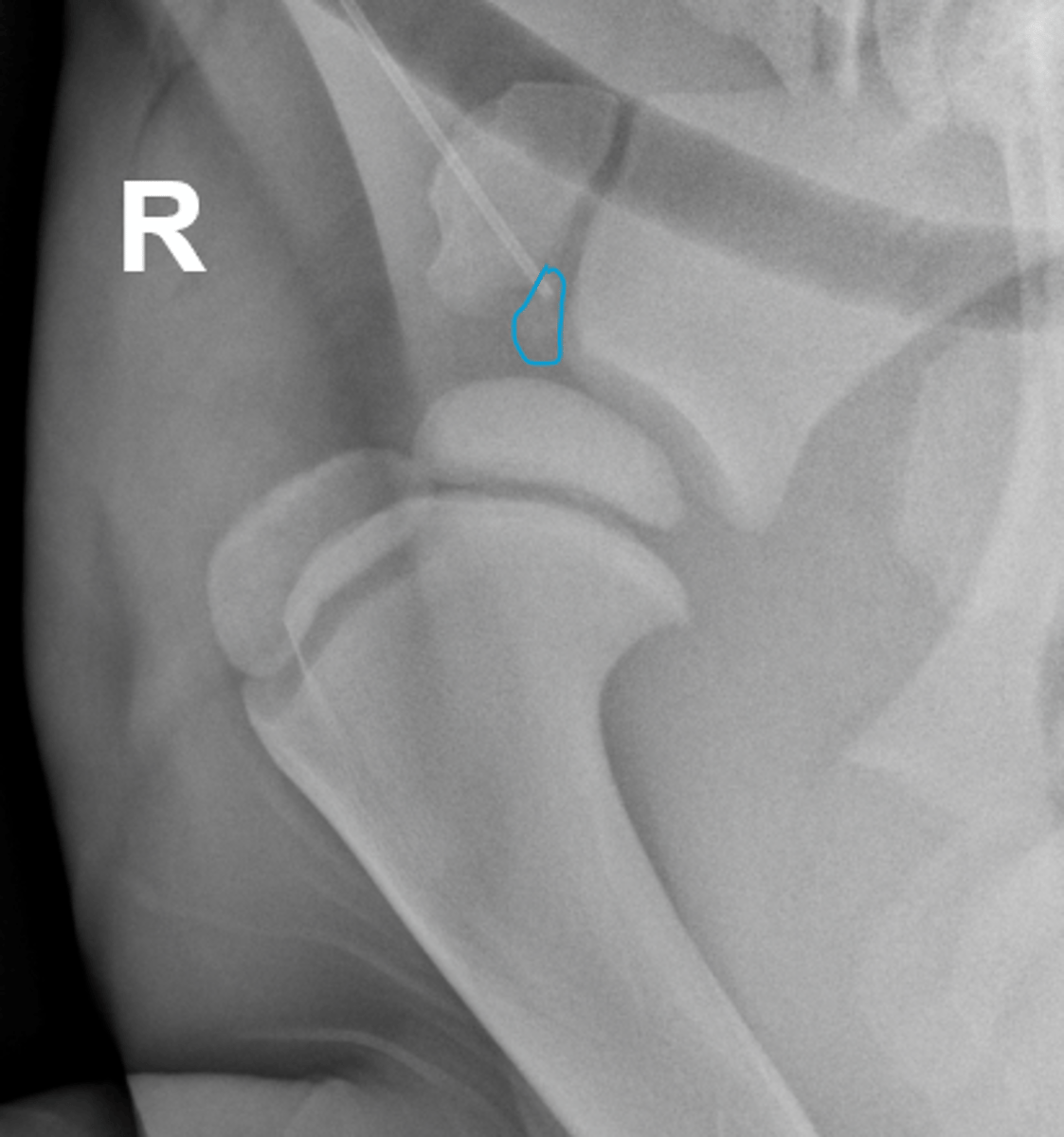
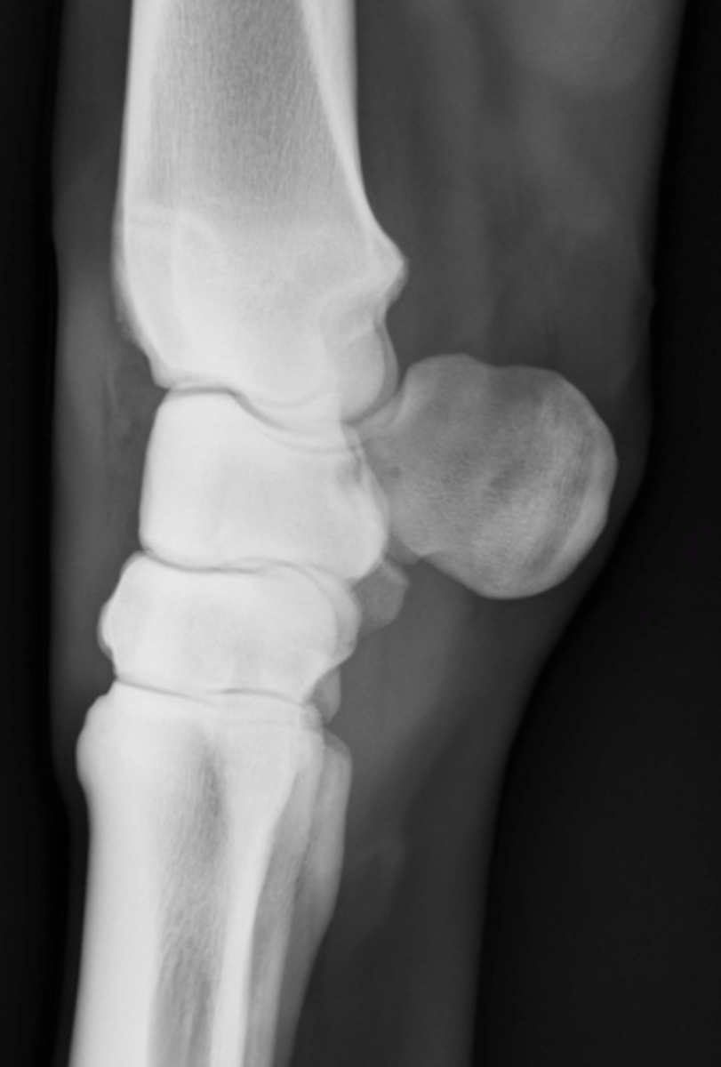
Foal (open physis)
What can you assume about the age of this horse?
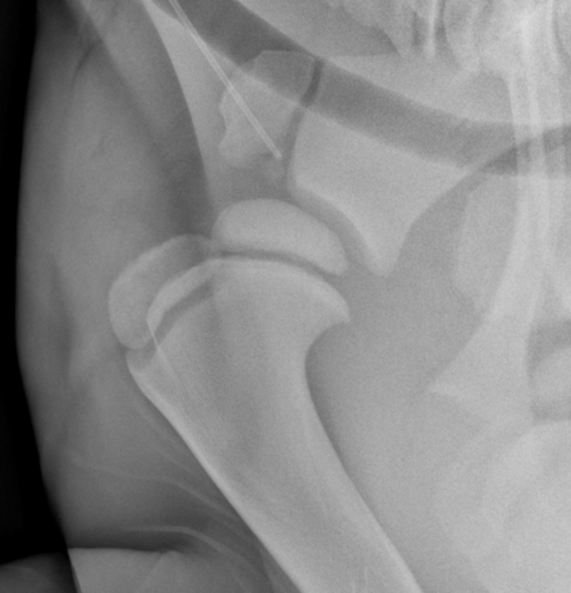
Cranial part of the glenoid cavity has yet to fuse (young animal)
How do you know that the circled more radiolucent structure is normal? (ignore catheter)
- Mediolateral
- Craniocaudal
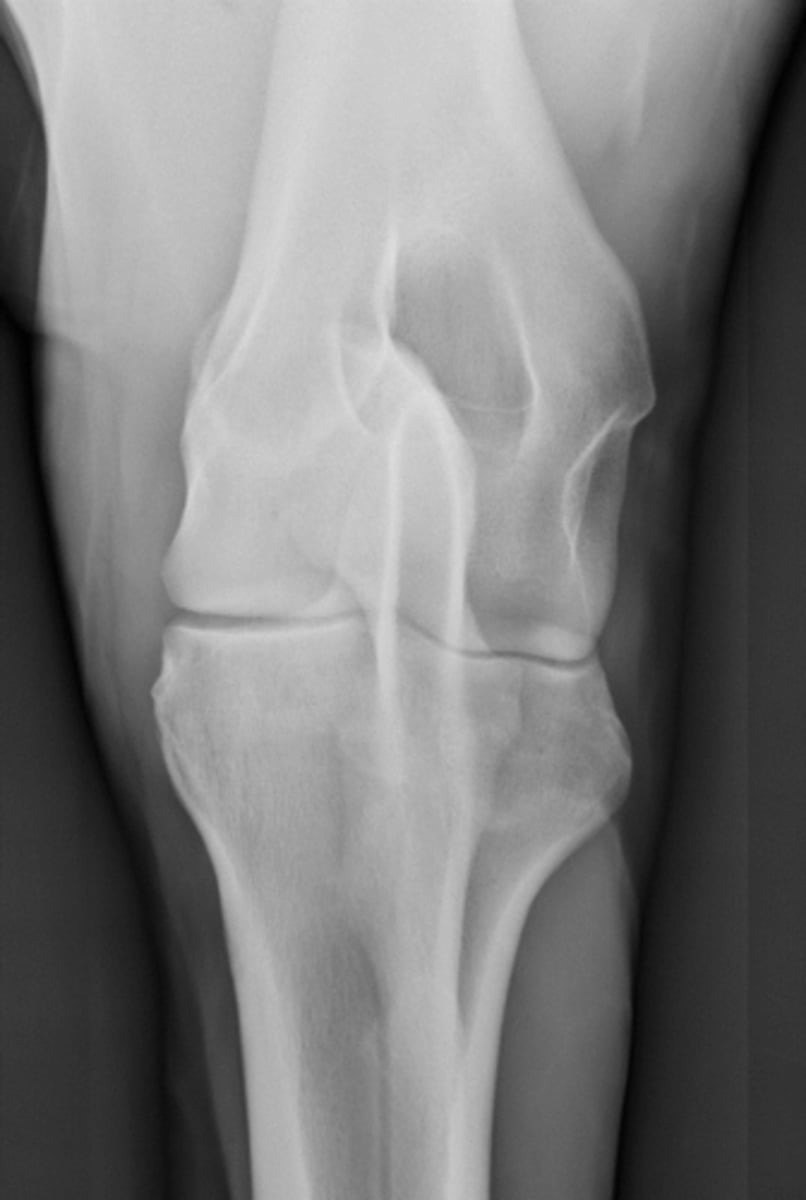
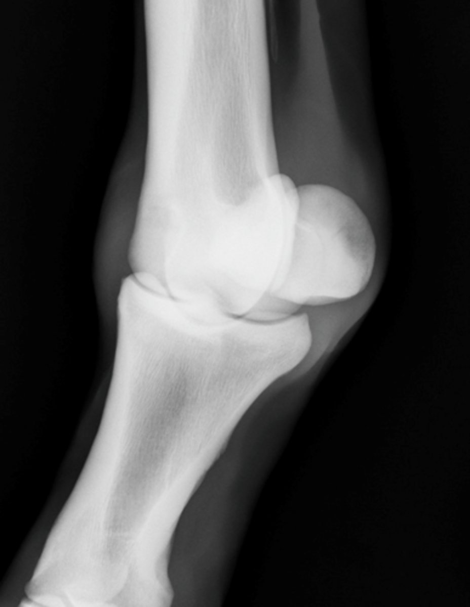
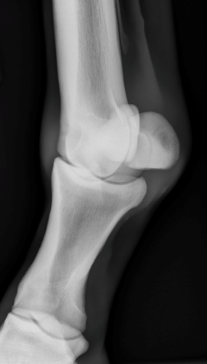
What are the standard views for the elbow?
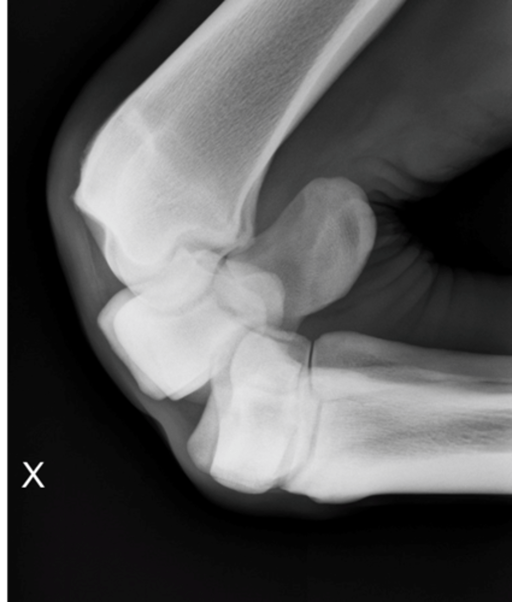
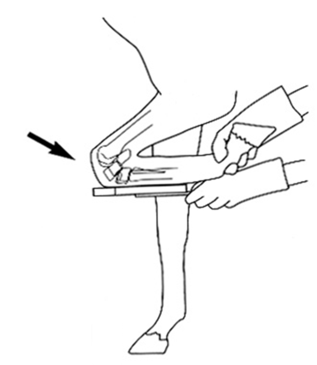
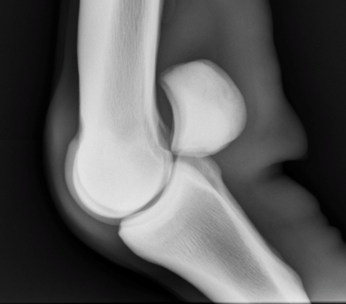
Mediolateral view of elbow
What view is being taken?

- Olecranon
- Anconeal process
- Elbow joint
- Radial tuberosity
What structures are highlighted by the ML elbow view?
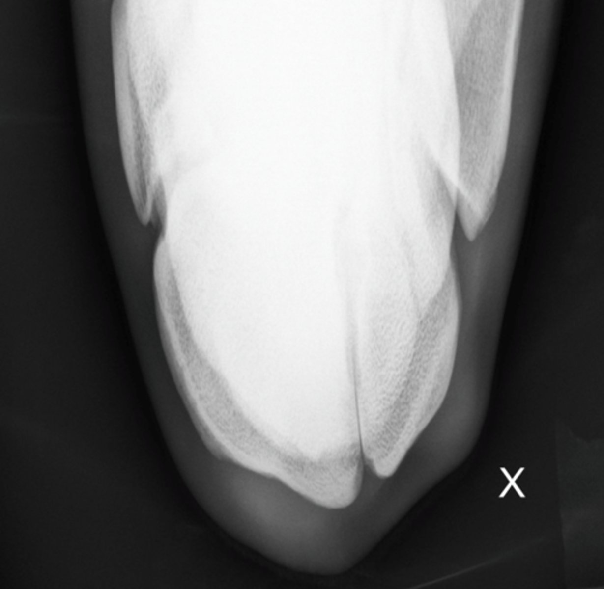
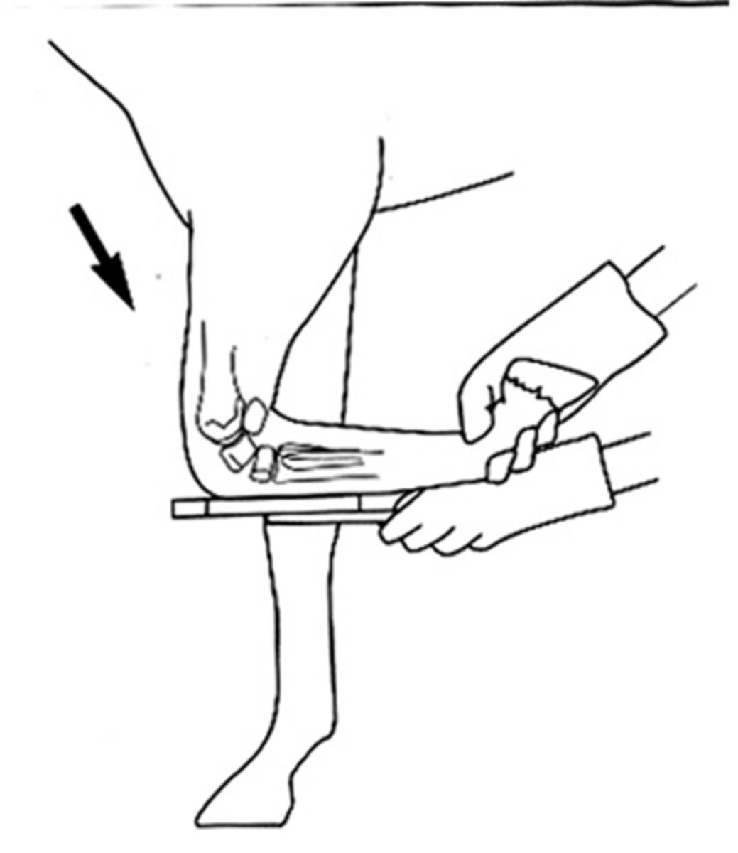
Craniocaudal view of elbow
What view is being taken?
- Medial and lateral humerus and radius
- Elbow joint
What does the craniocaudal view of the elbow highlight?
Standard:
- Lateromedial (LM)
- Dorso-palmar (DP)
- Dorsolateral-palmaromedial oblique (DLPMO)
- Dorsomedial-palmarolateral oblique (DMPLO)
- Flexed Lateromedial (Flexed LM)
Extra:
- Skyline (DPDDO)
- Distal radius: 80* proximal
- Proximal row: 55* proximal
- Distal row: 30* proximal
What are the standard and extra views that are taken with the carpus?
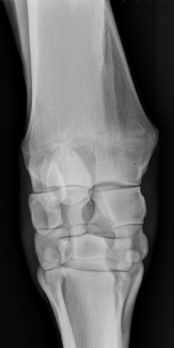
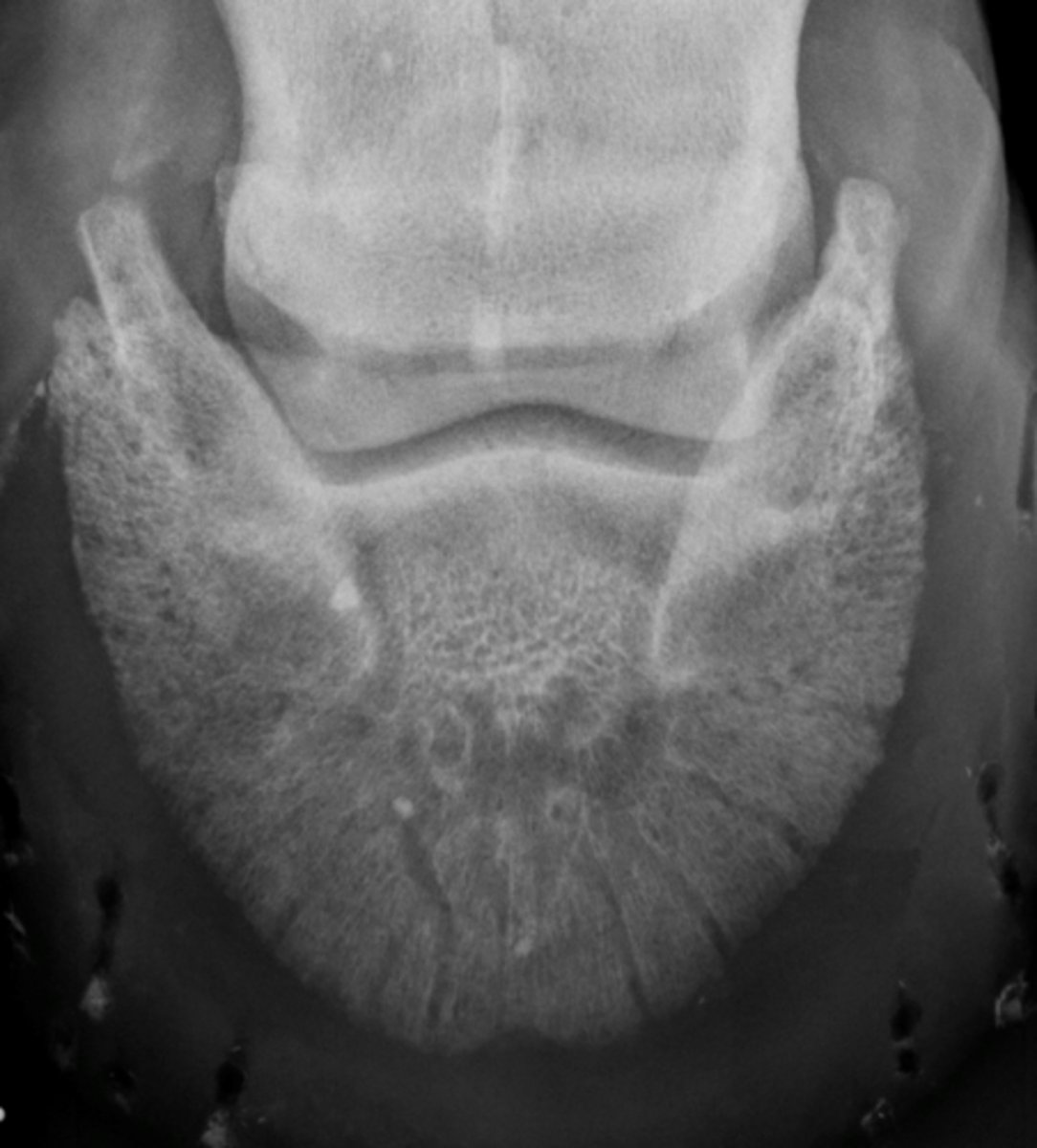
Dorsopalmar - Carpus
What view is being taken?
Dorsopalmar or palmarodorsal (don't usually because of X ray tube being under horse)
What view(s) could this be?
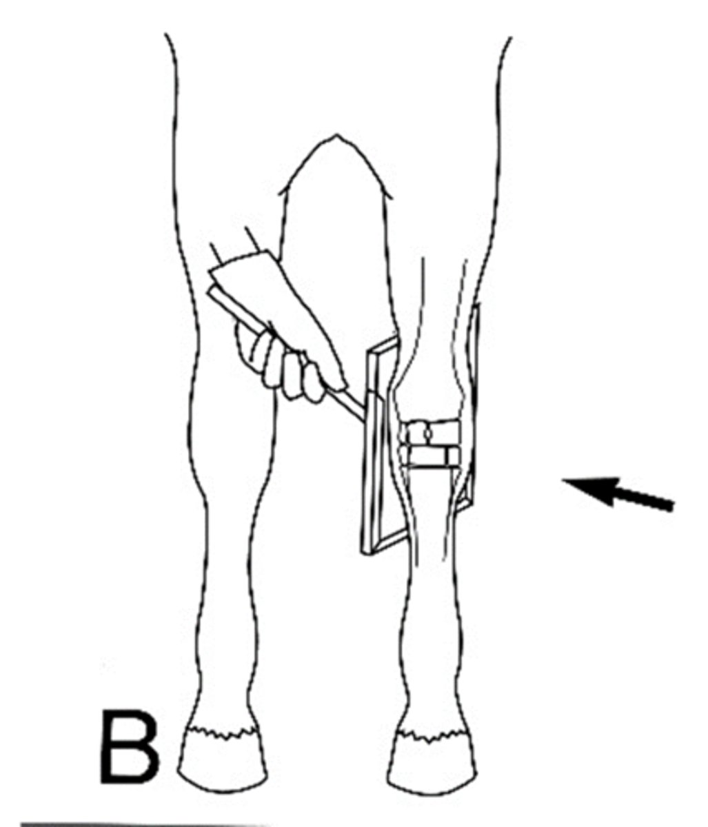
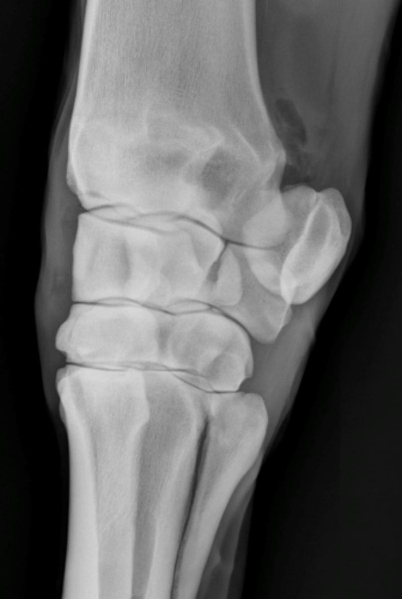
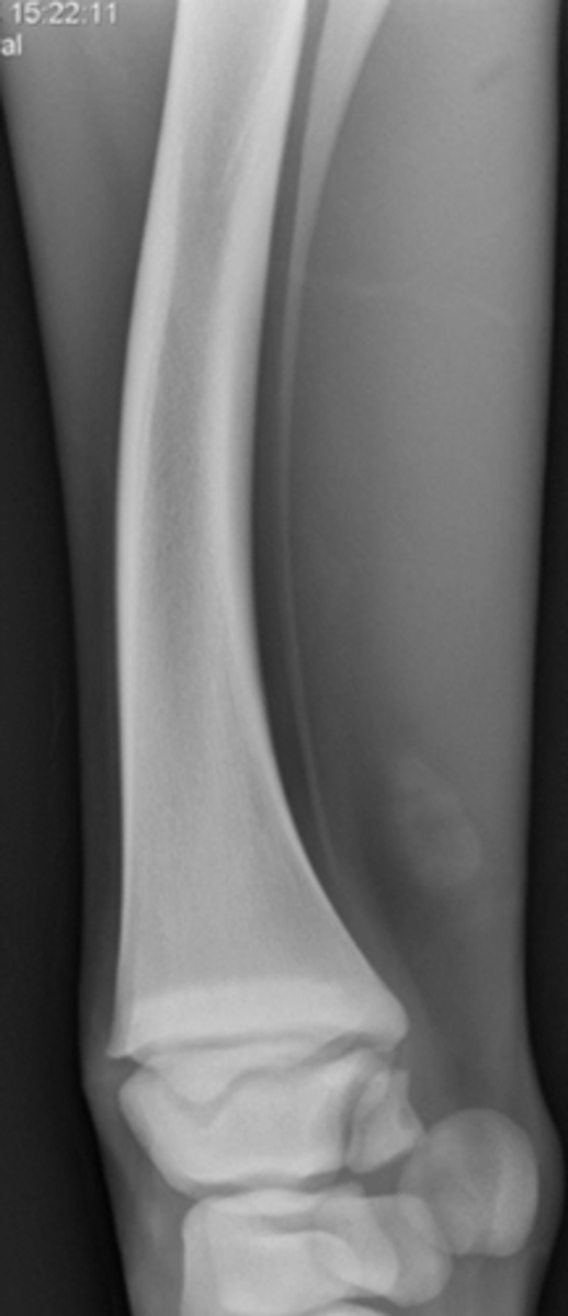
Lateromedial - Carpus
What view is being taken?
Accessory carpal bone
What is highlighted in a lateromedial view of the carpus?
D45*LPMO (DLPMO)
What view is being taken?
- Dorsomedial aspect of radius, radial & 3rd carpal bones
- Palmarolateral aspect of ulnar & 4th carpal bones
- Accessory carpal bone
- 4th articulates with MC3 & MC4
What is highlighted in a DLPMO view of the carpus?
D45*MPLO
What is the opposite view of the DLPMO taken of the carpus?
- Dorsolateral aspect of radius, intermediate and 3rd carpal bones
- Palmaromedial aspect of radial and 2nd carpal bones
- 2nd carpal bone articulates only with MC2
- May see 1st carpal bone (30%)
What is highlighted in a DMPLO view of the carpus?
Flexed Lateromedial
What view is being taken?
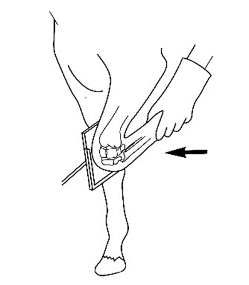
- Distal radius
- Proximal aspect of intermediate cb
- Distal aspect of radial cb
- Proximal aspect of the 3rd cb
What is highlighted in a flexed lateromedial view of the carpus?

Goes higher up - damage with corner fragments in race horses
How can you differentiate the intermediate carpal bone?
60%
What percentage of weight do the forelimbs hold?
D30*PDD Oblique - Carpus
What view is being taken?
Distal row: primarily 3rd carpal bone
What is highlighted in a D30PDD oblique view?
D55*Pr-DDiO - Carpus
What view is being taken?

Proximal row of carpal bones (Radial, intermediate, ulnar)
What is highlighted in a D55*Pr-DDiO view of the carpus?
D80*Pr-DDiO - Carpus
What view is being taken?
- Distal radius: 2 grooves
- Medial: Extensor carpi radialis
- Lateral: common digital extensor tendon
- Intertendinous eminence: common fracture site
What is highlighted in a D80*Pr-DDiO view of the carpus?
- Radial epiphysis: 24-36 m
- Lateral styloid process (ulna) = Fuses with epiphysis < 1 yr, Lucency in the caudodistal lateral radius
What are the two centers of ossification may show up separately in foals?
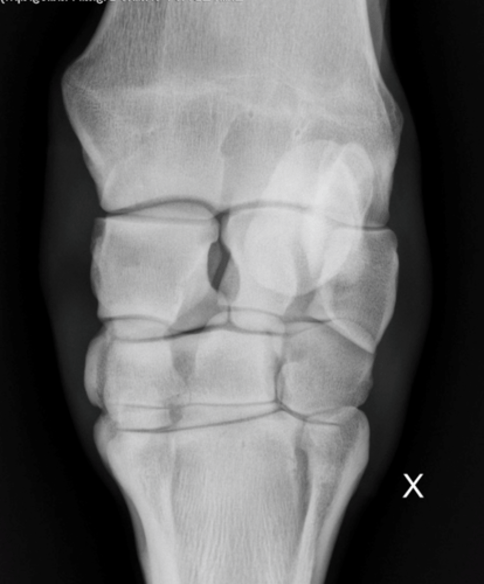
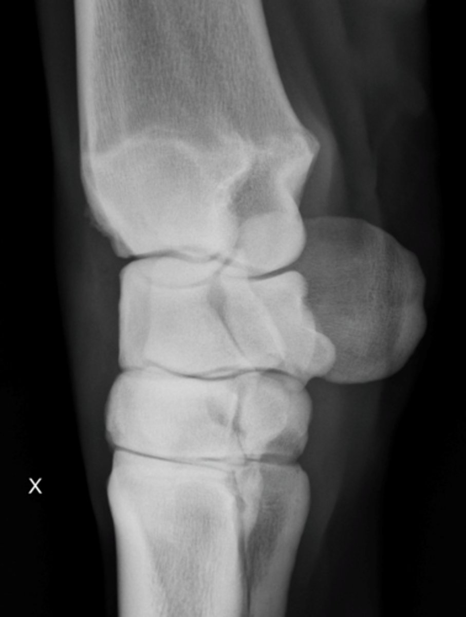
1st and 5th carpal bones
What "extra" bones are circled (not in all horses)?
Vestigeal Ulna - not typically a cause of lameness
What abnormality is shown?
Metacarpo-/metatarsophalangeal: Fetlock
What is the high motion joint of the distal limb?
- LM
- DP (D15oPr-PaDiO) *Confirmation
- DMPLO (D30oPr60oM-PaDiLO)
- DLPMO (D30oPr60oL-PaDiMO)
- Flexed LM
What are the standard views of the fetlock?
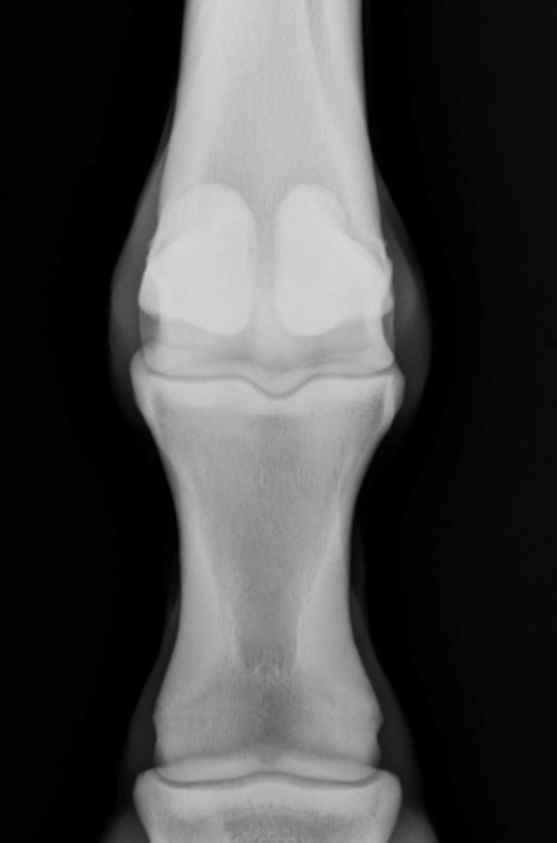
Dorsopalmar view - Fetlock
What view is being taken?
- Evaluate soft tissue, bone margins, joints, conformation
- Sesamoid bones (Lateral: thin, tall, Medial: wider)
What is highlighted in the DP view of the fetlock?
Lateromedial - Fetlock
What view is being taken?
DLMPO
What view is being taken?
Dorsomedial and palmarolateral aspect of limb
What is highlighted in a DLMPO view of the fetlock?
Dorsolateral and palmaromedial aspect of limb
What is highlighted in a DMPLO view of the fetlock?
Flexed LM - Fetlock
What view is being taken?
Sagittal ridge and palmar aspect of distal MC3
What is highlighted in the flexed LM view of the fetlock?
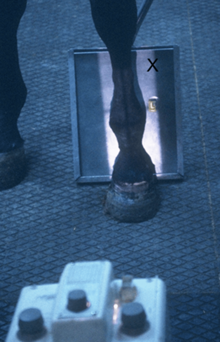
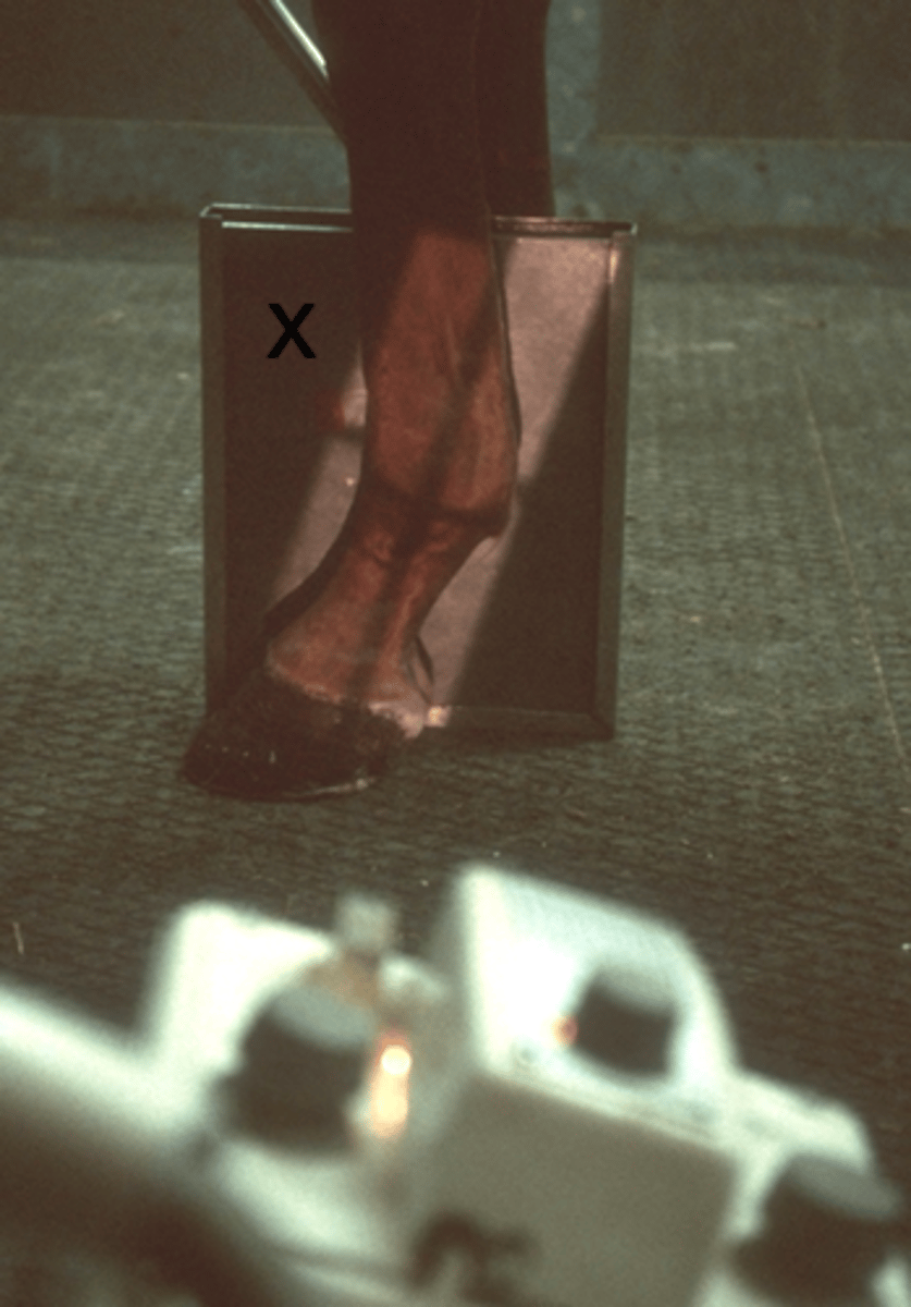
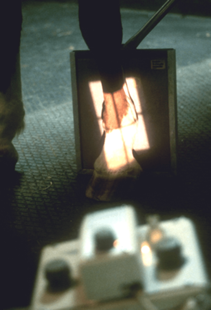
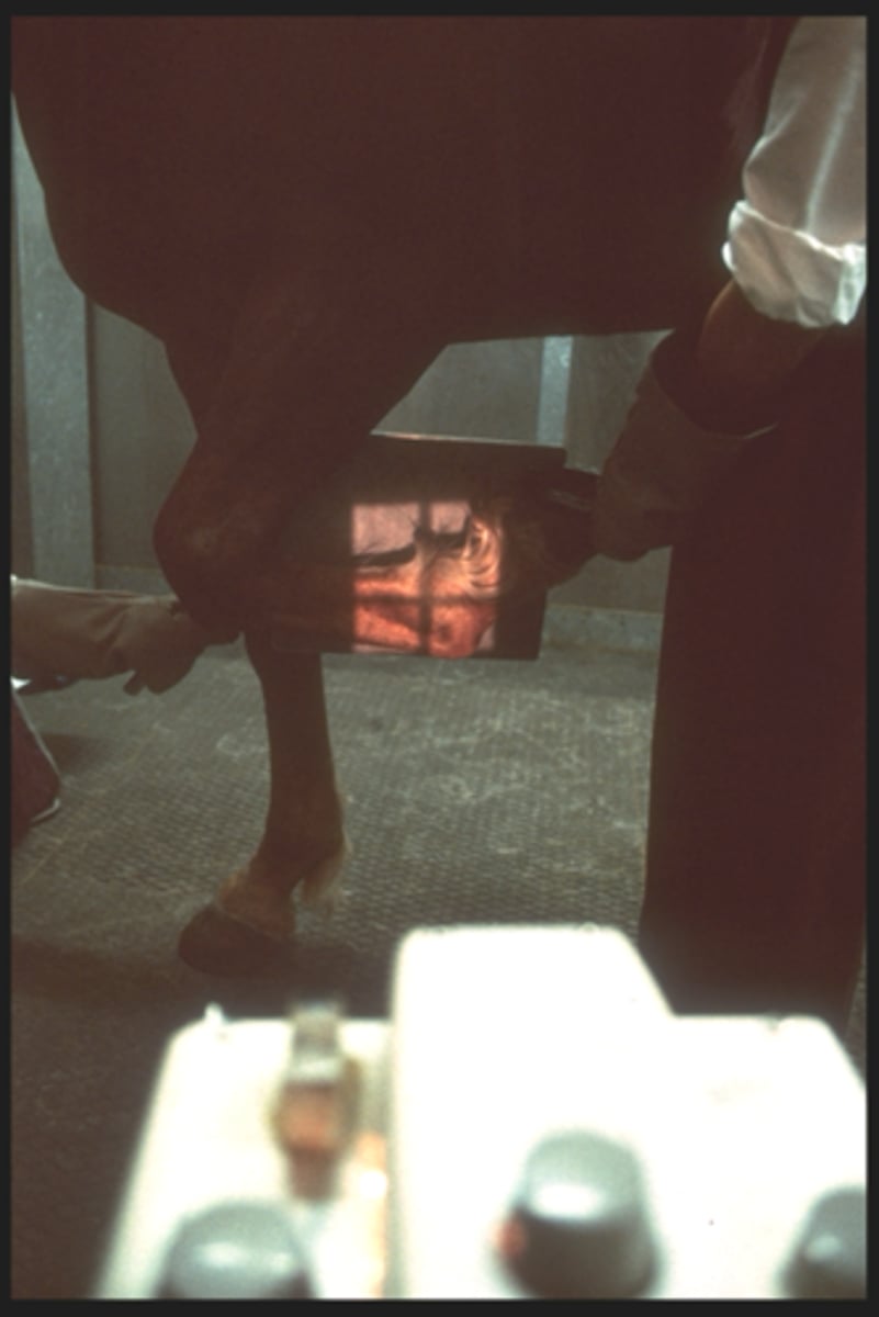
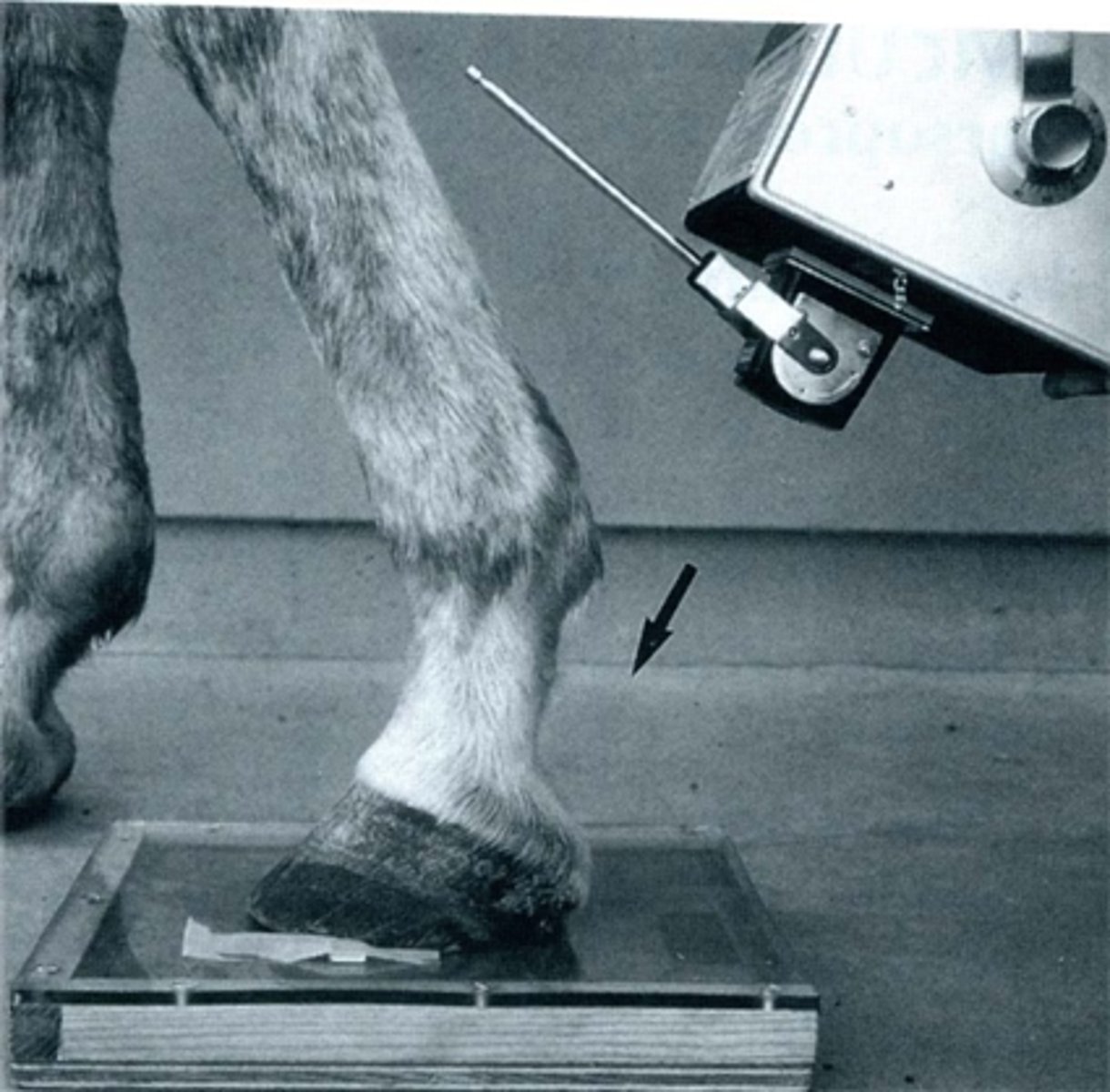
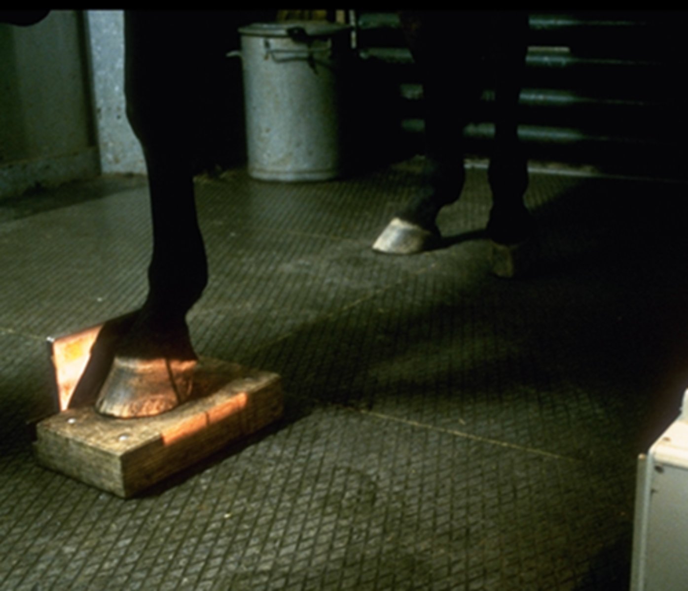
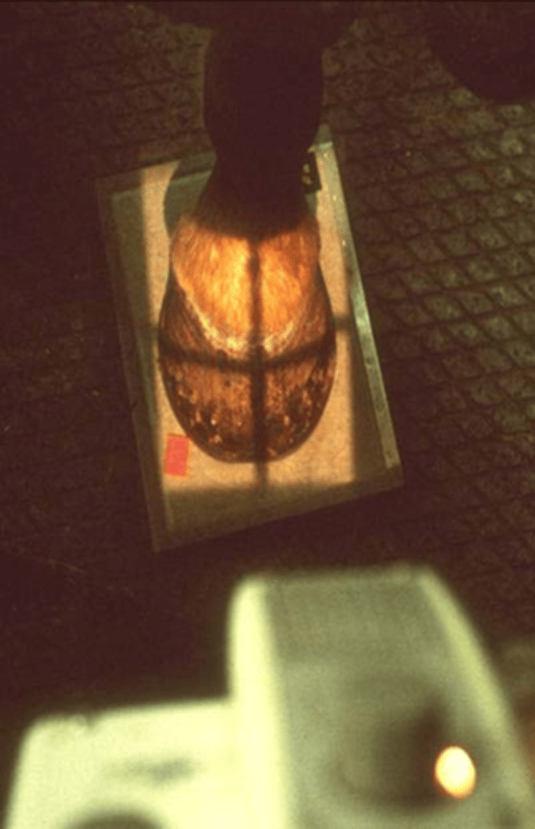
- Clean hoof wall, sole and sulci of frog
- Remove shoe
- Pack lateral, medial and central sulcus of the frog (play-doh = Don't have purple or blue = artifacts in MRI!)
- Wood block (LM)
- Cassette tunnel (DP & Obliques) = if horse has to stand on detector
What are special considerations when imaging the foot (distal phalanx, coffin joint)?
- LM
- 65o DP
- Skyline or tangential (Palmaro 45o proximal- palmarodistal view)
What are the standard views for a navicular series?
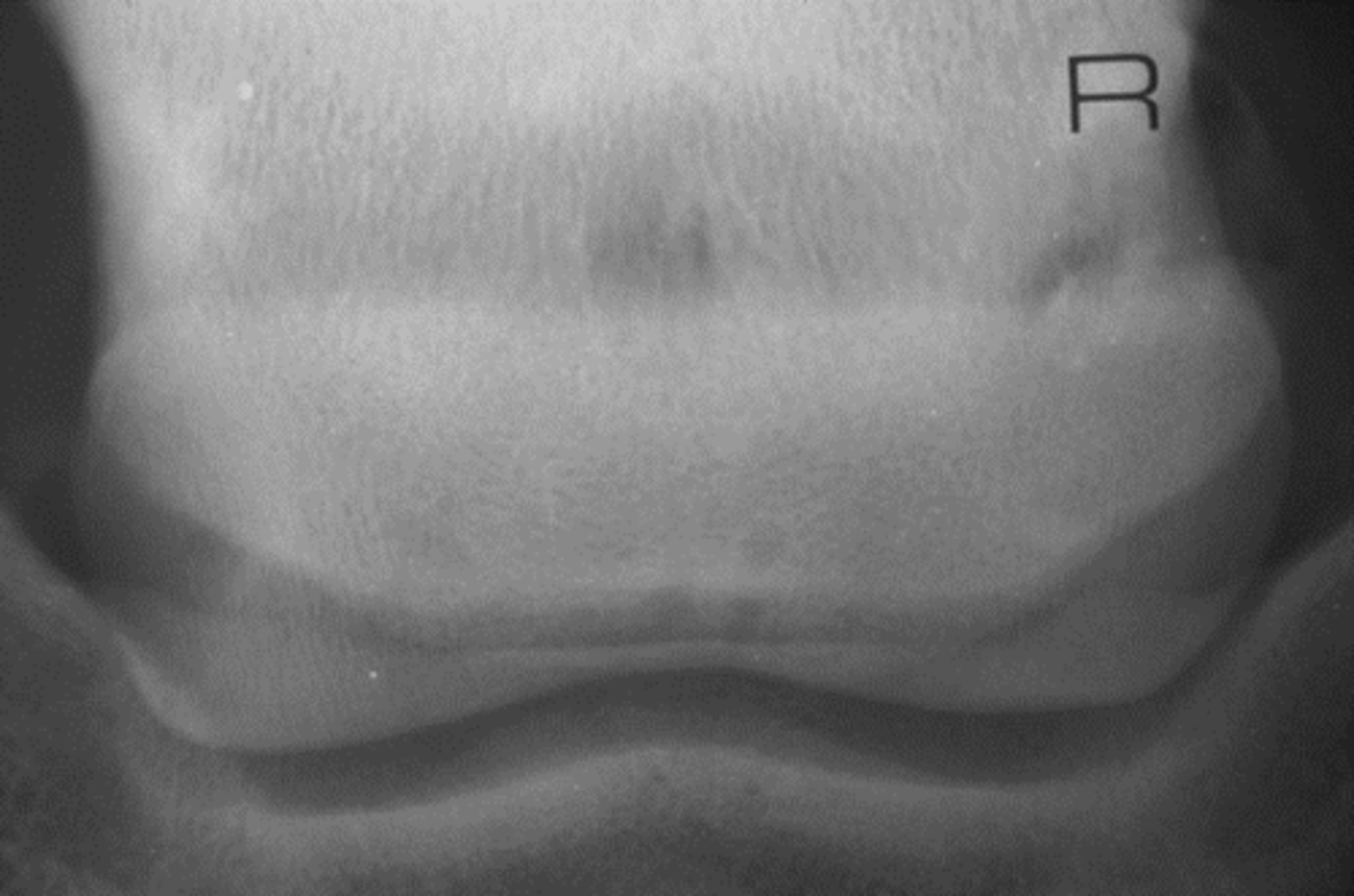
Lateromedial - Navicular
What view is being taken?

- Corticomedullary distinction
- Proximal & distal borders
What is highlighted in the LM view of the coffin joint (navicular series)?

65* DP
What view is being taken?
Evaluate body & distal border
What is highlighted in a 65* DP view in a navicular series?
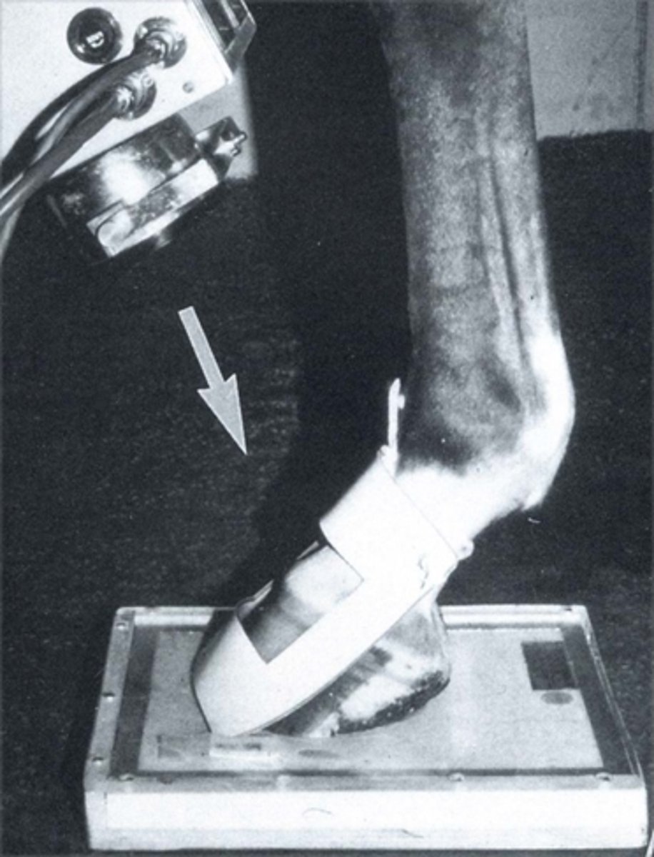
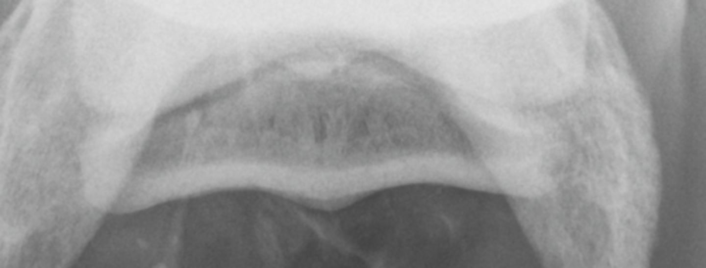
Navicular skyline: Palmaro 45o proximal- palmarodistal view
What view is being taken?
- Flexor cortex & corticomedullary distinction
- Midsagittal ridge lucency: normal
What is highlighted in a navicular skyline view?
- LM
- 65 DP
- DLPMO (D65Pr45L-PDiMO)
- DMPLO (D65Pr45M-PDiLO)
What are the standard views for the foot?
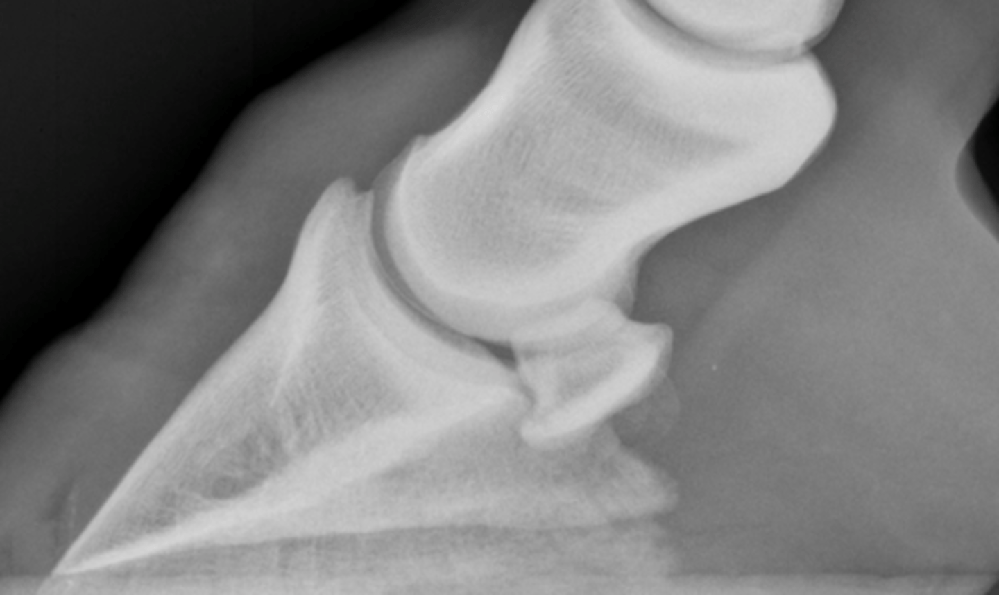
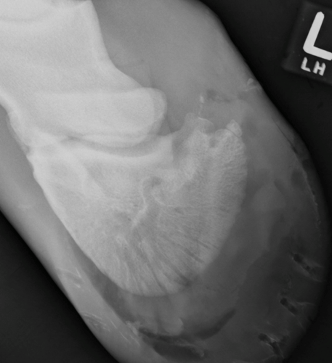
Lateromedial - Foot
What view is being taken?
- P3 position
- Hoof wall conformation
What is highlighted in LM views of the foot?
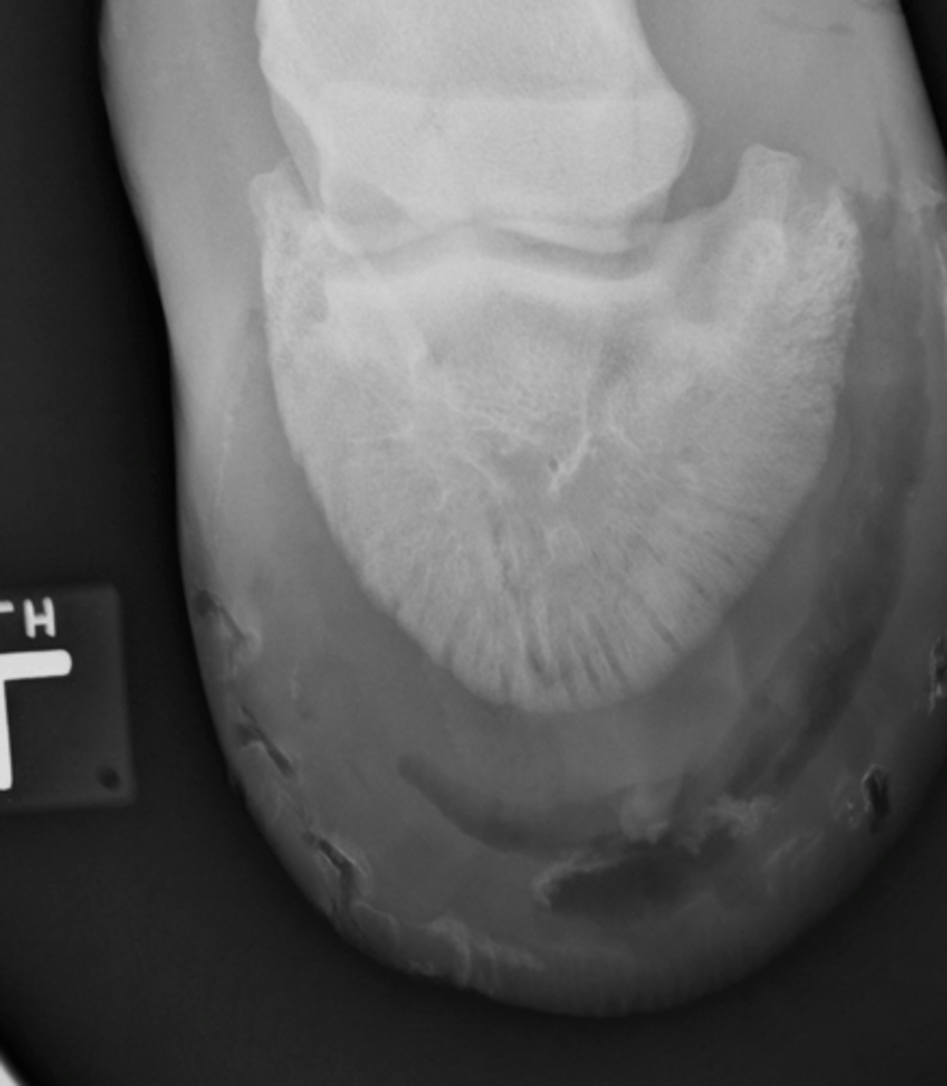
65* DP - Foot
What view is been taken?
P3
What is highlighted in a 65* DP foot view?
No periosteum, no medullary cavity
What is unique about P3?
Lateral wing P3
What is highlighted in a DLMPO view of the foot?
Medial wing P3
What is highlighted in a DMPLO view of the foot?