Exam 4.1
1/100
There's no tags or description
Looks like no tags are added yet.
Name | Mastery | Learn | Test | Matching | Spaced | Call with Kai | Chat |
|---|
No analytics yet
Send a link to your students to track their progress
101 Terms
The ventral rami of T1-T11 are also known as:
Brachial nerves
Cervical nerves
Intercostal nerves
Thoracic nerves
Spinal nerves
Intercostal nerves
Following an impulse through the nervous system (spinal cord to effector organ,) which of the following is the correct pathway?
spinal cord → nerve plexus → ventral rami → spinal nerve → ventral root → effector organ
spinal cord → nerve plexus → ventral root → spinal nerve → ventral rami → effector organ
spinal cord → spinal nerve → ventral root → ventral rami → nerve plexus → effector organ
spinal cord → ventral rami → spinal nerve → ventral root → nerve plexus → effector organ
spinal cord → ventral root → spinal nerve → ventral rami → nerve plexus → effector organ
spinal cord → ventral root → spinal nerve → ventral rami → nerve plexus → effector organ
Place the following structures in the correct order when considering the path of an impulse originating in the cerebral cortex as it travels from the spinal cord to the effector organ.
1. Nerve plexus 2. Spinal nerve 3. Ventral rami 4. Ventral root
1, 2, 3, 4
1, 3, 2, 4
2, 4, 3, 1
3, 2, 4, 1
4, 2, 3, 1
4, 2, 3, 1
The dorsal ramus of a typical spinal nerve innervates the:
Deep muscles of the back and the skin of the back
Major thoracic and abdominal organs
Muscles of the abdominal wall
Special senses of the thorax
Ventral and lateral trunk and the limbs
Deep muscles of the back and the skin of the back
The ventral ramus of a typical spinal nerve innervates the:
Deep muscles of the back and the skin of the back
Major thoracic and abdominal organs
Muscles of the abdominal wall
Special senses of the thorax
Ventral and lateral trunk and the limbs
Ventral and lateral trunk and the limbs
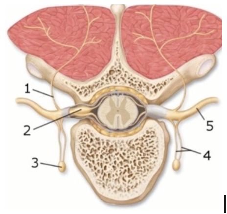
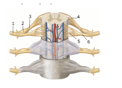
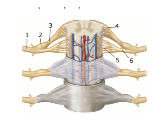
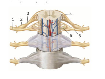
This figure demonstrates the branches of the spinal nerve. What structure(s) does number 1 indicate?
Dorsal ramus
Dorsal root ganglion
Rami communicans
Sympathetic trunk ganglion
Ventral ramus
Dorsal ramus
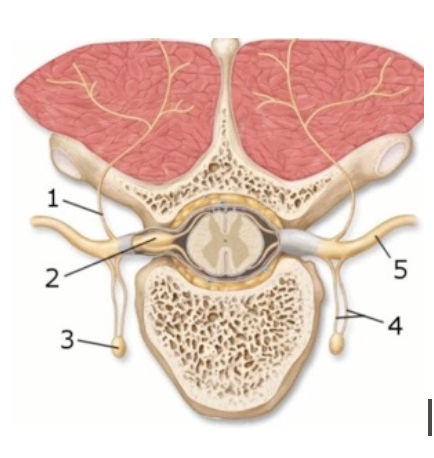
This figure demonstrates the branches of the spinal nerve. What structure(s) does number 2 indicate?
Dorsal ramus
Dorsal root ganglion
Rami communicans
Sympathetic trunk ganglion
Ventral ramus
Dorsal root ganglion
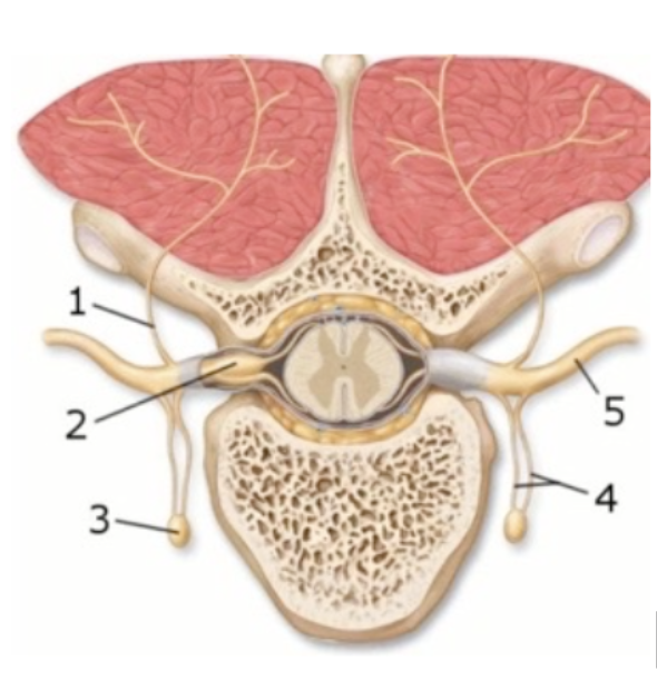
This figure demonstrates the branches of the spinal nerve. What structure(s) does number 3 indicate?
Dorsal ramus
Dorsal root ganglion
Rami communicans
Sympathetic trunk ganglion
Ventral ramus
Sympathetic trunk ganglion
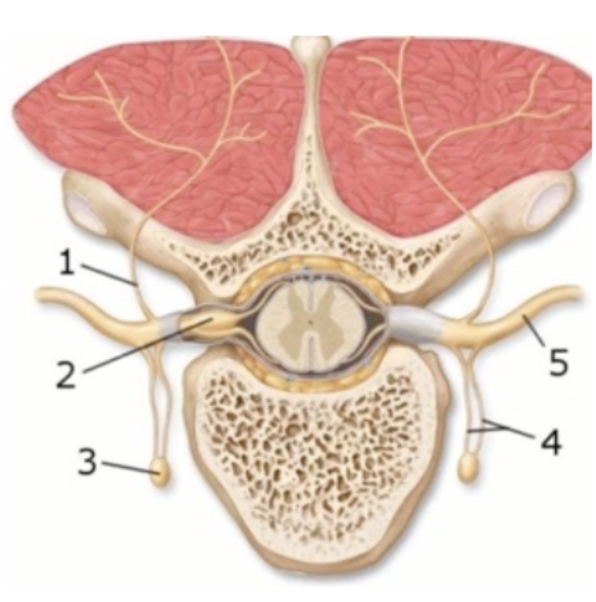
This figure demonstrates the branches of the spinal nerve. What structure(s) does number 4 indicate?
Dorsal ramus
Dorsal root ganglion
Rami communicans
Sympathetic trunk ganglion
Ventral ramus
Rami communicans
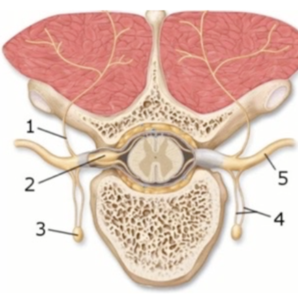
This figure demonstrates the branches of the spinal nerve. What structure(s) does number 5 indicate?
Dorsal ramus
Dorsal root ganglion
Rami communicans
Sympathetic trunk ganglion
Ventral ramus
Ventral ramus
A network of interwoven ventral rami of spinal nerves is best known as a:
Communicans
Ganglion
Dermatome
Plexus
Ramus
Plexus
Spinal nerves exiting the cord from the level of L4 to S4 form the ________.
Brachial plexus
Cervical plexus
Lumbar plexus
Sacral plexus
Thoracic plexus
Sacral plexus
Spinal nerves exiting the cord from the level of L1 to L4 form the ________.
Brachial plexus
Cervical plexus
Lumbar plexus
Sacral plexus
Thoracic plexus
Lumbar plexus
Spinal nerves exiting the cord from the level of C5 to T1 form the ________.
Brachial plexus
Cervical plexus
Lumbar plexus
Sacral plexus
Thoracic plexus
Brachial plexus
Spinal nerves exiting the cord from the level of C1 to C4 form the ________.
Brachial plexus
Cervical plexus
Lumbar plexus
Sacral plexus
Thoracic plexus
Cervical plexus
The spinal nerves that give rise to the sacral plexus are:
C1-C4
C5-T1
L1-L4
L4-S4
T1-T12
L4-S4
The spinal nerves that give rise to the cervical plexus are:
C1-C4
C5-T1
L1-L4
L4-S4
T1-T12
C1-C4
The spinal nerves that give rise to the brachial plexus are:
C1-C4
C5-T1
L1-L4
L4-S4
T1-T12
C5-T1
The spinal nerves that give rise to the lumbar plexus are:
C1-C4
C5-T1
L1-L4
L4-S4
T1-T12
L1-L4
The cervical plexus is formed by the:
Anterior rami of spinal nerves C1-C4
Anterior rami of spinal nerves C5-T1
Posterior rami of spinal nerves C1-C4
Posterior rami of spinal nerves C5-T1
Rami communicantes of spinal nerves C5-T1
Anterior rami of spinal nerves C1-C4
The brachial plexus is formed by the:
Anterior rami of spinal nerves C5-T1
Posterior rami of spinal nerves C1-C4
Posterior rami of spinal nerves C5-T1
Rami communicantes of spinal nerves C5- T1
Anterior rami of spinal nerves C5-T1
The ________ innervates the muscles of the anterior neck.
Brachial plexus
Cervical plexus
Lumbar plexus
Sacral plexus
Thoracic plexus
Cervical plexus
The plexuses that lie on either side of the neck are the _____.
Brachial plexus
Cervical plexus
Lumbar plexus
Sacral plexus
Thoracic plexus
Cervical plexus
The ________ contains all the nerves of the upper limbs.
Brachial plexus
Cervical plexus
Lumbar plexus
Sacral plexus
Thoracic plexus
Brachial plexus
The plexus that supplies the upper limbs is the _________.
Brachial plexus
Cervical plexus
Lumbar plexus
Sacral plexus
Thoracic plexus
Brachial plexus
The ________ contains the nerves of the anterior lower limbs.
Brachial plexus
Cervical plexus
Lumbar plexus
Sacral plexus
Thoracic plexus
Lumbar plexus
The plexus that supplies the anterior lower limbs is the _________.
Brachial plexus
Cervical plexus
Lumbar plexus
Sacral plexus
Thoracic plexus
Lumbar plexus
The ________ contains the nerves of the posterior lower limbs.
Brachial plexus
Cervical plexus
Lumbar plexus
Sacral plexus
Thoracic plexus
Sacral plexus
The plexus that supplies the posterior lower limbs is the _________.
Brachial plexus
Cervical plexus
Lumbar plexus
Sacral plexus
Thoracic plexus
Sacral plexus
Which spinal nerves contribute to the brachial plexus?
C1-C4
C5-T1
L1-L4
L4-S4
T1-T12
C5-T1
Which spinal nerves contribute to the cervical plexus?
C1-C4
C5-T1
L1-L4
L4-S4
T1-T12
C1-C4
Which spinal nerves contribute to the lumbar plexus?
C1-C4
C5-T1
L1-L4
L4-S4
T1-T12
L1-L4
Which spinal nerves contribute to the sacral plexus?
C1-C4
C5-T1
L1-L4
L4-S4
T1-T12
L4-S4
Which of the following lists the components of the brachial plexus in correct order starting with the most medial?
Cords, divisions, nerves, rami
Ganglia, rami, nerves, terminals
Rami, divisions, cords, trunks
Rami, trunks, divisions, cords
Trunks, divisions, rami, cords
Rami, trunks, divisions, cords
Within the axilla, there are several levels of organization of the branches of the anterior rami. Which of the following is a correct level of organization?
three cords--the posterior, medial, and lateral cords
three divisions--the superior, middle, and inferior divisions
five cords-- superior, inferior, medial, lateral, and middle divisions
five divisions--proximal, distal, ulnar, median, and radial divisions
five trunks-- superior, inferior, medial, lateral, and middle divisions
three cords--the posterior, medial, and lateral cords
Spinal nerves that innervate the anterior lower limbs form the ________.
Brachial plexus
Cervical plexus
Thoracic plexus
Sacral plexus
Lumbar plexus
Lumbar plexus
Spinal nerves that innervate the posterior lower limbs form the ________.
Brachial plexus
Cervical plexus
Thoracic plexus
Sacral plexus
Lumbar plexus
Sacral plexus
The function of lower motor neurons is to:
Excite motor portions of the thalamus
Excite or inhibit motor portions of the cerebral cortex
Excite or inhibit skeletal muscle cells
Excite or inhibit upper motor neurons
Excite skeletal muscle cells
Excite or inhibit upper motor neurons
The function of upper motor neurons is to:
Directly excite skeletal muscle cells
Excite motor portions of the thalamus
Excite or inhibit lower motor neurons
Excite or inhibit motor portions of the cerebral cortex
Excite or inhibit skeletal muscle cells
Excite or inhibit lower motor neurons
Motor pathways are _________ tracts that control _________.
Ascending, effectors
Ascending, receptors
Descending, effectors
Descending, receptors
Descending, effectors
The Golgi tendon reflex:
Helps to overcome pain
Aids in sending sensory information to the muscles
Prevents skeletal muscles from tensing excessively
Prevents muscles from contracting
Helps to gain balance through a complex series of muscular contractions
Prevents skeletal muscles from tensing excessively
A monosynaptic reflex that monitors and regulates skeletal muscle length is a _____ reflex.
Flexor
Golgi tendon
Hypoactive
Stretch
Withdrawal
Stretch
The largest and longest nerve in the body is the _________ nerve.
Femoral
Obturator
Radial
Saphenous
Sciatic
Sciatic
This nerve helps to plant the foot and receives sensory signals from the skin of the sole.
Femoral nerve
Deep fibular nerve
Genitofemoral nerve
Obturator nerve
Tibial nerve
Tibial nerve
Where the sciatic nerve splits, it branches directly into the:
Deep and superficial fibular nerves
Deep femoral and tibial nerves
Femoral and obturator nerves
Popliteal and sural nerves
Tibial and common fibular nerves
Tibial and common fibular nerves
Q16:
Which of the following nerves are part of the Muscular branch of the cervical plexus?
Auricular nerve
Cervical nerve
Occipital nerve
Phrenic nerve
Supraclavicular nerve
Phrenic nerve
Which of the following nerves are part of the Muscular branch of the cervical plexus?
Auricular nerve
Cervical nerve
Muscular nerve
Occipital nerve
Supraclavicular nerve
Muscular nerve
Which of the following nerves of the cervical plexus detects stimuli at the back of the scalp?
Auricular nerve
Cervical nerve
Occipital nerve
Phrenic nerve
Supraclavicular nerve
Occipital nerve
Which of the following nerves of the cervical plexus detects stimuli around the ear?
Auricular nerve
Cervical nerve
Occipital nerve
Phrenic nerve
Supraclavicular nerve
Auricular nerve
Which of the following nerves of the cervical plexus detects stimuli at the anterior neck?
Auricular nerve
Cervical nerve
Occipital nerve
Phrenic nerve
Supraclavicular nerve
Cervical nerve
Which of the following nerves of the cervical plexus detects stimuli at the shoulder?
Auricular nerve
Cervical nerve
Occipital nerve
Phrenic nerve
Supraclavicular nerve
Supraclavicular nerve
If a person suffers a thorax-crushing injury and is unable to breathe properly, you would suspect that this nerve was damaged.
Accessory nerve
Auricular nerve
Cervical nerve
Phrenic nerve
Sciatic nerve
Phrenic nerve
The nerve that innervates the deltoid muscle and receives sensory input from the superolateral arm is the:
Axillary nerve
Median nerve
Musculocutaneous nerve
Radial nerve
Ulnar nerve
Axillary nerve
Following an injury to his arm, a patient complains that he has no sensations from the thumb, index finger, middle finger, and part of the ring finger. What nerve do you suspect to be damaged?
Axillary nerve
Median nerve
Musculocutaneous nerve
Radial nerve
Ulnar nerve
Median nerve
Following an injury to his arm, a patient complains that he has lost the ability to flex his elbow and supinate his forearm. Which nerve do you suspect he has damaged?
Axillary nerve
Median nerve
Musculocutaneous nerve
Radial nerve
Ulnar nerve
Musculocutaneous nerve
Following an injury to his arm, a patient complains that he cannot extend the forearm, wrist, and digits. You suspect that he has damaged the:
Axillary nerve
Median nerve
Musculocutaneous nerve
Radial nerve
Ulnar nerve
Radial nerve
Following an injury to his arm, a patient complains that he has no sensations from his "pinky". You suspect that he has damaged the:
Axillary nerve
Median nerve
Musculocutaneous nerve
Radial nerve
Ulnar nerve
Ulnar nerve
A person suffering from carpal tunnel syndrome has lost function of the:
Axillary nerve
Median nerve
Musculocutaneous nerve
Radial nerve
Ulnar nerve
Median nerve
The two main nerves of the lumbar plexus are the:
Femoral and obturator nerves
Femoral and sciatic nerves
Obturator and pudendal nerves
Obturator and sciatic nerves
Pudendal and sciatic nerves
Femoral and obturator nerves
The nerve responsible for innervation (movement) of the quadriceps femoris muscle in the anterior thigh is the _____ nerve.
Genitofemoral
Femoral
Obturator
Sciatic
Tibial
Femoral
Which of the following is the correct simple spinal reflex arc?
Effector, afferent neuron, integration center, efferent neuron, receptor
Effector, efferent neuron, integration center, afferent neuron, receptor
Integration center, afferent neuron, receptor, efferent neuron, effector
Receptor, afferent neuron, integration center, efferent neuron, effector
Receptor, efferent neuron, integration center, afferent neuron, effector
Receptor, afferent neuron, integration center, efferent neuron, effector
Following a simple reflex arc from receptor to effector which of the following is the correct pathway?
Receptor → afferent fiber → efferent fiber → integration center → effector organ
Receptor → afferent fiber → integration center → efferent fiber → effector organ
Receptor → efferent neuron → afferent neuron → integration center → effector
Receptor → efferent fiber → integration center → afferent fiber → effector organ
Receptor → integration center → afferent fiber → efferent fiber → effector organ
Receptor → afferent fiber → integration center → efferent fiber → effector organ
Which of the following statements is most accurate?
Basic reflexes are always mediated by the brain
Basic reflexes are always autonomic
Basic reflexes are rapid, predictable, learned responses
Basic reflexes are somatic only
Basic reflexes are unlearned, built-in response that may be modified by learned behavior
Basic reflexes are unlearned, built-in response that may be modified by learned behavior
Reflexes are best described as:
Autonomic and spontaneous
Pre-programmed and voluntary
Rapid and involuntary
Slow and spontaneous
Spontaneous and self-initiating
Rapid and involuntary
What is the correct order for the events that occur during a reflex?
1. Effector responds
2. Impulse travels through sensory neuron to the CNS
3. Information is processed by interneurons
4. Motor neuron transmits impulse to effector
5. Stimulus activates a receptor
1, 2, 3, 4, 5
2, 4, 3, 1, 5
3, 1, 2, 4, 5
3, 2, 1, 4, 5
5, 2, 3, 4, 1
5, 2, 3, 4, 1
If the ventral root of a spinal nerve were cut, what would be the result in the tissue or region that nerve supplies?
A complete loss of sensation
A complete loss of sensation and movement
A complete loss of voluntary movement
Neither a loss of sensation nor movement but only of autonomic control
A complete loss of voluntary movement
Select the statement that is most correct.
The cell bodies of afferent ganglia are located in the spinal cord
The dorsal root ganglion is a motor-only structure
Ganglia are collections of neuron cell bodies in the spinal cord that are associated with efferent fibers
Ganglia associated with afferent nerve fibers contain cell bodies of sensory neurons
Ganglia associated with afferent nerve fibers contain cell bodies of sensory neurons
Which statement most accurately describes spinal nerves?
All spinal nerves are motor nerves
All spinal nerves are sensory nerves
Each spinal nerve is mixed in that it contains some sensory axons and some motor axons
Odd numbered spinal nerves are sensory and even numbered spinal nerves are motor
Spinal nerves are central axons contained within the vertebral canal
Each spinal nerve is mixed in that it contains some sensory axons and some motor axons
Which of the following is a branch off a spinal nerve?
Infeonrior ramus
Lateral ramus
Medial ramus
Rami communicans
Superior ramus
Rami communicans
Which of the following is a branch off a spinal nerve?
Dorsal ramus
Inferior ramus
Lateral ramus
Medial ramus
Superior ramus
Dorsal ramus
Which of the following is a branch off a spinal nerve?
Inferior ramus
Lateral ramus
Medial ramus
Ventral ramus
Superior ramus
Ventral ramus
The dorsal root of a spinal nerve contains:
A mix of sensory and motor axons
Autonomic nervous system axons only
Motor axons only
Sensory axons only
Sensory axons only
The ventral root of a spinal nerve contains:
A mix of sensory and motor axons
Autonomic nervous system axons only
Motor axons only
Sensory axons only
Motor axons only
Which of the following structures is most proximal to the spinal cord?
Dorsal nerve
Dorsal ramus
Dorsal root
Dorsal root ganglion
Dorsal rootlets
Dorsal rootlets
Which of the following structures is most distal to the spinal cord?
Dorsal nerve
Dorsal ramus
Dorsal root
Dorsal root ganglion
Dorsal rootlets
Dorsal ramus
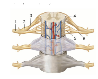
This figure shows an anterior view of the spinal cord and meninges. What structure does number 1 indicate?
Dorsal root
Dorsal rootlets
Spinal nerve
Ventral root
Ventral rootlets
Spinal nerve
This figure shows an anterior view of the spinal cord and meninges. What structure does number 5 indicate?
Dorsal root
Dorsal root ganglion
Dorsal rootlets
Ventral root
Ventral rootlets
Ventral rootlets
This figure shows an anterior view of the spinal cord and meninges. What structure does number 6 indicate?
Dorsal root
Dorsal root ganglion
Dorsal rootlets
Ventral root
Ventral rootlets
Ventral root
This figure shows an anterior view of the spinal cord and meninges. What structure does number 2 indicate?
Dorsal root
Dorsal root ganglion
Dorsal rootlets
Ventral root
Ventral rootlets
Dorsal root ganglion
This figure shows an anterior view of the spinal cord and meninges. What structure does number 3 indicate?
Dorsal root
Dorsal root ganglion
Dorsal rootlets
Ventral root
Ventral rootlets
Dorsal root
This figure shows an anterior view of the spinal cord and meninges. What structure does number 4 indicate?
Dorsal root
Dorsal root ganglion
Dorsal rootlets
Ventral root
Ventral rootlets
Dorsal rootlets
Which of the following has long preganglionic axons and therefore ganglia that are relatively far from the central nervous system?
Autonomic nervous system
Central nervous system
Parasympathetic nervous system
Somatic nervous system
Sympathetic nervous system
Parasympathetic nervous system
2)Which of the following contains short preganglionic axons that branch extensively?
Autonomic nervous system
Central nervous system
Parasympathetic nervous system
Somatic nervous system
Sympathetic nervous system
Sympathetic nervous system
3)Which of the following statements concerning the sympathetic system is most accurate true?
Sympathetic division has long preganglionic and short postganglionic fibers
Sympathetic division has minimal branching of preganglionic fibers
Sympathetic fiber origin is thoracolumbar
Sympathetic ganglia are close to visceral organs served
Sympathetic fiber origin is thoracolumbar
Which of the following statements concerning the sympathetic system is most accurate true?
Sympathetic division has extensive branching of preganglionic fibers
Sympathetic division has long preganglionic and short postganglionic fibers
Sympathetic fiber origin is craniosacral
Sympathetic ganglia are close to visceral organs served
Sympathetic division has extensive branching of preganglionic fibers
Which of the following statements concerning the sympathetic system is most accurate true?
Sympathetic division has minimal branching of preganglionic fibers
Sympathetic division has short preganglionic and long postganglionic fibers
Sympathetic fiber origin is craniosacral
Sympathetic ganglia are close to visceral organs served
Sympathetic division has short preganglionic and long postganglionic fibers
Which of the following statements concerning the sympathetic system is most accurate true?
Sympathetic division has long preganglionic and short postganglionic fibers
Sympathetic division has minimal branching of preganglionic fibers
Sympathetic fiber origin is craniosacral
Sympathetic ganglia are within a few centimeters of the CNS
Sympathetic ganglia are within a few centimeters of the CNS
Which of the following statements concerning the parasympathetic system is most accurate true?
Parasympathetic division has extensive branching of preganglionic fibers
Parasympathetic division has short preganglionic and long postganglionic fibers
Parasympathetic fiber origin is craniosacral
Parasympathetic ganglia are within a few centimeters of the CNS
Parasympathetic fiber origin is craniosacral
Which of the following statements concerning the parasympathetic system is most accurate true?
Parasympathetic division has minimal branching of preganglionic fibers
Parasympathetic division has short preganglionic and long postganglionic fibers
Parasympathetic fiber origin is thoracolumbar
Parasympathetic ganglia are within a few centimeters of the CNS
Parasympathetic division has minimal branching of preganglionic fibers
Which of the following statements concerning the parasympathetic system is most accurate true?
Parasympathetic division has extensive branching of preganglionic fibers
Parasympathetic division has long preganglionic and short postganglionic fibers
Parasympathetic fiber origin is thoracolumbar
Parasympathetic ganglia are within a few centimeters of the CNS
Parasympathetic division has long preganglionic and short postganglionic fibers
Which of the following statements concerning the parasympathetic system is most accurate true?
Parasympathetic division has extensive branching of preganglionic fibers
Parasympathetic division has short preganglionic and long postganglionic fibers
Parasympathetic fiber origin is thoracolumbar
Parasympathetic ganglia are close to visceral organs served
Parasympathetic ganglia are close to visceral organs served
Which of the following is a distinguishing characteristic of the sympathetic division of the ANS?
Acetylcholine as a postganglionic neurotransmitter
Localized responsiveness
Long postganglionic axons
Long preganglionic axons
Preganglionic neuron cell bodies in cranial region and sacral region of the spinal cord
Long postganglionic axons
Which of the following is a distinguishing characteristic of the sympathetic division of the ANS?
Localized responsiveness
Long preganglionic axons
Norepinephrine as a postganglionic neurotransmitter
Preganglionic neuron cell bodies in cranial region and sacral region of the spinal cord
Short postganglionic axons
Norepinephrine as a postganglionic neurotransmitter
Which of the following is a distinguishing characteristic of the sympathetic division of the ANS?
Acetylcholine as a postganglionic neurotransmitter
Localized responsiveness
Long preganglionic axons
Preganglionic neuron cell bodies in the T1- L2 regions of the spinal cord
Short postganglionic axons
Preganglionic neuron cell bodies in the T1- L2 regions of the spinal cord
Which of the following is a distinguishing characteristic of the sympathetic division of the ANS?
Acetylcholine as a postganglionic neurotransmitter
Localized responsiveness
Preganglionic neuron cell bodies in cranial region and sacral region of the spinal cord
Short postganglionic axons
Short preganglionic axons
Short preganglionic axons
Which of the following is a distinguishing characteristic of the sympathetic division of the ANS?
Acetylcholine as a postganglionic neurotransmitter
Long preganglionic axons
Preganglionic neuron cell bodies in cranial region and sacral region of the spinal cord
Short postganglionic axons
Systemic responsiveness
Systemic responsiveness
Which of the following is a distinguishing characteristic of the parasympathetic division of the ANS?
Norepinephrine as a postganglionic neurotransmitter
Preganglionic neuron cell bodies in the T1- L2 regions of the spinal cord
Short postganglionic axons
Short preganglionic axons
Systemic responsiveness
Short postganglionic axons
Which of the following is a distinguishing characteristic of the parasympathetic division of the ANS?
Acetylcholine as a postganglionic neurotransmitter
Long postganglionic axons
Preganglionic neuron cell bodies in the T1- L2 regions of the spinal cord
Short preganglionic axons
Systemic responsiveness
Acetylcholine as a postganglionic neurotransmitter
Which of the following is a distinguishing characteristic of the parasympathetic division of the ANS?
Long postganglionic axons
Norepinephrine as a postganglionic neurotransmitter
Preganglionic neuron cell bodies in cranial region and sacral region of the spinal cord
Short preganglionic axons
Systemic responsiveness
Preganglionic neuron cell bodies in cranial region and sacral region of the spinal cord
Which of the following is a distinguishing characteristic of the parasympathetic division of the ANS?
Long postganglionic axons
Long preganglionic axons
Norepinephrine as a postganglionic neurotransmitter
Preganglionic neuron cell bodies in the T1- L2 regions of the spinal cord
Systemic responsiveness
Long preganglionic axons