(II) Gynecological & breast malignancies
 Call Kai
 Learn
Learn Practice Test
 Spaced Repetition
 Match
Match Flashcards
 Knowt Play
1/201

There's no tags or description
Looks like no tags are added yet.
Name | Mastery | Learn | Test | Matching | Spaced | Call with Kai | Chat |
|---|
No analytics yet
Send a link to your students to track their progress
202 Terms
-_________= pregnancy/the period of time between conception and birth
-_________= cells that help form a layer around a developing embryo and eventually become a major part of the placenta (and produce hCG)
-_________= disorders related to abnormal proliferation of placental trophoblasts
Gestation
Trophoblasts
GTD
is hydatidiform mole (HM)/molar pregnancy benign, premalignant, or malignant?
premalignant
occurs after an aberrant fertilization between egg and sperm
molar pregnancies
molar pregnancy happens when abnormal proliferation of placental __________ causes a tumor to form
trophoblasts
what tissue do molar pregnancies come from
arises from fetal and not maternal tissue
how are molar pregnancies categorized?
categorized as partial or complete molar pregnancy
molar pregnancies are common at what ages?
at extremes of maternal age (< 20yo or > 35yo)
what are the metastatic risks of molar pregnancy?
Risk of gestational trophoblastic neoplasia (GTN)
What is the chromosomal composition of a partial molar pregnancy?
Triploid (XXX, XXY, or XYY)
How does a partial molar pregnancy typically occur?
From fertilization of a normal ovum by two sperm
Is there a fetus present in a partial molar pregnancy? if so, what can be detected?
Yes, a fetus may be present
cardiac activity may be detected.
How is a partial molar pregnancy often misdiagnosed?
It is often misdiagnosed as incomplete or missed abortion.
How do hCG levels in partial molar pregnancies compare to complete molar pregnancies?
hCG levels are generally lower in partial molar pregnancies.
Complete molar pregnancy is haploid or diploid? Which chromosomes are lost?
Diploid - most cases due to fertilization of an ovum in which maternal chromosomes are lost and results in two sets of paternal DNA
T/F: complete molar pregnancy has a fetus present
false
No fetus is present
what marker is different in complete molar pregnancy from patrial?
complete = Marked elevation of hCG
Higher risk of gestational trophoblastic neoplasia compared to partial molar pregnancy
What should be considered in any reproductive-age female with abnormal vaginal bleeding?
Molar pregnancy
What is a common initial manifestation of a molar pregnancy?
Initial missed period and positive pregnancy test
What is a common symptom of molar pregnancy in the first trimester?
Most cases develop vaginal bleeding
How does the uterine size in molar pregnancy compare to the gestational age?
Uterine size is greater than gestational age
How is molar pregnancy typically detected?
Picked up on physical exam or ultrasound in 1st trimester
What causes the symptoms of nausea and vomiting in molar pregnancy?
Thought to be related to high hCG levels
In which type of molar pregnancy are symptoms generally more severe?
Complete molar pregnancy
What rare complications can develop if a molar pregnancy reaches the second trimester?
Hyperthyroidism and preeclampsia
What are the common serum hCG levels in molar pregnancy?
OVER 50,000 - 100,000 mIU/mL
What does a complete molar pregnancy show on transvaginal ultrasound?
'SNOWSTORM' appearance (or swiss cheese/ cluster of grapes)
No fetus/amniotic fluid, + heterogeneous mass with anechoic spaces,
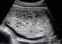
What does a partial molar pregnancy show on transvaginal ultrasound?
Fetus present but often growth restricted, + enlarged placenta with cystic spaces
partial or complete molar preg?
Fetus present on TVUS but often growth restricted, + enlarged placenta with cystic spaces
partial
How is molar pregnancy diagnosed?
clinically and then confirmed on histology after uterine evacuation.
What is the follow-up procedure for a partial molar pregnancy with a fetus and cardiac activity?
Follow with serial hCG and ultrasound
What is the most common treatment option for molar pregnancy?
Surgical uterine evacuation (dilation and evacuation - D&E)
What is an alternative treatment for molar pregnancy in those who have completed childbearing?
Hysterectomy
What should be monitored post-operatively after treatment for molar pregnancy?
Weekly Serial hCG measurements until levels are undetectable or plateaued
When is prophylactic chemotherapy considered for molar pregnancy?
In cases of complete molar pregnancy if post-op hCG is unavailable and at high risk of GTN
What factors increase the risk of GTN in cases of complete molar pregnancy?
-Age > 40
-hCG > 100,000 mIU/L
-uterine size greater than gestational age
Is prophylactic chemotherapy indicated in cases treated with hysterectomy?
No
is gestational trophoblastic neoplasia (GTN) malignant or benign
what type of pregs can GNT be found in
malignant!
(is a neoplasms consisting of abnormal proliferation of trophoblastic tissue)
molar or nonmolar pregnancy
What happens to retained trophoblastic tissue in the uterus?
It can turn cancerous.
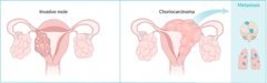
What is an invasive mole?
A type of GTN that only occurs after a molar pregnancy.
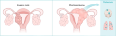
What is choriocarcinoma?
An aggressive type of gestational trophoblastic neoplasia.
Prior molar pregnancy, advanced maternal age, and Asian/American Indian ancestry are what?
What are the most common sites of metastatic disease in GTN?
r/fs of GTN
The lungs (mc) and vagina (30%).
what is lab value is elevated in GTN?
what's the imaging of choice?
what imaging evals for lung cancer associated with GTN?
Elevated hCG
TVUS (poorly defined intrauterine mass with vascular flow on Doppler.)
chest XRAY
other conditions that result from GTN (5)
-hyperthyroidism (tachy, tremor)
-ovarian theca lutein cyst
-hyperemesis
-AUB
What are some symptoms of metastasis in GTN?
Dyspnea
chest pain
cough
hemoptysis
vaginal bleeding/discharge
hCG of invasive mole and choriocarcinoma
hCG PSTT and ETT
invasive mole and choriocarcinoma = more elevated in hCG.
PSTT/ETT = less elevated
How is a clinical diagnosis of GTN made?
clinical based on elevated hCG after pregnancy with ultrasound showing changes of GTN.
Is tissue diagnosis required prior to treatment for GTN?
nar
Why is biopsy not pursued in GTN treatment?
risk of significant hemorrhage.
What is the treatment for low-risk GTN patients who desire future childbearing?
Methotrexate +/- chemotherapy.
What is the treatment for high-risk GTN patients or those who do not desire future childbearing?
Hysterectomy +/- chemotherapy.
What is the most common type of vulvar cancer?
Squamous cell carcinoma (SCC)
What are two other other types of vulvar cancer?
Basal cell carcinoma (BCC) and melanoma
What are the two proposed pathways for vulvar cancer?
HPV-associated
HPV-independent
Which HPV genotypes are most commonly associated with vulvar cancer?
HPV 16 and 33
What causes HPV-independent cases of vulvar cancer?
Chronic inflammatory or autoimmune processes
ex. vulvar dystrophy or lichen sclerosus
What is the average age of diagnosis for vulvar cancer?
Mid 60s
What are the common presentations of vulvar cancer?
Localized plaque, nodule, or mass
Where are most vulvar cancer lesions located?
Labia majora
Which areas are less commonly affected by vulvar cancer?
Labia minora, perineum, clitoris
What is the typical appearance of vulvar cancer lesions?
Firm and white/red/skin colored
What type of surface do vulvar cancer lesions often have?
Ulcerated or friable surface
What symptom is often present in vulvar cancer?
Pruritus
What can be a sign of vulvar cancer related to lymph nodes?
Enlarged lymph nodes of groin
How is vulvar cancer diagnosed?
biopsy of lesion(s).
What procedure may be necessary to evaluate for subclinical lesions in vulvar cancer?
Colposcopy may be necessary.
What is the primary treatment for vulvar cancer?
surgical excision of lesion(s) +/- lymph nodes.
Is primary vaginal cancer common?
No, primary vaginal cancer is rare.
What are the most common sources of metastatic lesions to the vagina?
Endometrial, cervical, ovarian, and breast cancers.
What type of cancer is most commonly classified as primary vaginal cancer?
SCC
What are other 3 types of primary vaginal cancer besides SCC?
Melanoma, sarcoma, and adenocarcinoma.
What is the most common cause of primary vaginal cancer?
HPV 16 or 18.
What is the average age of diagnosis for vaginal cancer?
60 years old
What is the most common symptom of vaginal cancer?
Vaginal bleeding
What type of vaginal bleeding is commonly associated with vaginal cancer?
Postmenopausal or postcoital
Are many patients with vaginal cancer symptomatic or asymptomatic?
asymptomatic
What additional symptoms may be noted in patients with vaginal cancer?
Vaginal mass, discharge, or urinary/GI symptoms
Is pelvic pain common in vaginal cancer?
Pelvic pain usually absent unless disease extends beyond the vagina
What findings may be observed on exam for vaginal cancer?
Mass, plaque, or ulcer
What is the most common site for vaginal cancer?
Posterior wall of upper 1/3 of vagina
What should be done during a speculum exam for vaginal cancer?
MOVE speculum during exam and inspect during removal to ensure entire area visualized
What type of specimen should be taken during the initial exam for vaginal cancer management?
Cytology specimen
What is necessary to confirm a diagnosis of vaginal cancer?
Biopsy
Where can a biopsy for vaginal cancer be performed?
In-office or under anesthesia
What procedure may be necessary to evaluate for subclinical lesions in vaginal cancer?
Colposcopy
What imaging studies are used for staging vaginal cancer?
Chest and skeletal x-rays
Which imaging techniques can assist with treatment planning for vaginal cancer?
CT/MRI
What pelvic structures may a vaginal tumor extend to?
Bladder, urethra, rectum
What is the typical treatment for most patients with vaginal cancer?
Surgical excision +/- radiation
What treatment is used for more advanced vaginal cancer?
Chemotherapy
-Squamous intraepithelial lesions (SIL)
-VIN 1/2/3 vs. LSIL or HSIL documentation
-~45-50 years old
-HPV most common cause
-Asx - occ. pruritus or dysuria
-Biopsy +/- colposcopy to confirm
Vulvar intraepithelial neoplasia
LSIL tx for vulvar intraepithelial-
no tx needed unless symptomatic
SIL vulvar intra neoplasm tx
excision vs. ablation or topical imiquimod
-Squamous cell atypia without invasion
-VaIN 1/2/3 vs. LSIL or HSIL documentation
•~40-60 years old
-HPV most common cause
-Asx - occ. spotting or discharge
-Picked up on pap - colposcopy/bx to confirm
Vaginal intraepithelial neoplasia
LSIL vaginal intra neoplasm tx
surveillance, spontaneous regression
HSIL vaginal intra neoplasm
excision or ablation
What is cervical intraepithelial neoplasia (CIN)?
A premalignant condition of the cervix.
What are the two grades of cervical intraepithelial neoplasia?
Low grade (LSIL) and high grade (HSIL).
What were the previous classifications of CIN?
CIN 1, CIN 2, and CIN 3.
What is the typical age range for cervical intraepithelial neoplasia (CIN)?
~25-35 years old.
What is the most common cause of cervical intraepithelial neoplasia (CIN)?
Human papillomavirus (HPV).