Kines 227 Exam 1 - Intro to Structural Kinesiology
1/114
There's no tags or description
Looks like no tags are added yet.
Name | Mastery | Learn | Test | Matching | Spaced | Call with Kai |
|---|
No analytics yet
Send a link to your students to track their progress
115 Terms
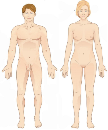
Anatomical position
Standing upright, feet parallel to the floor, eyes looking forward, arms at side, palms facing forward, thumbs pointing away from the body
Superior
Above, towards the head
Inferior
Below, towards the feet
Proximal
Closer to an attached area or the trunk
Distal
Further from an attached area or the trunk
Medial
Toward the midline
Lateral
Away from the midline
Anterior (ventral)
Front of the body
Posterior (dorsal)
Back of the body
Deep
Towards the inside
Superficial
Towards the surface
Sagittal plane
Separates the body into right and left
Frontal (coronal) plane
Separates the body into anterior and posterior
Transverse plane
Divides the body into superior and inferior regions
Midsagittal plane
Plane down the middle separating the body into equal right and left halves
Anteroposterior axis
Perpendicular to the frontal plane; ex. jumping jacks
Transverse axis
Perpendicular to the sagittal plane; ex. bicep curls or nodding yes
Vertical axis
Perpendicular to the transverse plane; ex. shaking your head no
Frontal
Forehead
Occipital
Base of skull
Orbital
Eye
Otic
Ear
Nasal
Nose
Buccal
Cheek
Oral
Mouth
Mental
Chin
Clavicular
Collar bone
Pectoral
Chest
Sternal
Breastbone
Costal
Rib
Scapula
Shoulder blade
Vertebral
Spinal column
Lumbar
Lower back
Coxal
Hip
Sacral
Between hips
Gluteal
Buttock
Acromial
Point of shoulder
Axillary
Armpit
Brachial
Arm
Olecranon
Point of elbow
Antecubital
Front of elbow
Antebrachial
Forearm
Carpal
Wrist
Palmar
Palm
Dorsal
Back of hand
Digital
Finger
Femoral
Thigh
Patella
Kneecap
Popliteal
Back of knee
Sural
Calf
Crural
Leg
Talus
Ankle
Dorsum
Top of foot
Tarsal
Instep
Plantar
Bottom of foot
Digital
Toe
Long bone
Bone that is longer than they are wide; ex. femur
Short bone
Bone that is as wide as it is long; ex. cuneiforms in the foot
Flat bone
Bone that is flat…ex. sternum
Irregular bone
Bone shape that doesn’t fit into another category; ex. vertebra
Sesamoid bone
Bones that sit in the middle of a tendon; ex. knee cap
Pneumatized bone
Bone with air filled categories; ex. ethmoid or sphenoid in the skull
Condyle
Large rounded projection that articulates with another bone
Facet
Small flat surface on a bone that articulates with another bone
Head
Rounded proximal end of a bone that articulates with another bone
Epicondyle
Projection above a condyle for muscle attachment
Ramus
Part of an irregular bone; thicker than a process for muscle attachment
Spine (bone)
Sharp projection on a bone for muscle attachment
Trochanter
Very large projection on a bone for muscle attachment
Tubercle
Small projection on a bone for muscle attachment
Tuberosity
Large projection on a bone for muscle attachment
Axial skeleton
Includes the skull, spinal column, and ribs
Appendicular skeleton
Includes the upper and lower limbs, meaning the pectoral girdle and pelvic girdle
Articulation
Where a bone meets another bone, cartilage, or teeth; vary in mobility and stability
The relationship between mobility and stability
Most mobile, means least stable. Immobile means most stable.
Factors that influence joint stability
Shape of articular surfaces
More ligaments = more stable joint
Muscle tone keeps tension on tendons to stabilize them
Diarthroses
Freely mobile joints
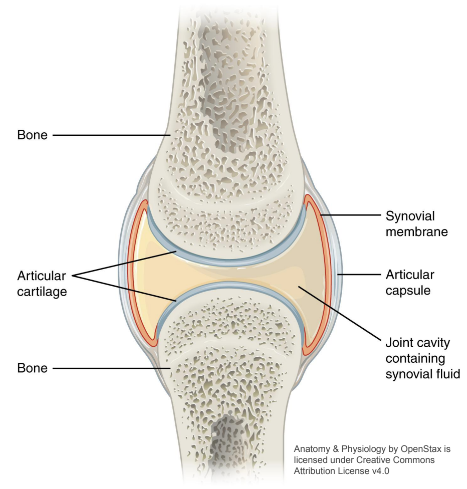
Synovial Joint
Articulating bones are separated by a fluid filled joint cavity including an articular capsule, synovial membrane which secretes synovial fluid, articular cartilage, intrinsic/extrinsic ligaments, nerves and blood vessels (but vessels are not within the joint cavity)
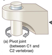
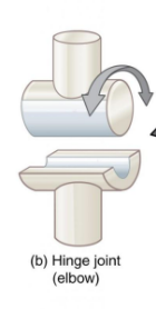
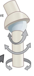
Uniaxial joint
Joint involved in rotation and angular motion (flexion/extension, abduction/adduction). Examples are pivot joints and hinge joints.
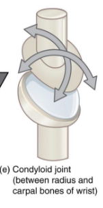
Biaxial joint
Joint involved in angular motion (flexion/extension, abduction/adduction). Example is a condylar joint
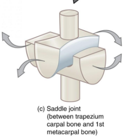
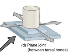
Multiaxial joint
Joint involved in angular motion (flexion/extension, abduction/adduction), rotation (medial and lateral), and circumduction. Examples are plane joints, saddle joints, and ball and socket joints.
Pivot Joint
Hinge joint
Saddle joint
Plane joint
Condyloid joint
Ball and socket joint
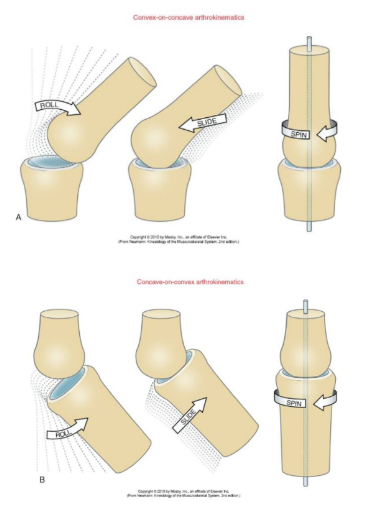
Joint arthrokinematics
Motion between articular surfaces. Most synovial joints are comprised of one bone with a concave articular surface, and one with a convex articular surface. This helps them to roll, glide, and spin so they don’t dislocate.
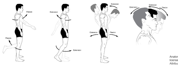
Flexion
Movement that decreases the angle between two bones or parts of the body. Occurs in the sagittal plane on the transverse axis of the body. Examples are the neck, shoulder, elbow, wrist, digits, spine, hip, and knee.
Extension
Movement that increases the angle between two bones or parts of the body. Occurs in the sagittal plane on the transverse axis of the body. Examples are the neck, shoulder, elbow, wrist, digits, spine, hip, and knee.

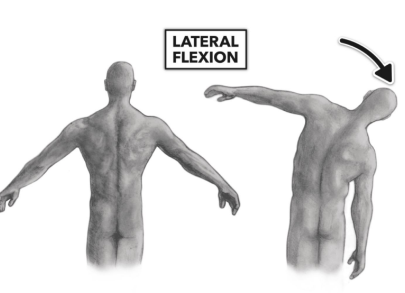
Lateral flexion
Bending the vertebral column to the side. Occurs in the frontal plane along the anteroposterior axis of the body. Occurs in the vertebral column (cervical, thoracic, and lumbar regions)
Rotation
Turning around a central vertical axis. Occurs in the transverse plane on the vertical axis.

Medial rotation
Rotation of the limbs when the anterior surface is turned medially towards the midline.

Lateral rotation
Rotation of the limbs (shoulder and hip) when the anterior surface is turned laterally away from the midline.

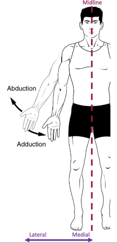
Abduction
Movement of the limb/digit away from the midline. Occurs in the frontal plane on the anteroposterior axis. Occurs in the shoulder, digits, and hip.

Adduction
Movement of the limb/digit towards from the midline. Occurs in the frontal plane on the anteroposterior axis. Occurs in the shoulder, digits, and hip.
Pronation
Rotation of the forearm (radius) medially, causing the palm to face downwards. Occurs in the transverse plane on the vertical axis.
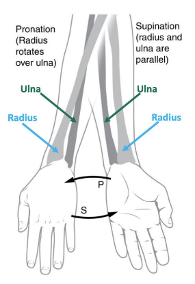
Supination
Rotation of the forearm (radius) laterally, causing the palm to face upwards. Occurs in the transverse plane on the vertical axis.

Opposition
Movement of the pad of the thumb/pinky to touch another finger
Reposition
Reversal of opposition