dental embryology, histology, and anatomy Ch 1 and 15 quiz
1/69
There's no tags or description
Looks like no tags are added yet.
Name | Mastery | Learn | Test | Matching | Spaced | Call with Kai |
|---|
No analytics yet
Send a link to your students to track their progress
70 Terms
dentition
used to describe the natural teeth in the jaws
a person has two dentitions during a lifetime: primary and permanent dentition
primary dentition (baby teeth)
primary school ~ first school = primary dentition ~ first set of teeth
20 teeth in total
8 incisors
4 canines
8 molars
NO premolars
permanent dentition (adult teeth)
second dentition to develop
32 teeth in total
8 incisors
4 canines
8 pre molars
12 molars
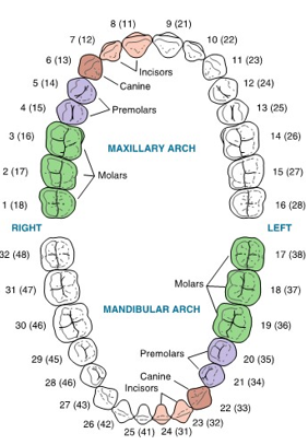
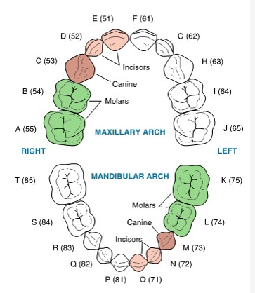
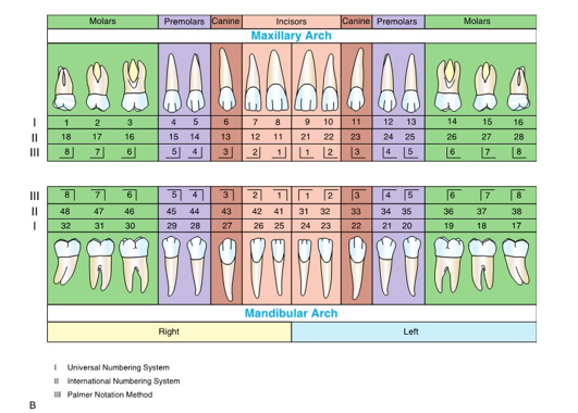
universal numbering system (UNS)
united states
Starts on patient’s upper right side, 1-16, then dropping down to mandible starting at 17-32
primary dentition follows the same pattern, using letters A-T

international numbering system (INS)
teeth are designated from each other by using a two digit code
the first digit corresponds to the specific quadrant the tooth lies in, with the second number indicating the position of the tooth in the quadrant
for example: the right central maxillary incisor would be tooth “11”
primary dentition starts first quadrant at 5

palmer notation method
known as the military tooth numbering system
teeth are designated from each other with a right-angle symbol indicating the quadrants and arch with tooth number placed inside
dentition periods
There are two dentitions, but three ___ ___
primary
mixed
permanent
mixed dentition period
~6 to 12 years
eruption of permanent mandibular first molar (6-year molar)
primary and permanent
fastest and most noticeable
primary dentition period
~6 months to 6 years
eruption of primary mandibular central incisor
primary
beginning
permanent dentition period
After ~ 12 years
shedding of last primary tooth
usually permanent
slowest and least noticeable dentition period
dental anatomy
is the area of the dental sciences dealing with the morphology or form of the teeth, both the crown and root.
alveolus
Alveolar process means bone.. it needs an alveoLUS because the tooth needs less bone (hole) for the tooth to go in
surrounds and supports each tooth
the bone of the tooth socket
located in the alveolar process (tooth bearing part of each jaw)

occulsion
is the method by which the teeth of the mandibular arch come into contact with those of the maxillary arch
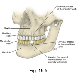
mandible
man down! (lower jaw)

D-A-Q-T system
is based on the tooth within its quadrant
D for dentition (primary, permanent)
A for arch (maxillary or mandibular)
Q for quadrant (left or right)
and T for tooth type (molar, premolar, canine, incisor, etc)
ex: (D) permanent, (A) mandibular, (Q) left (T) first premolar

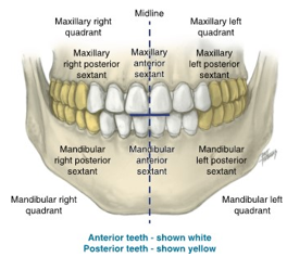
sextants
further divide each dental arch into three parts according to the relationship with the midline
each arch is divided into left and right
central six incisors and canine are considered “anterior” or “front facing” sextant
remaining molars and considered either left or right posterior sextant
crown
Each tooth consists of a __ and one or more roots
can be seen on the top of the tooth
each __ has dentin covered by enamel
Dentin meets enamel at the dentinoenamel junction (DEJ)
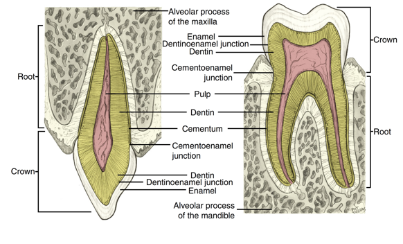
root
Each tooth consists of a crown and one or more __
The __ consists of dentin surrounded by cementum
The cementum meets the enamel at the cementoenamel junction (CEJ)

pulp cavity
consists of a pulp chamber, pulp canal or canals, an apical foramen, and possibly a pulp horn
covered by dentin
consists of nerves, blood vessels, and the connective tissues of the tooth

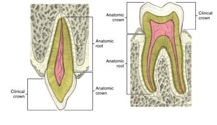
anatomic crown
anatomy never changes
is the entire part of the tooth that is covered by the enamel
stays constant except for attrition and other physical wear

clinical crown
is the part of the anatomic crown that is visible and not covered by the gingiva
height is determined by the location of the marginal gingiva
depends on health of patient
“what is visible?”

apical
the tip of the tooth’s root
the hole that the pulp and nerves use to enter the alveolar process
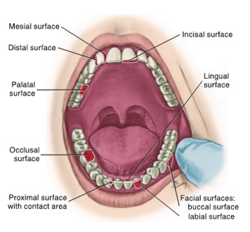
masticatory surface
Is the chewing surface on the most superior surface of the crown
incisal surface for anterior teeth
occlusal surface on posterior teeth
both have lineal elevations (ridges) which are named according to location
cusps
The masticatory surfaces of both canines and posterior teeth have at least one major elevation; a __
__ contribute to a significant part of the tooth’s surface
mesial
the surface of the specific tooth closest to the midline

distal
the surface of the specific tooth farthest from the midline

proximal
together, both the mesial and distal surfaces between adjacent teeth are considered the __
interproximal space
the area between adjacent tooth surfaces

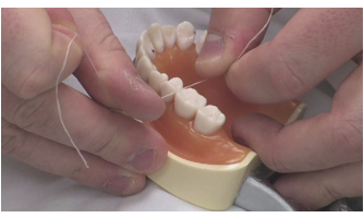
contact areas
The area where the crowns of adjacent teeth in the same arch physically touch on each proximal surface is the ___ ____, or as referred to by clinicians, the __
(The clicking sound you hear when flossing between teeth and the resistance felt are due to strong __)
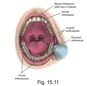
embrasures
when two teeth in the same arch come into contact, the curvatures next to the contact areas form spaces considered __.
line angle
is formed by the lines created at the junction of two crown surfaces and the name is derived by combining the names of the two surfaces
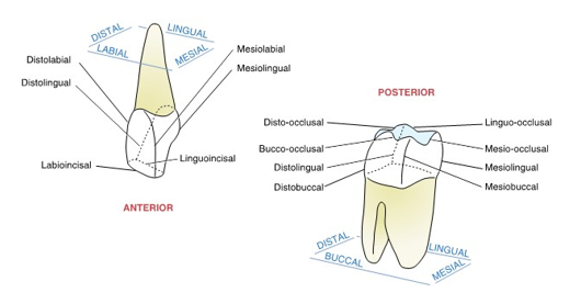
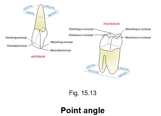
point angle
is another way to determine the specific area of the crown
the junction of three surfaces of the crown, the point angle takes its name from those three surfaces
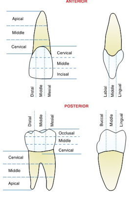
cutting a tooth into thirds
a crown can be divided both horizontally and vertically into three parts, or thirds, to designate specific tooth areas
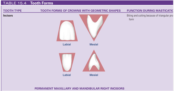
incisors
instruments for biting and cutting food during mastication (triangular proximal form of their crowns)
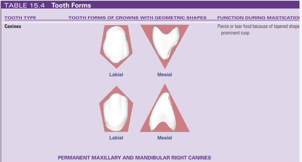
canines
because of the tapered shape and the prominent cusp of their crowns, function to pierce or tear food during mastication
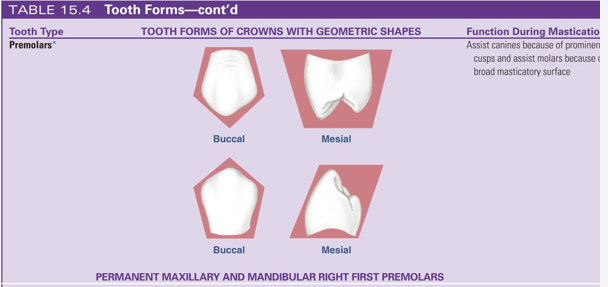
premolars
only found in permanent dentition
assist canines in piercing and tearing food because of prominent cusps of their crowns during mastication
assist molars in grinding food due to wide masticatory surface (occlusal surface)
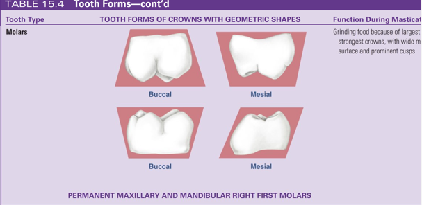
molars
The __ function in grinding food during mastication, assisted by the premolars
The wide masticatory surface, the occlusal surface of the ___ , with the prominent cusps, functions during mastication
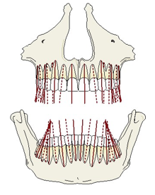
root axis line (RAL)
an imaginary line representing the long axis of a tooth, drawn in a way to bisect the root (and thus the crown) in the cervical area into two halves
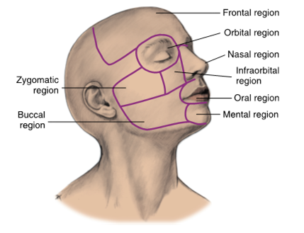
the regions of the face
frontal
orbital
nasal
infraorbital
zygomatic
buccal
oral
mental regions

lymph nodes
are bean-shaped, contain lymphatic vessels
located in certain areas of the face and head and when palpable, should be noted in the patient record
frontal regions of face
(forehead)
includes the forehead and areas above (superior) eyes

orbital
in the __ region of the face, the eyeball and all its supporting structures are contained in the orbit of the skull, which is the bony eye socket
infraorbital region
is located inferior (below) to the orbital region and lateral to the nasal region

zygomatic region
overlies the bony support for the cheek, the zygomatic arch
extends from just below (inferior) lateral margin of the eye toward the middle part of the external ear
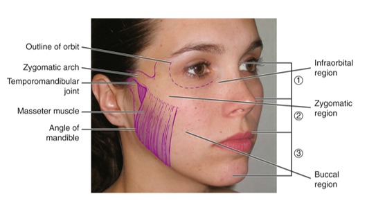
buccal region
is composed of the soft tissue of the cheek
forms the side of the face and is a broad area of the face between the nose, mouth, and ear
masseter muscle
angle of mandible

submandibular lymph node drainage (not on quiz)
The forehead and the anterior part of the face drain into the ___ lymph nodes
Buccal lymph nodes may be present along the course of the previous vessel
parotid lymph node drainage (not on quiz)
the lateral part of the eyelids and lateral part of face drain into __ lymph nodes
submental lymph nodes (not on quiz)
lower lip and the skin of the chin are drained into __ lymph nodes
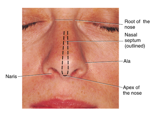
nasal region
the main feature of the nasal region of the face is the external nose
the root of the nose is located between the eyes
the tip of the nose is the apex
naris = holes of nose that surrounded by ala
oral region
has many structures within it such as lips and oral cavity
the upper and lower lips are fleshy fold that mark the gateway of the oral cavity proper

labial commissure
the corners of the mouth where both lips meet
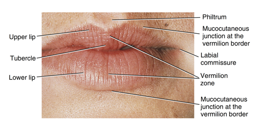
philtrum
the area of the upper lip that has a slight groove located inferior to the nasal septum
midline of upper lip

vermillion zone
has a darker reddish appearance than the surrounding skin
outlines from the surrounding skin by a transition zone, the mucocutaneous junction at the vermillion border

tubercle of the upper lip
the philtrum terminates in a thicker area of the midline of the upper lip known as the __

the mental region
The chin is the major feature of the __ region of the face
The bone underlying the __ region is the mandible (lower jaw)

mandibular symphysis
marks the midline of the mandible
prominent midlines may result in “butt chin”
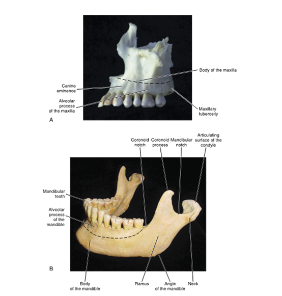
ramus
Lateral aspect of the mandible
stout flat plate of the __ extends superiorly and posteriorly from the body of the mandible on each side

coronoid process
The anterior border of the ramus is a thin sharp margin that terminates in the ___ ____
(crown always in front/ on top)

coronoid notch
The main part of the aNterior border of the ramus forms a concave curve, the ___ ___

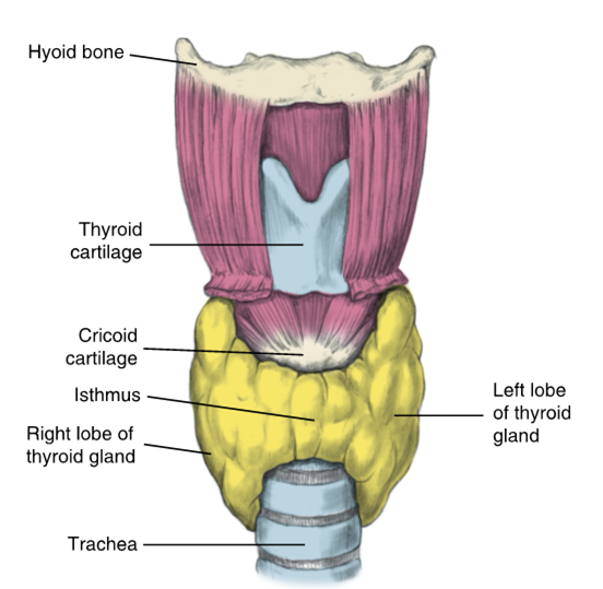
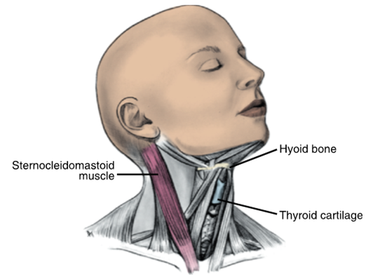
thyroid gland
Butterfly-shaped organ
endocrine gland
prominence of the larynx, which is considered the “voice box”
The vocal cords, as ligaments of the larynx, are attached to the posterior surface of the ___ cartilage

face and neck
can be examined by visualization and palpation
a certain degree of variation in surface features can be considered within a normal range
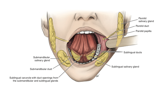
parotid salivary gland
has a small part that can be palpated in the buccal region as well as in the zygomatic region
largest of three major salivary glands located inferior to the external ear

mucocutaneous junction
located at the vermillion border of the lips
(where the skin and the mucus border meet)

mandibular condyle
head articulates with the TMJ
(con people are always behind)

mandibular notch
located between mandibular condyle and coronoid process

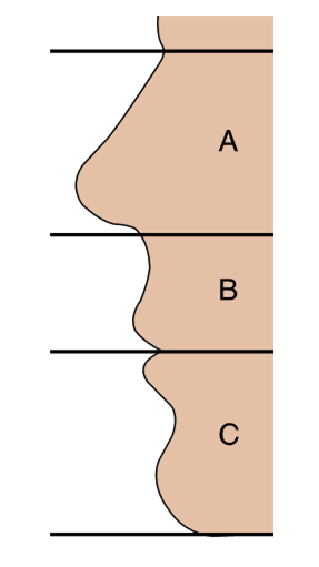
golden proportions
a set of guidelines with three divisions illustrating the considerations of vertical facial dimension
regions of neck (triangles of neck)
extend from the skull and lower jaw down to the clavicles and sternum
sternocleidomastoid muscle (SCM) is easily palpated on each side with the bordered dividing the neck into posterior and anterior regions
parathyroid glands
endocrine glands
cannot be palpated
regulate calcium
submandibular and sublingual salivary gland
can be palpated in a patient in the neck region
submandibular gland is located inferior to mandible
sublingual gland is located inferior to tongue (lingua)
hyoid bone
controls position of tongue