Interpretation of ECG & Select Dysrythmias | NURS230 (V2)
 Call Kai
 Learn
Learn Practice Test
 Spaced Repetition
 Match
Match Flashcards
 Knowt Play
1/82

There's no tags or description
Looks like no tags are added yet.
Name | Mastery | Learn | Test | Matching | Spaced | Call with Kai | Chat |
|---|
No analytics yet
Send a link to your students to track their progress
83 Terms
what are the different types of ECGs?
3 lead
12 lead
continuous
single application
holter monitoring
stress testing
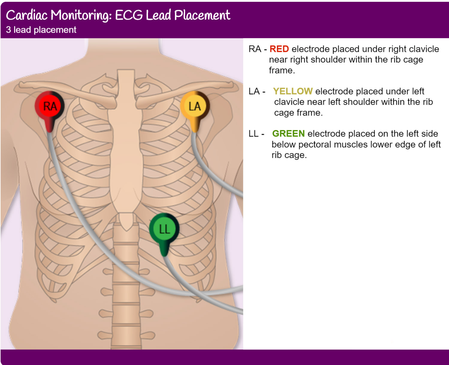
what is the 3 lead ECGs and where are the electrode placed?
a simplified version of a standard 13-lead ECG that uses only three electrodes to record the electrical activity of the heart
RED (RA) - placed under the right clavicle near the right shoulder within the rib cage frame
YELLOW (LA) - placed under the left clavixle near the left shoulder within the rb cage frame
GREEN (LL) - placed under the left side below the pectroal muscles lower edge of left ribcage
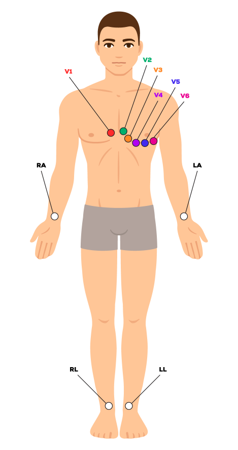
what is the 12-lead ECG and where are the electrode placed?
V1 - fourth intercostal space on the right sternum
V2 - fourth intercostal space at the left sternum
V3 - Midway between placement of V2 and V4
V4 - fifth intercostal space at the midclavicular line
V5 - anterior axillary line on the same horizontal level as V4
V6 - mid-axllary line on the same horizontal level as V4 and V5
RA (right arm) - anywhere between the right shoulder and the right elbow
RL (right leg) - lateral calf muscle
LA (left arm) - anywhere between the left shoulder and the left elbow
LL (left leg) - lateral calf muscle
what is the difference between continuous, single application, holter monitoring, and stress testing ECG?
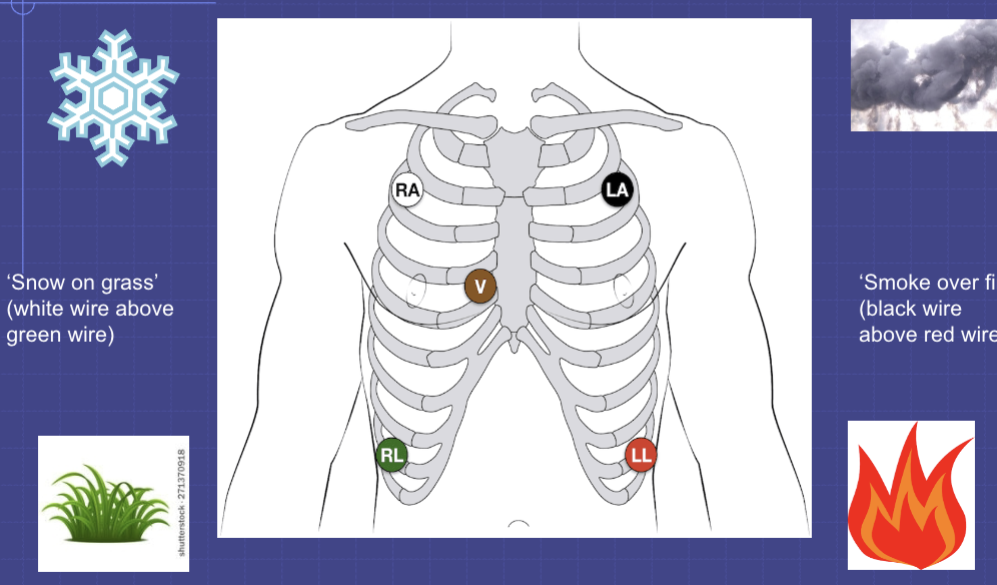
what does the ‘snow on grass,’ ‘smoke over fire,’ and ‘poop comes forom the tummy’ mean in term of ECG?
it is the placement of the electrodes for a 5-lead monitor
white at the second right intercostal space
black at the second left intercostal space
green at the eighth right intercostal space
red at the eighth left intercostal space
brown at the fourth intercostal space on the right sternal border
what are the fourh properties of cardiac cells and a brief description of each?
automaticity - ability to self-regulate
excitability - ability to start the electrical impulse
conductivity - ability to transfer the electrical impulse
contracbility - ability to contract
what happen when the SA node is no longer working properly?
the AV node take over but can only fire at a lower rate of 40-60 BPM (contrasting to the 60-100 BPM of the SA node)
what happen when the SA and AV node is no longer working properly
the Bundle of His and Purkinje Fibers can take over after both the SA, then the AV node is knocked out, but can only maintain the HR at a rate of 20-40 BPM
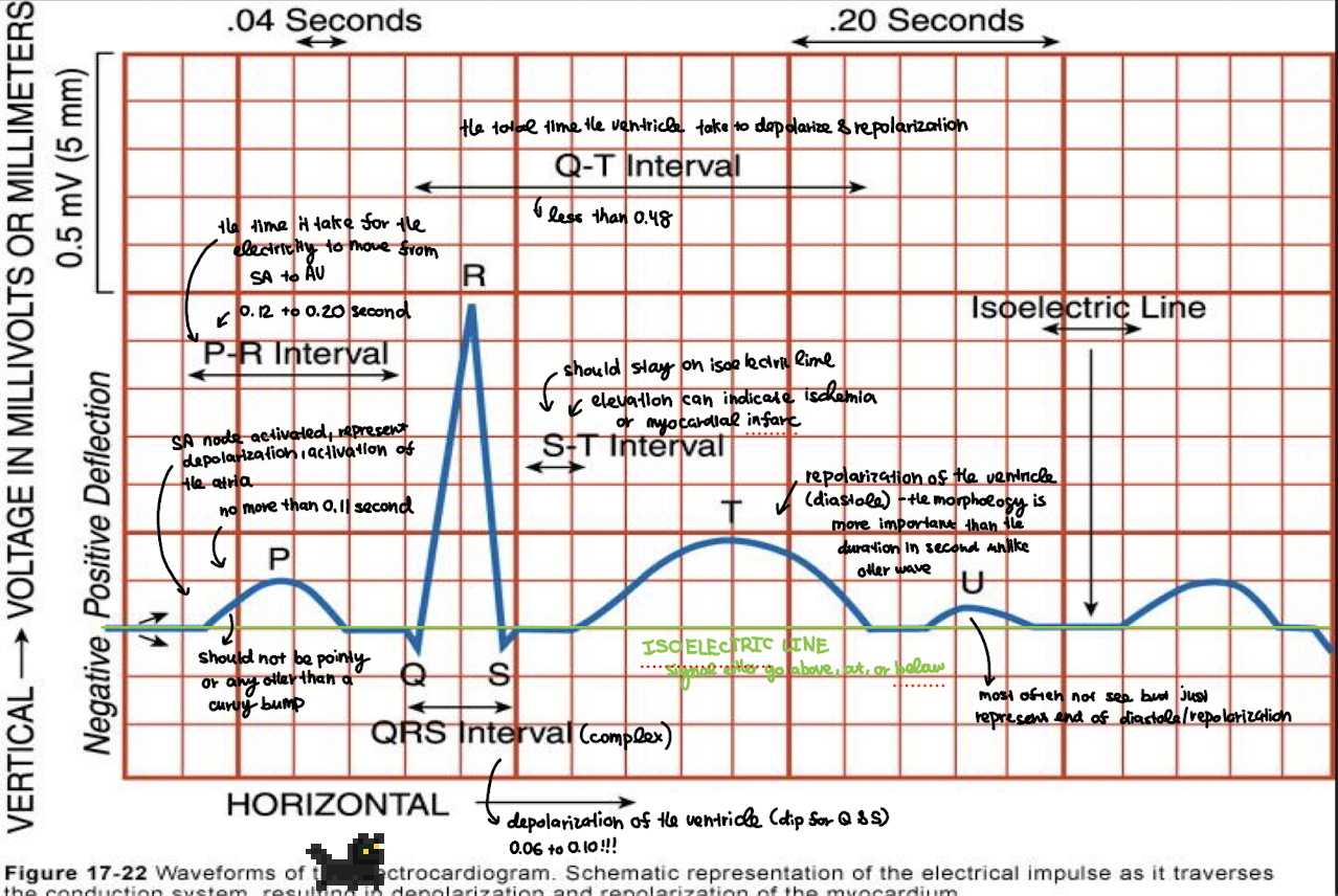
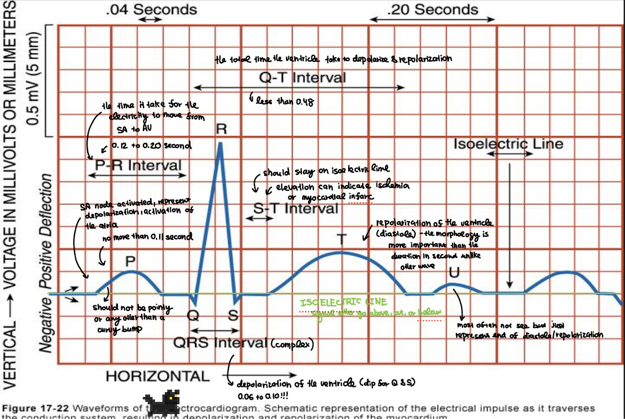
what does P wave represent? what is the normal duration?
atrial depolarization - electrical activity that triggers atrial contraction (should have a normal shape of upright and rounded!)
the normal duration is no more than 0.11 second
what is the normal duration for P wave?
no more than 0.11 second
what does the PR interval represent? what is the normal duration?
time from the start of atrial depolarization (P wave) to the start of ventricular depolarization (QRS complex) - reflects the AV node conductions time?
the normal range is 0.12-0.20 second
what is the normal duration for PR interval?
0.12-0.20 second
what does PR segment represent?
the flat line between the end of the P wave and the start of QRS complex; represent the delay in the AV node before ventricular depolarization
what does the QRS complex representation? what is the normal duration?
ventricular depolarization - elecrrical activity that triggers ventricular contraction
the normal duration is between a 0.06-0.10 second
what is the normal duration for a QRS complex?
0.06-0.10 second
what does the ST interval (AKA segment) represent?
flat line between he end of the QRS complex and the start of the T wave; represents the ventricles being; normally isoelectric (flat on the isoelectric line with no depression or elefation) —> elevation or depression suggsts ischemia or injury to the heart tissues
true or false: ST interval should be isoelectric as elevation or depression can significant ischemia or injury to the heart tissues
true
what does the T wave represent?
ventricular repolarization - electrical recovery of the ventricles
what does the U wave represent?
small wave after the T wave (not always visible); thought to represent depolarization of the Purkenje fibers or papillary muscles - prominent in hypokalemia
what does the QT interval represent? what is the normal duration?
the total time the ventricle take to depolarize and depolarize
the normal duration is less than 0.48 second
what is the normal duration for the QT interval
less than 0.48 seconds
what is the T-wave morphology to look out for? (what does being tall/peaked, flat/inverted, or sudden changes represent)
Normal: Smooth, slightly asymmetrical, upright in most leads.
Tall / Peaked T waves: May indicate hyperkalemia — monitor potassium levels, prepare for treatment.
Flat or Inverted T waves: May indicate hypokalemia, ischemia, or MI — report changes.
Sudden changes in T waves are clinically important and should be promptly reported.
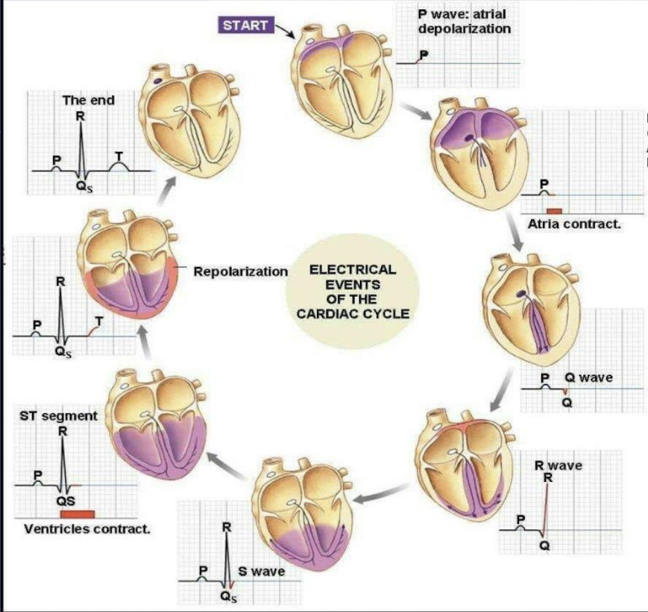
what does the Q, R, and S, wave in the complex each represent with the addition of the ST segments?
Q wave = the initial ventricular depolarization, specifically the signal traveling down the intraventricular septum from left to right
R wave = reflect the major ventricular depolarization, the bulk of both ventricle depolarizing through the Purkinje fibers
S wave = the final phase of ventricular depolarization, specificlaly the activation of the upper parts of the ventricles → depolarization is complex and the ventricle contracts at the end of the QRS complex
ST segment = ventricles are fullu depolarized and conractions
how do you determine atrial and ventricular rhythm & regularity on an ECG?
Atrial Rhythm:
Look at P–P intervals (distance between P waves).
If evenly spaced → regular atrial rhythm.
If uneven → irregular atrial rhythm.
Ventricular Rhythm:
Look at R–R intervals (distance between R waves).
If evenly spaced → regular ventricular rhythm.
If uneven → irregular ventricular rhythm.
Tip for both: Use a piece of paper or calipers to mark and compare spacing.
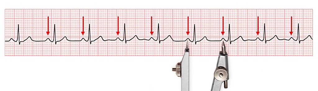
what does it mean if there is not a P wave for every QRS complex?
Atria are not triggering each ventricular beat.
Electrical signal may start from AV node or ventricles (junctional or ventricular rhythm).
Could also indicate heart block — atrial impulses aren't reaching the ventricles.
Always abnormal → should be reported and monitored.
what does it mean if P waves come after the QRS complex?
The ventricles are depolarizing before the atria.
Electrical impulse is coming from the AV node or ventricles, not the SA node.
This is retrograde atrial depolarization → seen in junctional rhythms or AV dissociation.
Always abnormal and should be reported/monitored.
what does it mean if P waves are absent on an ECG?
The atria are not depolarizing normally.
Impulse may not be coming from the SA node.
Seen in (focus on A-Fib and V-Fib):
Atrial fibrillation (most common) - no P waves + irregular rythm
Ventricular fibrillation + no P waves + no organized rythm (EMERGENCY)
The heart is beating so fast (like in Supraventricular tachycardia) that the QRS complex engulf the P wave in the reading
Junctional rhythm (AV node is pacemaker)
Sinus arrest / SA node failure
Ventricular rhythm
Should be assessed and reported.
true or false: the PR interval cannot be measured if the P wave is missing
true
true or false: sometimes the heart beats so fast that the QRS complex enguft the P waves in the reading
true
what does it mean if one or more QRS complexes are missing on an ECG?
Missing QRS complexes = ventricles did not contract because the impulse didn’t reach them.
This usually means a heart block or conduction issue.
One missing QRS (occasionally):
→ May indicate a dropped beat from a 2nd-degree AV block (Mobitz Type I or II).Two or more QRS complexes missing in a row:
→ Suggests a more serious block, such as:Mobitz Type II (2nd-degree AV block)
3rd-degree (complete) heart block
→ Ventricles are not getting signals from atria → can lead to low cardiac output or asystole.
Always abnormal — should be reported immediately.
how many second does each small box on an ECG paper represent?
0.04 second
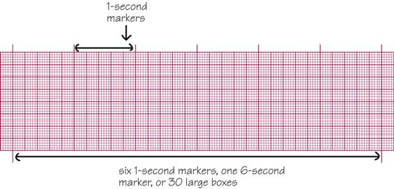
how many second does each large box on an ECG paper represent?
0.20 second
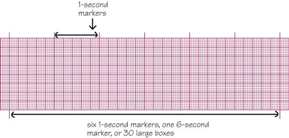
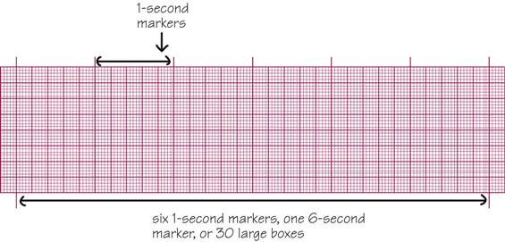
how many second does 5 large boxes on an ECG paper represent?
1 second
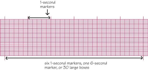
every ________ second (________ large boxes) is marked with a verticle line
3 seconds; 15 large boxes
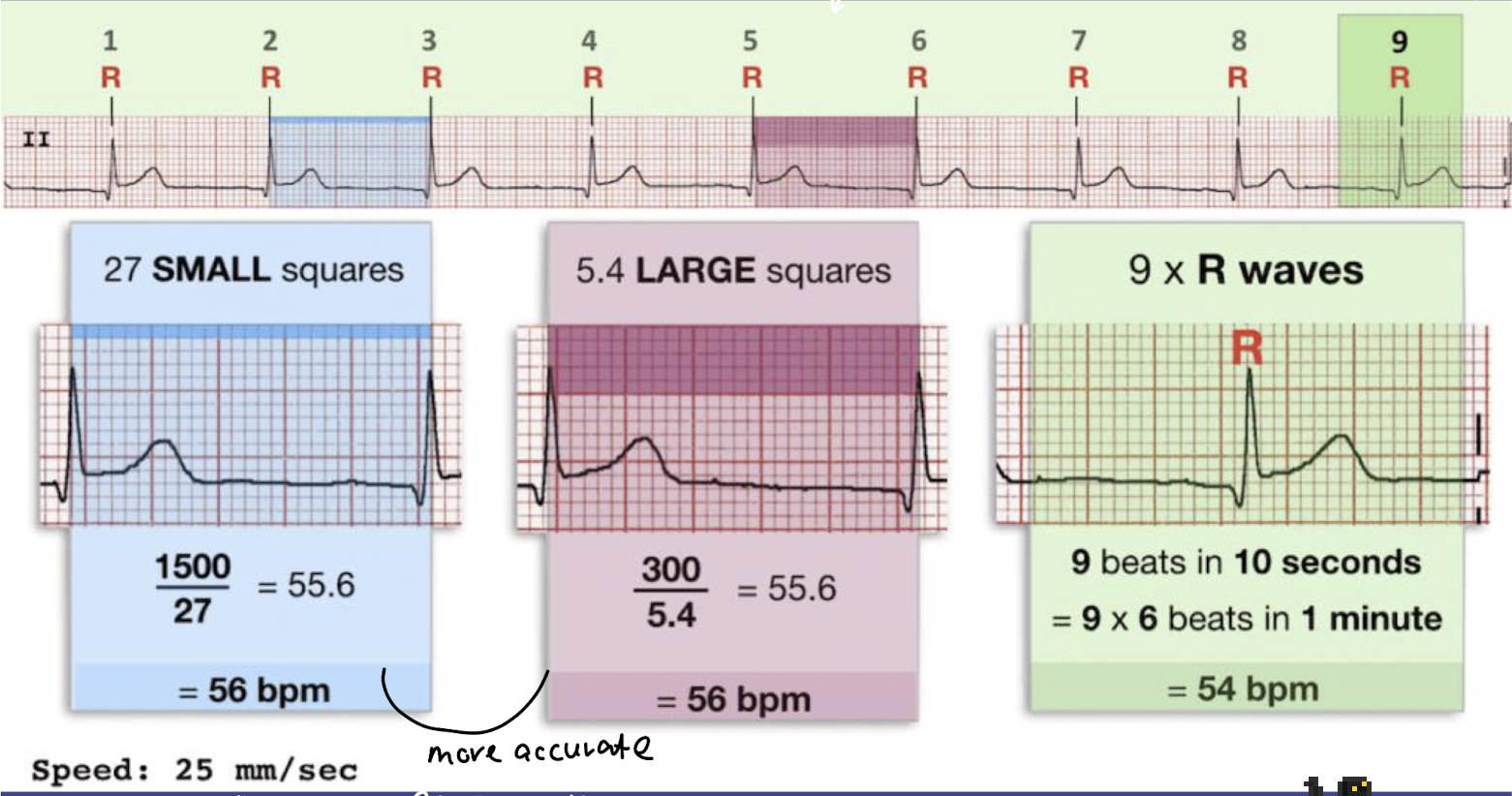
how can you calculate the heart rate using the small box methods?
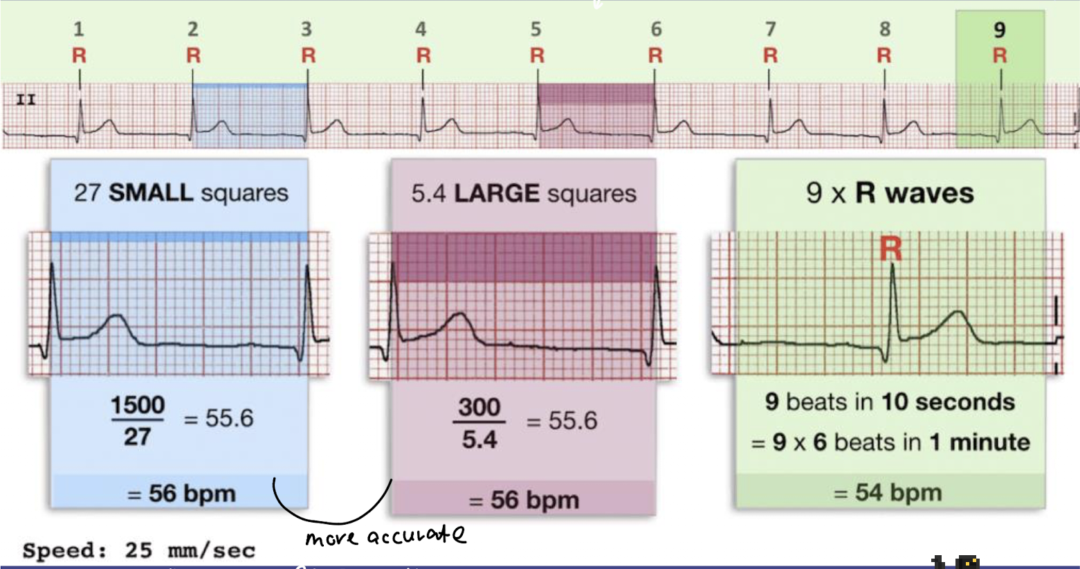
AKA Rule of 1500
count the number of small boxes between two R waves
use the formular: 1500 divided by the number of small boxes
example: 20 small boxes = 1500/20 = 75 bpm
only work if the rhythm is regular
how can you calculate the heart rate using the small box methods?
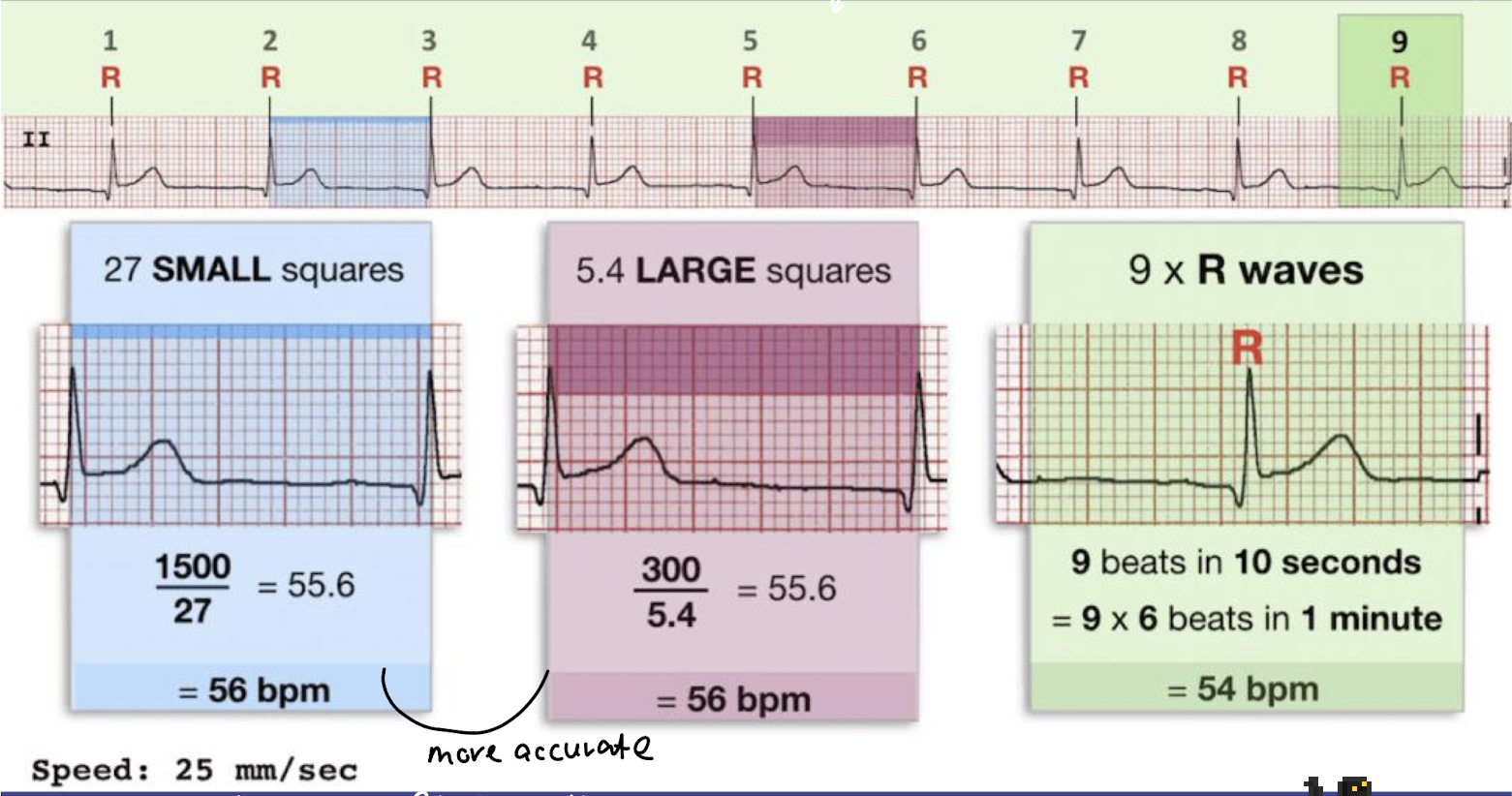
AKA the Rule of 300
Count the number of large boxes between two R waves.
Use the formula: 300 ÷ number of large boxes = heart rate (bpm).
Example: 4 large boxes → 300 ÷ 4 = 75 bpm.
Only work with regular rhythm
true or false: the six-second method, small box method, and large box, can all we use whether the rythm is regular or irregular
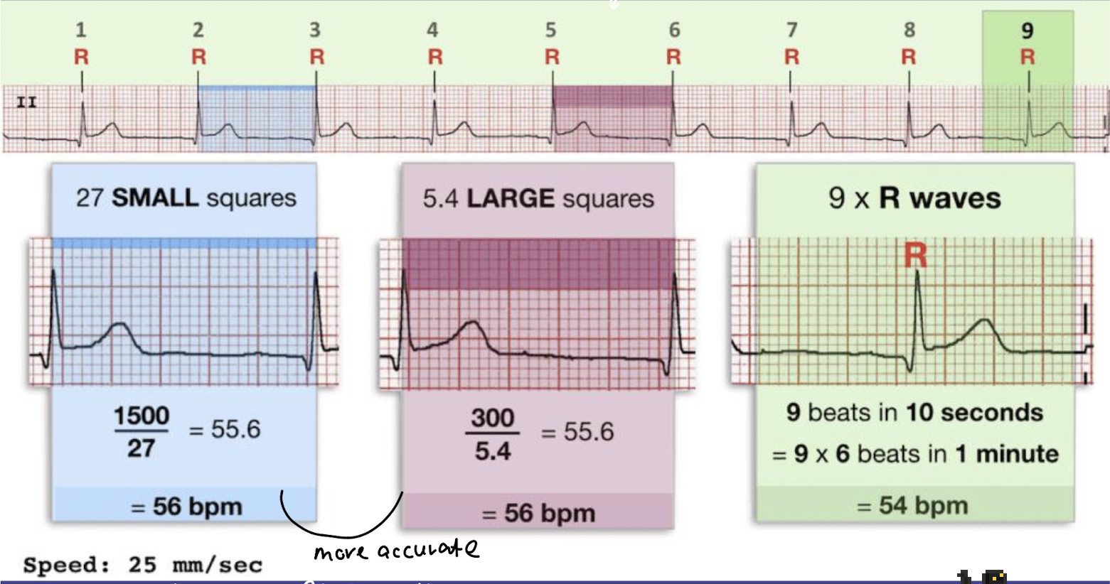
false; only the six-second methods can be use when the rythm is either regular or irregular
how do you calculate heart rate using the 6-second method?
Use this when the rhythm is irregular or regular.
Count the number of R waves (QRS complexes) in a 6-second strip.
A 6-second strip is marked by 3 hash marks or 30 large boxes.
Then multiply by 10.
Heart Rate = Number of QRS complexes × 10
what does a wide QRS complex (over 0.10 secons) mean?
Meaning: Impulse starts in ventricles or conduction is delayed.
Possible Diagnosis:
Ventricular rhythm / Ventricular tachycardia
Bundle branch block
Nursing action: Check pulse, BP, possible emergency if pulseless VT/VF.
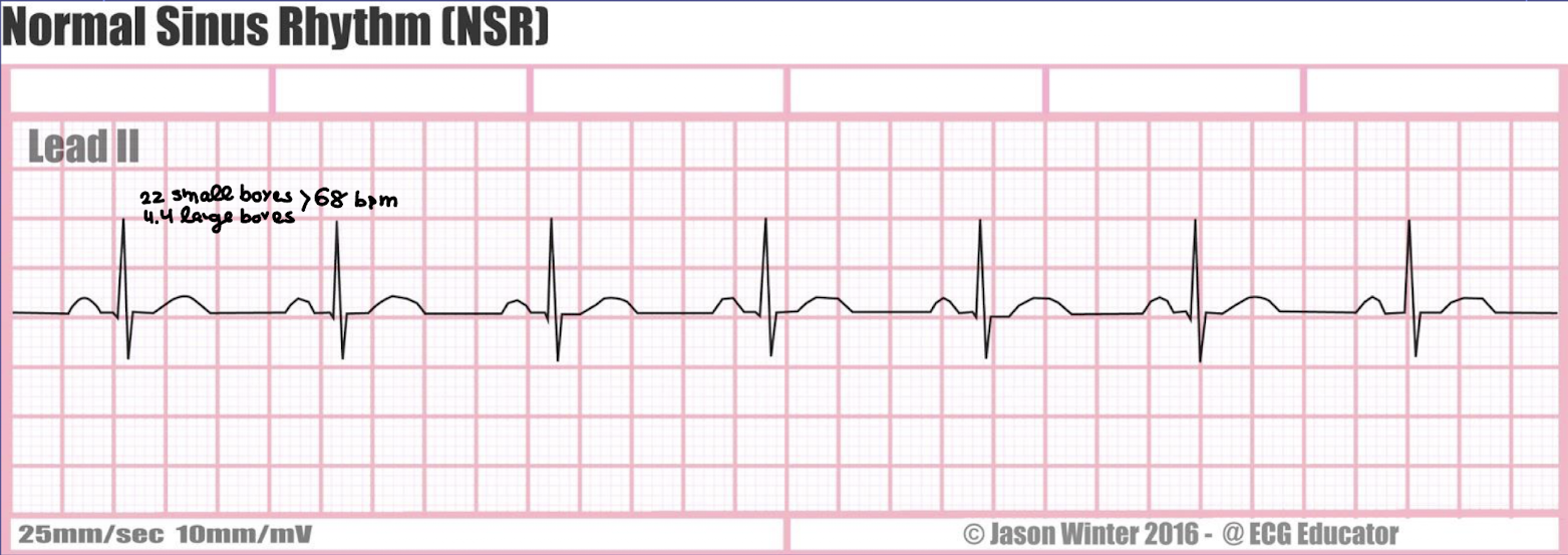
what is the rate, rhythm, P waves characteristics, PR interval duration, and QRS duration of a normal sinus rhythm
rate: 60-100 beats/min
rhythm: regular
P waves: present, consistent configurations, one P wave before each QRS complex with the spacing between each other P waves being regular
PR interval: 0.12-0.20 second and constant
QRS interval: 0.04-0.10 second and constant
what is the rate, rhythm, P waves characteristics, PR interval duration, and QRS duration of a normal sinus rhythm
rate: less than 60 bpm
rhythm: regular
P waves: present, consistent configurations, one P wave before each QRS complex with the spacing between each other P waves being regular
PR interval: 0.12-0.20 second and constant
QRS interval: 0.04-0.10 second and constant
what is the main etiology of sinus bradycardia?
physical fitness
excessive vagal stimulation
hypoxia
hypothermia
disease (e.g, hypothyroidism, hypothermia, beta-blockers, CCBs, MI, increased vagal tone, etc)
medications
what are the clinical presentations of sinus bradycardia and how is it managed?
Clinical Presentations
Often asymptomatic if mild
Symptoms (if HR too low or perfusion affected):
Dizziness or lightheadedness
Syncope (fainting)
Fatigue
Weakness
Confusion
Hypotension
Shortness of breath or chest pain (rare)
Management / Nursing Actions
If asymptomatic: Usually no treatment, just monitor
If symptomatic:
Assess ABCs (Airway, Breathing, Circulation) and vital signs
Oxygen therapy if needed
Identify & treat underlying cause (medications, MI, hypothyroidism, electrolyte imbalance)
Medications:
Atropine (first-line for symptomatic bradycardia)
If atropine is ineffective administer an IV infusion of dopamine or epinephrine as per protocol
Advanced interventions:
Temporary pacing if severe, unresponsive bradycardia
Permanent pacemaker for chronic sick sinus syndrome
Nursing Tip: Always assess perfusion and symptoms, not just heart rate.
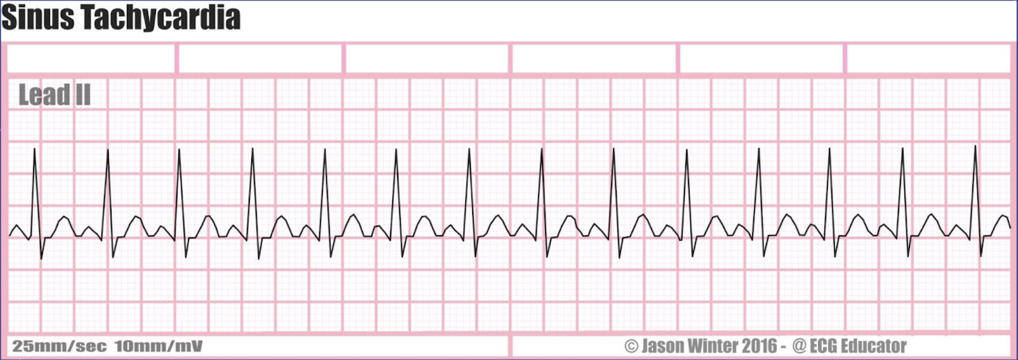
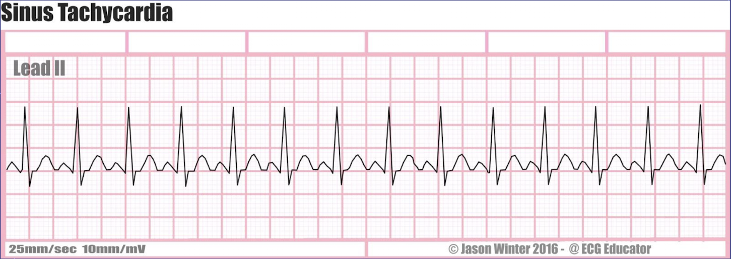
what is the rate, rhythm, P waves characteristics, PR interval duration, and QRS duration of sinus tachycardia?
rate: 100-180/min
rhythm: regular
P waves: present, consistent configurations, one P wave before each QRS complex with the spacing between each other P waves being regular
PR interval: 0.12-0.20 second and constant
QRS interval: 0.04-0.10 second and constant
what are the common causes of sinus tachycardia?
respond to exercise
caffeine
SNS stimulation
fever
anemia
medications (e.g., decongestants, CNS stimulant, caffeine, illici substances, etc)
hyper/hypovlemia
usually it is caused as compensatory mechanism
true or false: sinus tachycardia is concerning but we typically look about for a sustained tachycardia as they are much more concerning
true
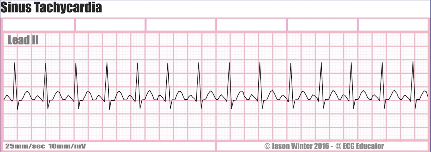
what is the clinical manifestations of sinus tachycardia and what are the management for it?
Clinical Manifestations
Often asymptomatic if mild
Symptoms (if HR high or sustained):
Palpitations / rapid pulse
Dizziness or lightheadedness
Shortness of breath
Chest pain or discomfort
Fatigue or weakness
Anxiety
Management / Nursing Actions
Identify & treat underlying cause (most important!)
Fever, pain, hypovolemia, anemia, hyperthyroidism, anxiety, medications, hypoxia
Monitor vital signs and cardiac rhythm
Supportive care:
Oxygen if hypoxic
IV fluids if hypovolemia
Medications (if persistent or symptomatic):
Beta-blockers (e.g., metoprolol) or calcium channel blockers
Nursing Tip: Treat the cause, not just the heart rate
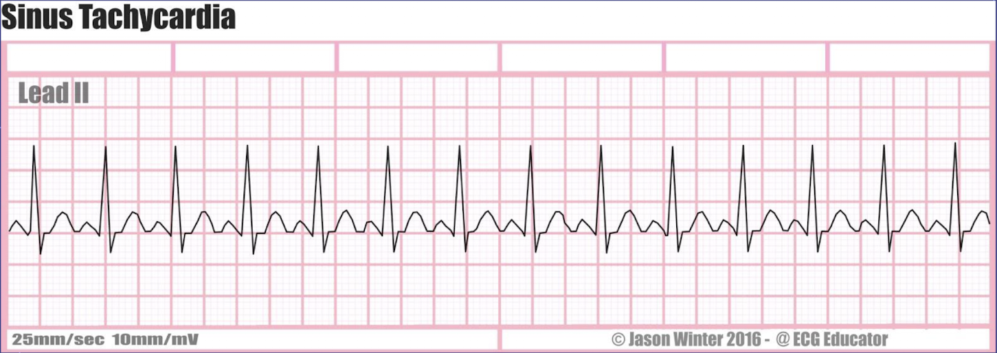
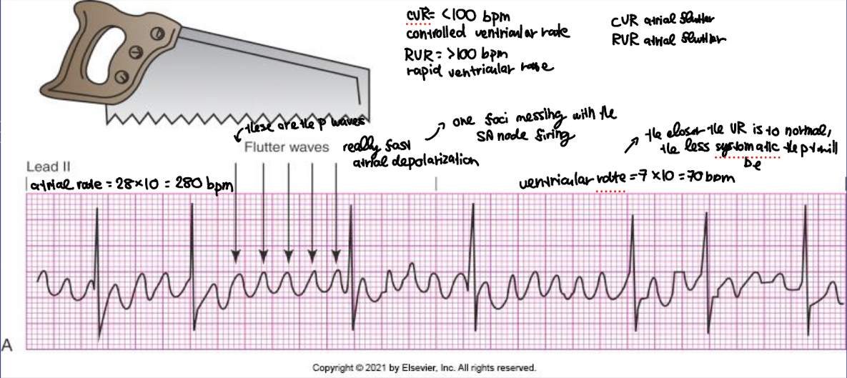
what is the rate, rhythm, P waves characteristics, PR interval duration, and QRS duration of atrial flutter?
rate: 250-350/min; ventricular rate varies
rhythm: irregular rhythm → rapid atrial depolarization
P waves: present but have a ‘saw tooth” appearance
PR interval: varies
QRS interval: varies (usually normal - 0.06 to 0.10 if there is no bundle branch block)
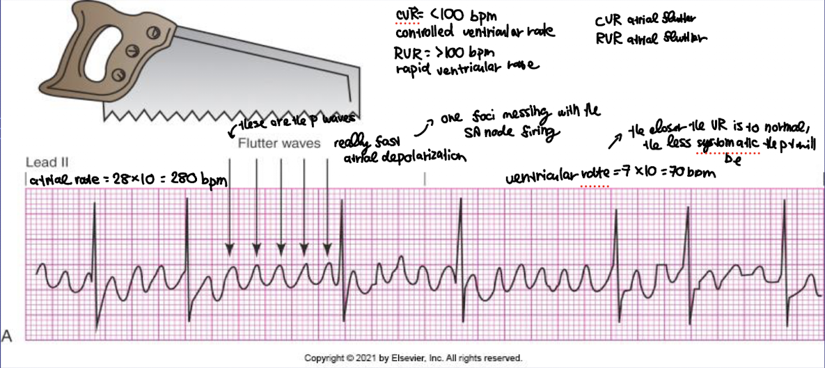
how do you calculate the ventricular and atrial rate using ECG?
1. Ventricular Rate (using QRS complexes)
Steps:
Count the number of QRS complexes in a 6-second strip.
Multiply by 10 → ventricular rate (bpm).
Works for regular and irregular rhythms.
For regular rhythms, you can also use:
Large box method: 300 ÷ number of large boxes between 2 R waves
Small box method: 1500 ÷ number of small boxes between 2 R waves
2. Atrial Rate (using P waves or F waves)
Steps:
Count the number of P waves (or flutter F waves) in a 6-second strip.
Multiply by 10 → atrial rate (bpm).
For regular atrial rhythms, you can also measure P–P intervals:
Large box method: 300 ÷ number of large boxes between 2 P waves
Small box method: 1500 ÷ number of small boxes between 2 P waves
true or false: in atrial flutter, one foci of the SA node is messing up the entire SA node firing
true
how do the nurse know if the atrial flutter have a controlled ventricular rate (CVR) or a rapid ventricular rate (RVR)?
CVR = less than 100 bpm
RVR = more than 100 bpm
what is the common etiology of atrial flutter?
rheumatic or ischemic heart disease
controlled heart fialure
AV valve disease
Pulmonary embolism
alcoholism
pericarditis
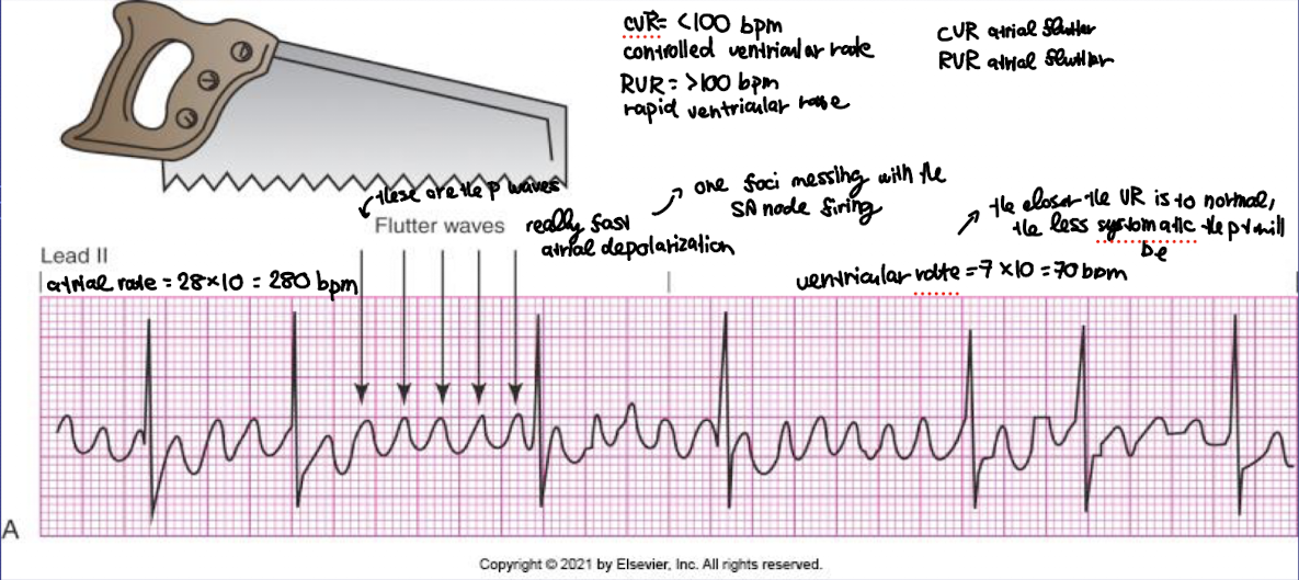
what is the clinical manifestation and management of atrial flutter?
Clinical Manifestations
Often asymptomatic if the ventricular rate is controlled (less than 100 bpm)
Symptoms (if rapid ventricular response or decreased cardiac output):
Palpitations / rapid pulse
Dizziness or lightheadedness
Fatigue or weakness
Shortness of breath
Chest pain or discomfort
Anxiety
Nursing note: Look for irregular ventricular response and signs of decreased perfusion.
Management / Nursing Actions
Assess ABCs and vital signs
Monitor cardiac rhythm
Treat underlying cause (e.g., hyperthyroidism, heart disease, infection)
Medications:
Rate control: Beta-blockers (e.g., metoprolol), Calcium channel blockers (e.g., diltiazem)
Rhythm control / cardioversion: Amiodarone or synchronized cardioversion if unstable
Anticoagulation: To prevent stroke or a traveling blood clot in general (e.g., warfarin, DOACs)
Nursing tips:
Observe for hypotension, syncope, or signs of heart failure (if prolonged due to the weakning of the heart muscle over time due to its excessively fast rate)
Educate patient on medication adherence and stroke prevention
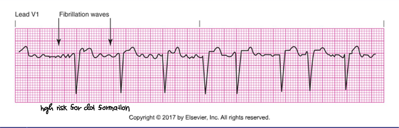
what is the rate, rhythm, P waves characteristics, PR interval duration, and QRS duration of atrial fibrillation?
rate: atrial of more than 400 bpm and ventricular rate varies-
→ caused by multiple atrial foci firing simultaneously (atrial chaos)
rhythm: irregular rhythm → the atria is not contracting and is actually quivering
P waves: absence of countable P waves
PR interval: no measureable
QRS interval: usually normal (0.06-0.10) because ventricular conduction through the AV nodes and bundle branches is intact however the ventricular rhythm will be irregular
what type of heart rhythm lead to a high risk for clot formation?
A-Fib
A-Flutter
what is premature atrial contraction (PAC) and is there a need for treatment?
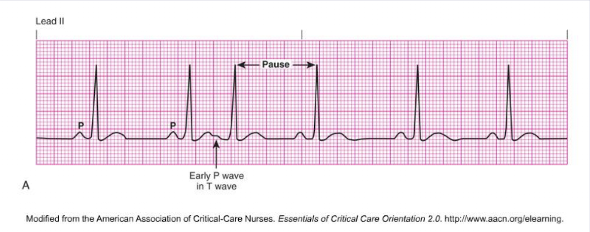
premature ectopic beat from atrial tissues followed by a non-compensatory pause
P waves often looks different
it is mostly harmless just an indicated need for monitoring for patient with weak or diseased heart → it is often referred to as palpitations with no change in cardiac output - no treatment are indicated
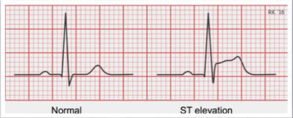
what does an ST segment elevation indicate?
myocardial stress
may also indicate ventricular damage, from multiple causes such angina, CAD, or evolving myocardial infarction where the heart muscle is not getting enough oxygen
what is the rate, rhythm, P waves characteristics, PR interval duration, and QRS duration of supraventricular tachycardia (SVT)?
rate: 100-250 bpm
rhythm: regular; R-R are consistent
P waves: may not be able to visualize P waves as they are often hidden in the preceding T wave due to the fast rate
PR interval: may not be able to measure, depending on whether you can see the P wave
QRS interval: usually narrow but still within 0.06-0.10
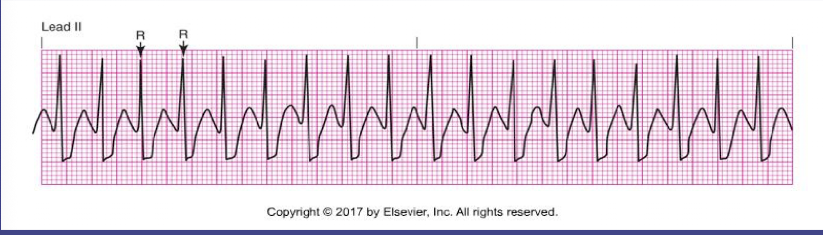
what does supraventrcicular tachycardia mean?
generic term for any tachycardia that originates from a site above the ventricles
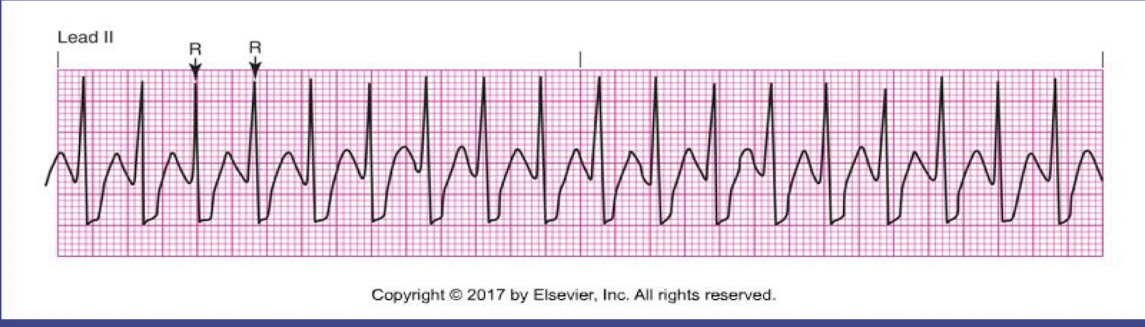
true or false: the onset of supraventricular tachycardia is usually sudden
true
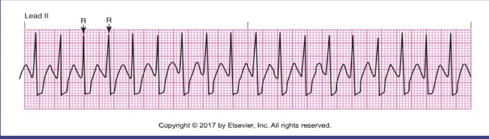
what is the clinical manifestations and management of supraventricular tachycardia?
Clinical presentation:
Palpitations
Dizziness or lightheadedness
Shortness of breath
Chest pain or anxiety
May cause hypotension if prolonged
Management:
Vagal maneuvers (e.g., Valsalva) for stable patients
Adenosine IV if vagal maneuvers fail
Beta-blockers or calcium channel blockers for recurrent episodes
Electrical cardioversion if unstable and symptomatics
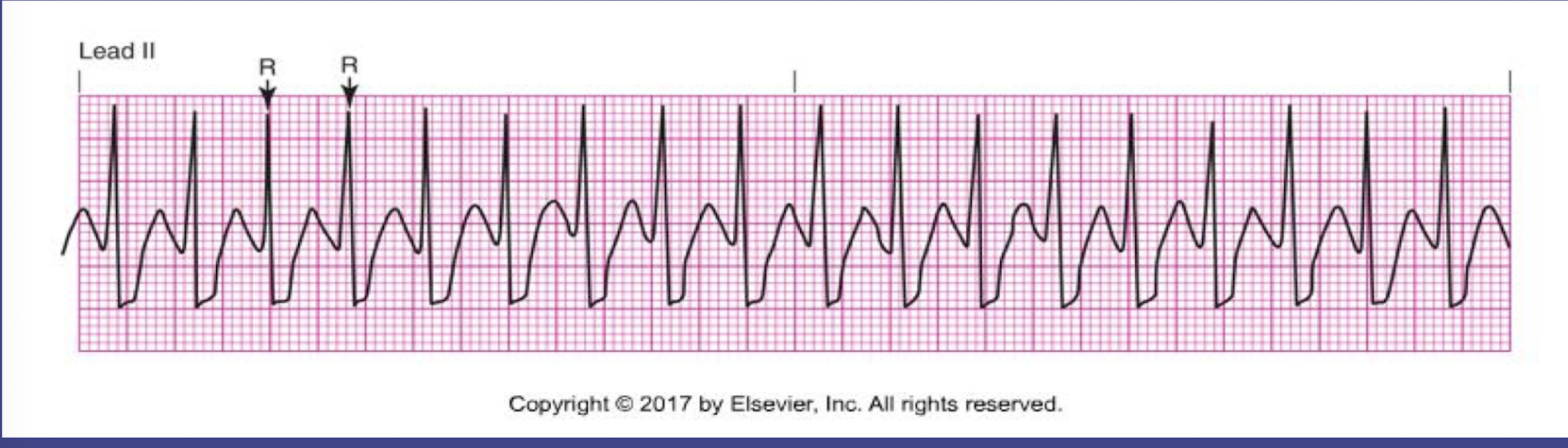
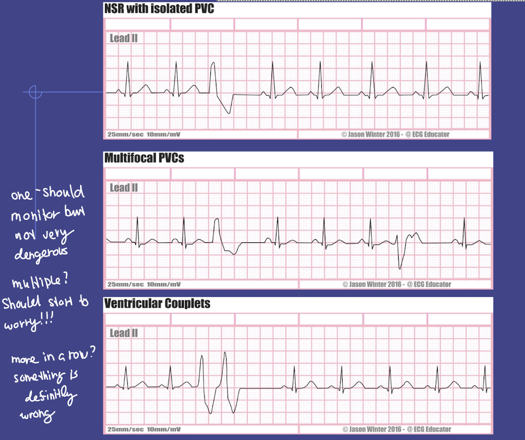
what is the rate, rhythm, P waves characteristics, PR interval duration, and QRS duration of premature ventricular contraction (PVCs)?
rate: varies
rhythm: irregular; R-R are not consistent as it is ectopic beats followed by full compensatory pause
P waves: none
PR interval: none
QRS interval: wide, disorted (over 0.12 second)
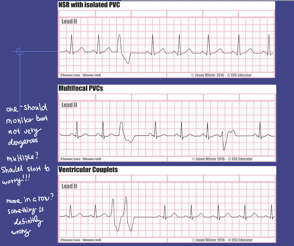
3 or more in a row = ‘non-sustained ventricular tachycardia’
true or false: PVCs is more deadly than PACs
true
what does 3 PVC in a row mean?
non-sustained ventricular tachycardia
what is the main cause of PVC?
increased irriation of ventricular cells
Cardiac Causes
Myocardial ischemia or infarction
Heart failure / cardiomyopathy
Valvular heart disease
Electrolyte / Metabolic Causes
Hypokalemia (low potassium)
Hypomagnesemia (low magnesium)
Hypoxia
Medications / Stimulants
Digoxin toxicity
Caffeine, nicotine, alcohol
Sympathomimetic drugs (e.g., epinephrine, albuterol)
Other Causes
Stress or anxiety
Fever
Hyperthyroidism
Nursing Tip: Always assess vital signs, electrolytes, oxygenation, and cardiac history in patients with PVCs.
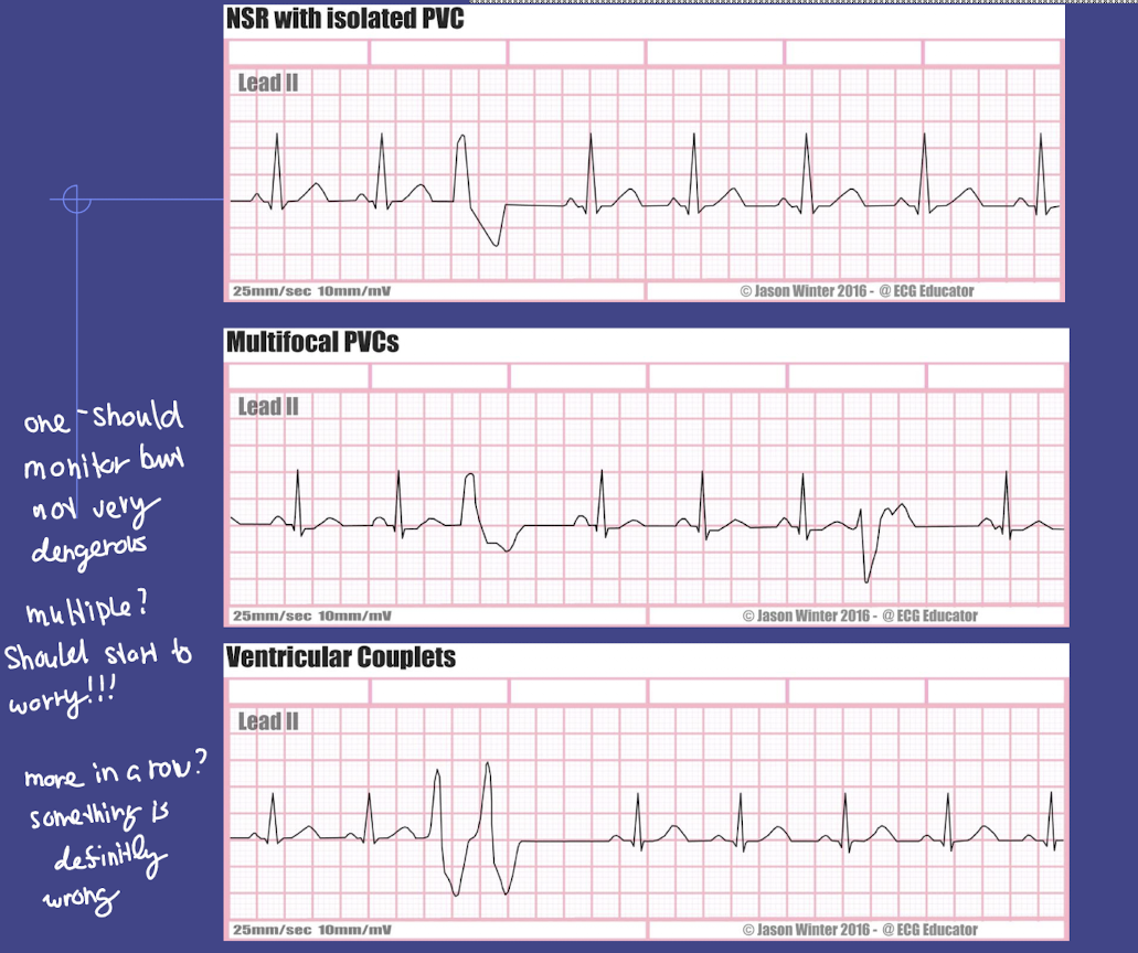
what is the clinical significance if PVCs occur more than 6 times per minute
Indicates increased ventricular irritability
May progress to more serious ventricular arrhythmias (VTach or VFib)
Nursing action: Monitor vital signs, electrolytes, and cardiac rhythm; assess for ischemia or hypoxia
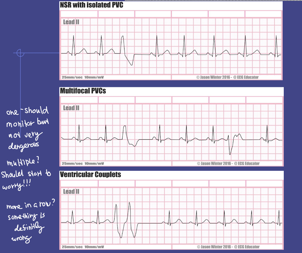
what does it mean if PVCS occur in pairs (couplets)?
Two consecutive PVCs → indicates higher risk of ventricular tachycardia
Nursing action: Continuous ECG monitoring; notify provider if sustained
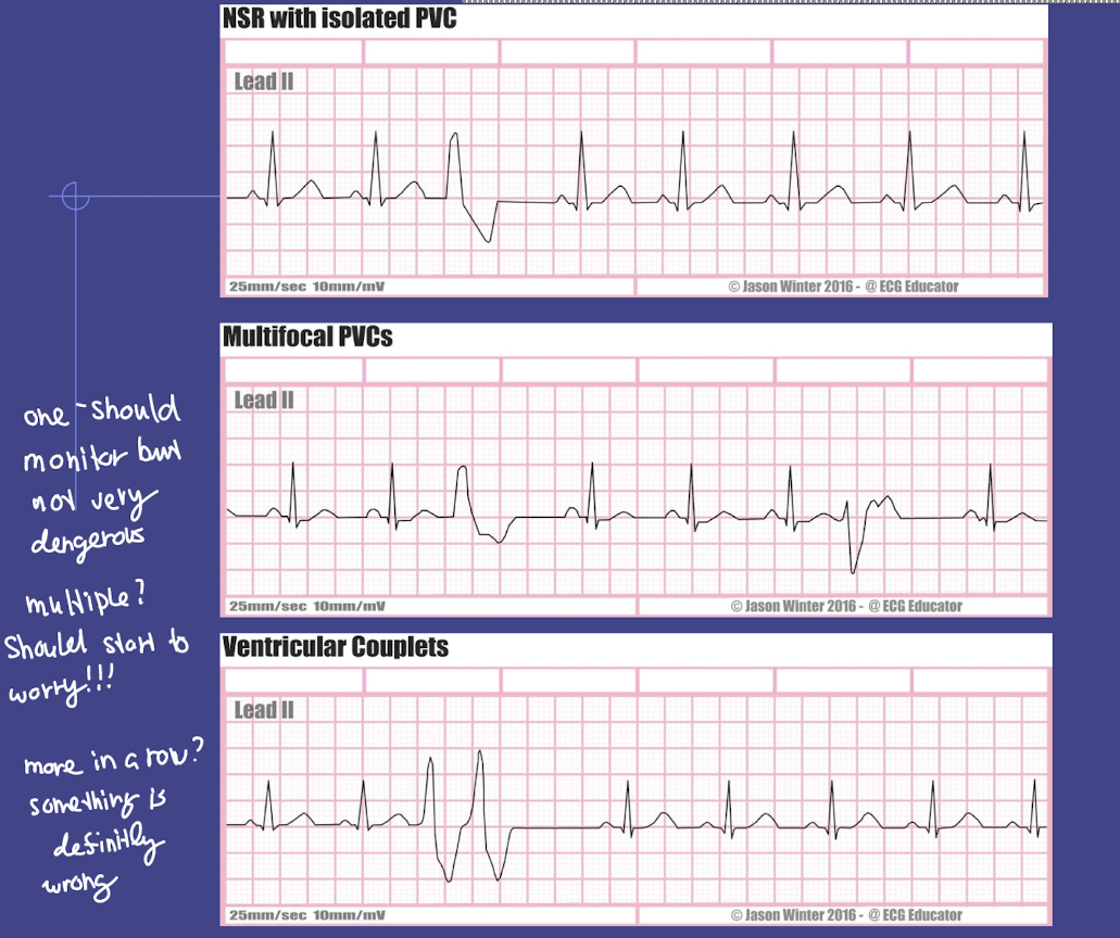
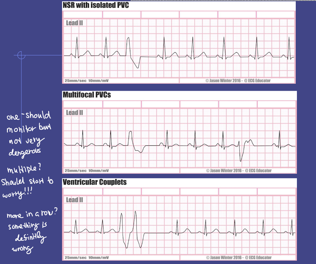
what does multifocal PVCs mean and why is it important?
PVCs originate from different ventricular sites → QRS shapes vary
Indicates more severe ventricular irritability
Higher risk for VTach or VFib
Nursing action: Monitor, check electrolytes, medications, ischemia
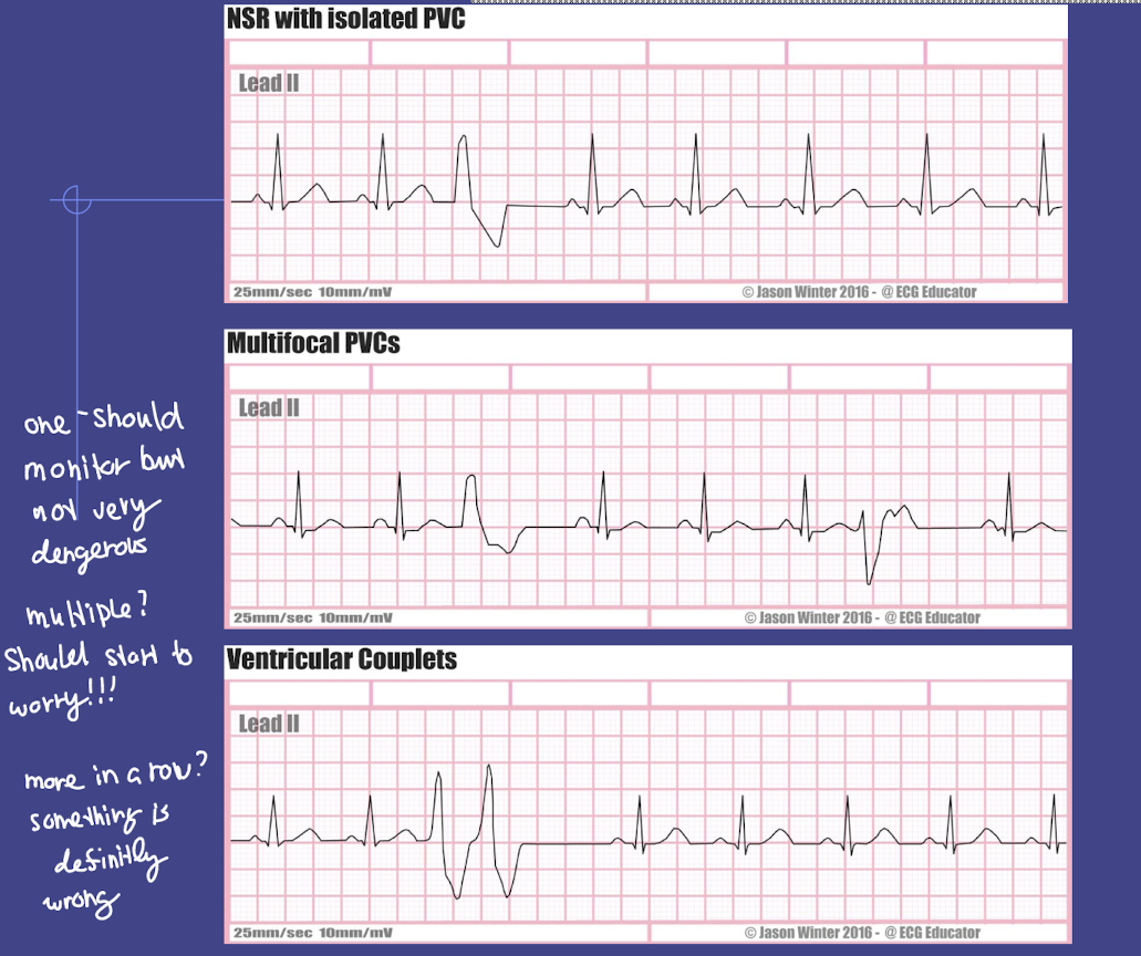
what is the R-on-T phenomenon?
Definition: A PVC occurs during the T wave of the preceding beat
Why it’s dangerous: The ventricles are vulnerable during repolarization → can trigger ventricular tachycardia or ventricular fibrillation
Nursing action: Immediate monitoring, assess for symptoms, prepare for advanced cardiac life support (ACLS) interventions if unstable
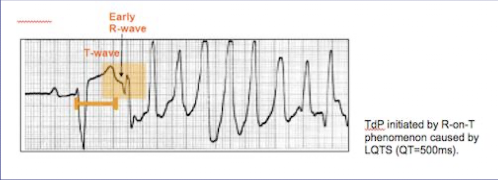
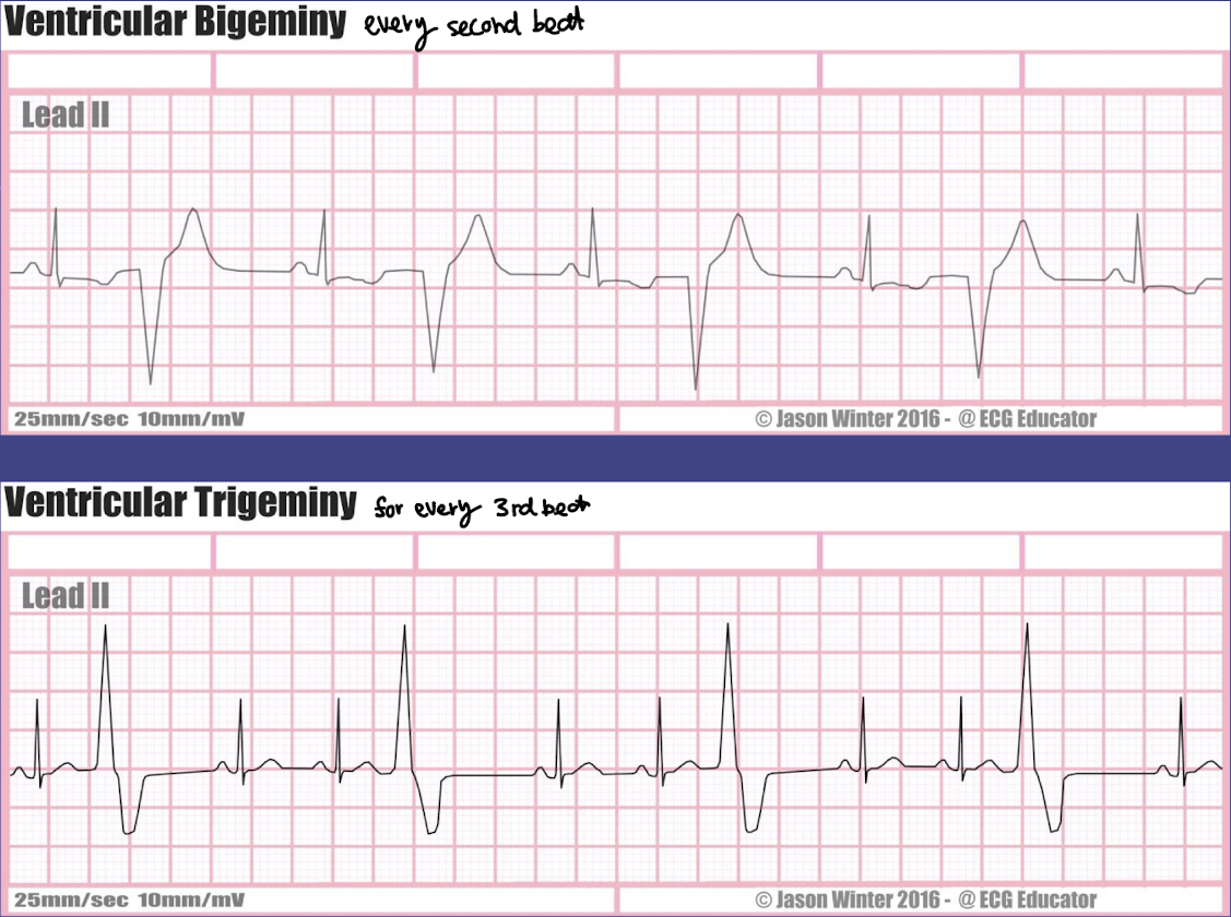
what is ventricular bigeminy, clinical significant, and nursing action?
Definition: Every other beat is a PVC (normal beat → PVC → normal beat → PVC …)
Clinical significance:
Indicates ventricular irritability
May progress to ventricular tachycardia or fibrillation if frequent or symptomatic
Nursing actions:
Monitor vital signs and ECG
Assess for chest pain, dizziness, or syncope
Check electrolytes, oxygenation, and medications
Notify provider if PVCs increase in frequency or become multifocal / R-on-T
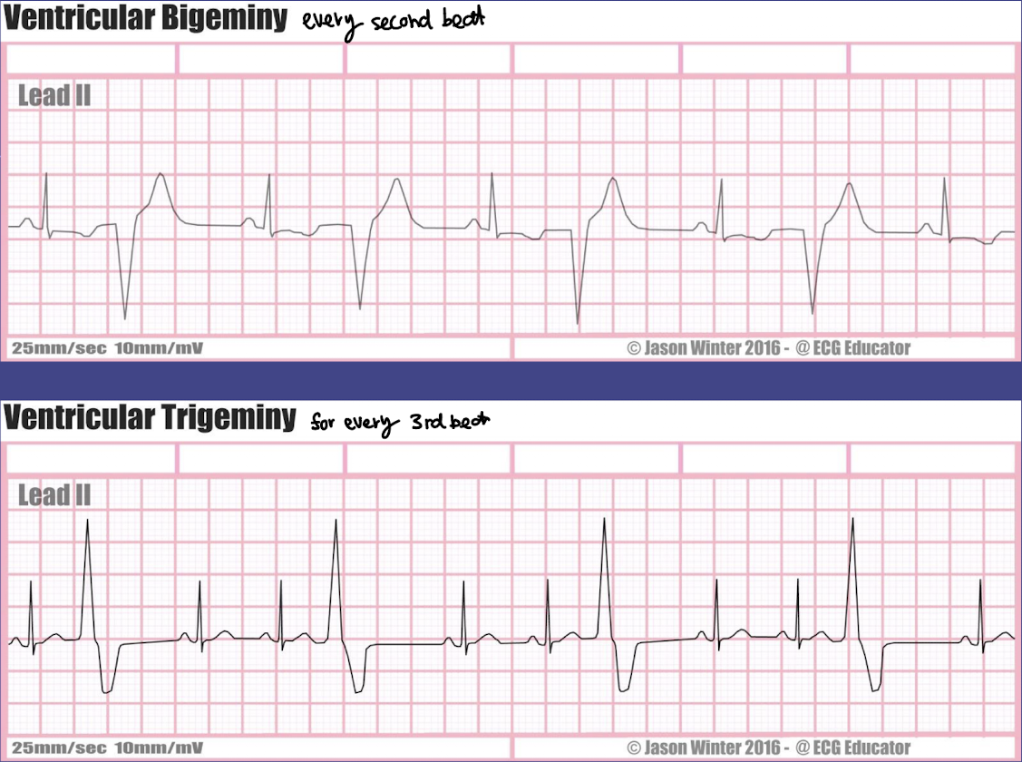
what is ventricular trigeminy, its clinical signficant, and the nursing action?
Definition: Every third beat is a PVC (two normal beats → PVC → two normal beats → PVC …)
Clinical significance:
Shows ventricular irritability, usually less frequent than bigeminy but still abnormal
Can progress to more serious arrhythmias if underlying cause not addressed
Nursing actions:
Same as bigeminy: monitor ECG, vital signs, and patient symptoms
Assess electrolytes, oxygenation, and cardiac history
Notify provider if PVCs increase in frequency, become multifocal, or show R-on-T phenomenon
what are the differences between SVT, sinus tachycardia, and ventricular tachycardia?
SVT (Supraventricular Tachycardia)
Origin: Above ventricles (atria or AV node)
Rate: 150–250 bpm
Rhythm: Regular
P waves: Often hidden in T wave
QRS duration: Narrow (0.06–0.10 sec)
Clinical significance: Rapid rate → dizziness, hypotension
Nursing action: Vagal maneuvers, adenosine, beta-blockers, cardioversion if unstable
Sinus Tachycardia
Origin: SA node
Rate: 100–150 bpm
Rhythm: Regular
P waves: Normal, before each QRS
QRS duration: Narrow (0.06–0.10 sec)
Clinical significance: Usually physiological; rarely dangerous
Nursing action: Treat underlying cause; monitor
Ventricular Tachycardia (VT)
Origin: Ventricles
Rate: 100–250 bpm
Rhythm: Usually regular
P waves: Usually absent or dissociated
QRS duration: Wide (>0.12 sec)
Clinical significance: Life-threatening; may progress to VF → cardiac arrest
Nursing action: Immediate ACLS if unstable, antiarrhythmic drugs, possible defibrillation
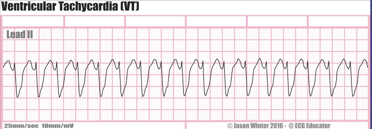
what is the rate, rhythm, P waves characteristics, PR interval duration, and QRS duration of ventricular tachycardia (VT)?
rate: more than 100 bpm
rhythm: regular
P waves: usually absent or dissociated
PR interval: can’t be measure
QRS interval: wide (more than 0.10 second)
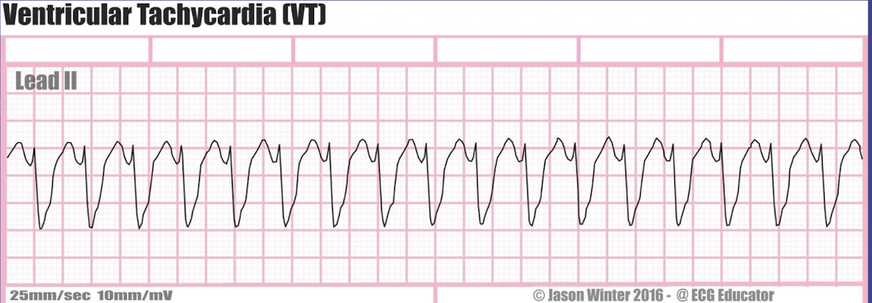
what are the clinical significance and management of ventricular tachycardia (VT)?
Clinical Significance
Life-threatening arrhythmia
Ventricles beat rapidly and ineffectively → reduced cardiac output
Can lead to:
Hypotension
Syncope or dizziness
Chest pain
Pulseless VT → ventricular fibrillation → cardiac arrest
Often associated with heart disease, MI, electrolyte imbalance, or drug toxicity
Management / Nursing Actions
1. Assess patient immediately
Check pulse, blood pressure, LOC, and ECG
2. If pulseless VT (emergency)
Start CPR immediately
Defibrillation per ACLS protocol
Administer IV antiarrhythmic drugs (e.g., amiodarone, lidocaine)
3. If VT with pulse (hemodynamically stable)
Continuous cardiac monitoring
IV antiarrhythmics
Prepare for synchronized cardioversion if unstable
4. Identify and treat underlying cause
Electrolyte imbalances (K⁺, Mg²⁺)
Ischemia / MI
Drug toxicity
Nursing Tip: VT is always a serious arrhythmia — rapid recognition and intervention are critical.
why is VT more symptomatic than SVT?
SVT (Supraventricular Tachycardia)
Origin: Above ventricles
Ventricular contraction: Normal → cardiac output okay
Symptoms: Usually mild (palpitations, dizziness)
VT (Ventricular Tachycardia)
Origin: Ventricles
Ventricular contraction: Ineffective → cardiac output drops
Symptoms: Severe (hypotension, syncope, chest pain, pulseless)
Key point: VT reduces cardiac output → more dangerous as it can lead to cardiac arrest; SVT usually tolerated better.
what are the general causes of VTach?
MI
hypokalemia
hypomagnesemia
valvular disease
drug toxicity
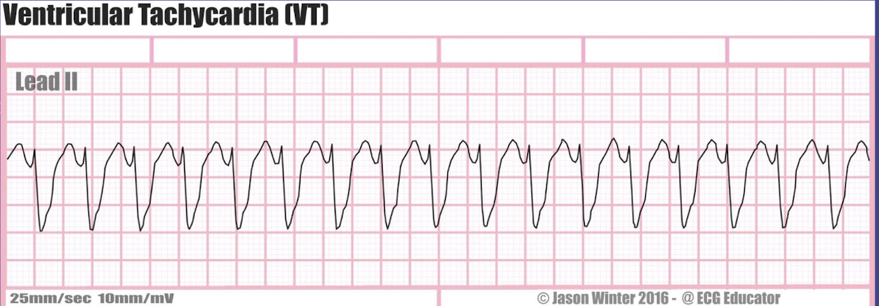
during which heart rhythms might a patient be pulseless?
Pulseless VT → ventricular tachycardia without effective cardiac output
Ventricular fibrillation (VF) → chaotic ventricles, no cardiac output
Asystole → no electrical activity, no cardiac output
Nursing tip: These are emergencies → start CPR and follow ACLS protocol immediately
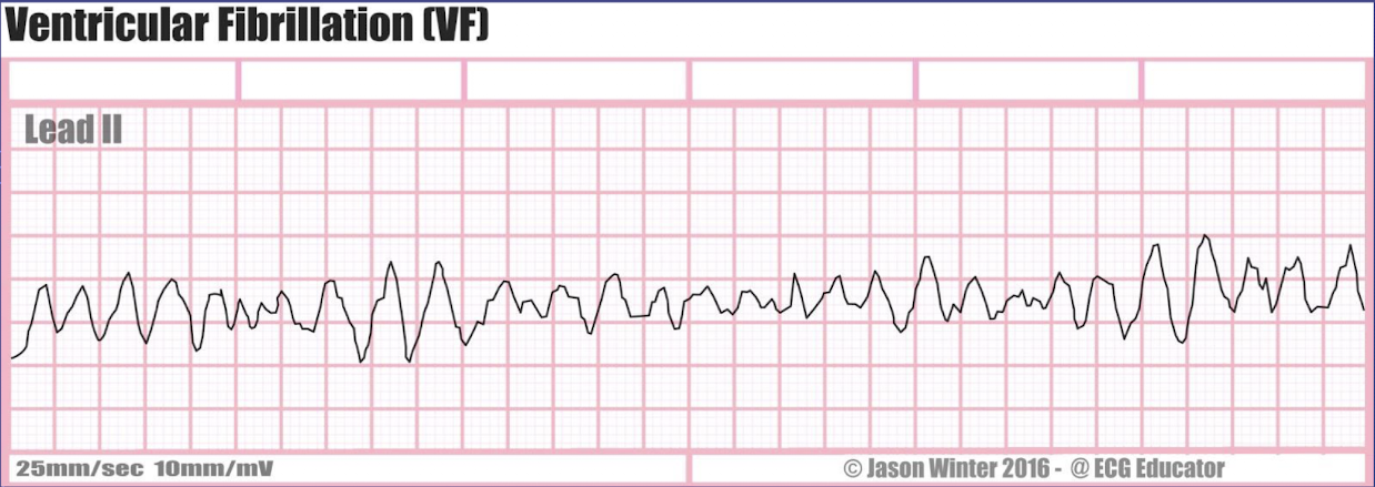
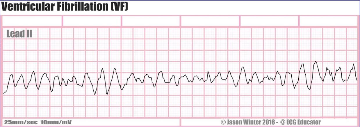
what is the rate, rhythm, P waves characteristics, PR interval duration, and QRS duration of ventricular fibrillation (V-Fib)?
rate: cannot reliably identify the heart rate
rhythm: irregular
P waves: cannot be identified
PR interval: cannot be measured
QRS interval: cannot be identifed
what are the common causes of ventricular fibrillations (V-Fib)?
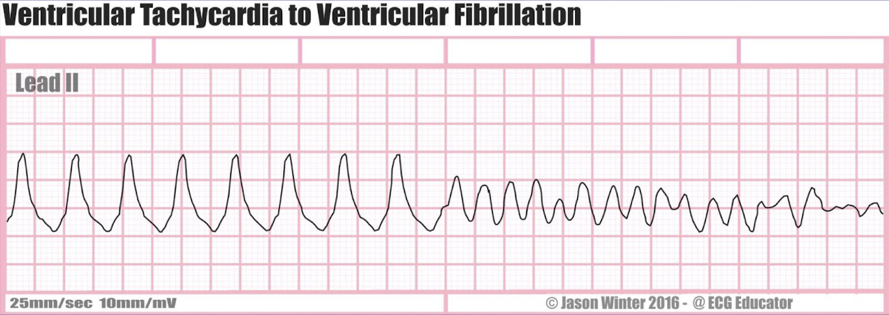
patient with V-Tach initially that progresses into V-Fib
MI
hypokalemia
hypo magnesia
shock
trauma
what are the heart rhythm that are commonly caused by hypokalemia (low potassium, K+)?
Premature ventricular contractions (PVCs)
Ventricular tachycardia (VT)
Ventricular fibrillation (VF)
U waves on ECG
Flattened T waves
what are the heart rhythm that are commonly caused by hypomagnesemia (low magniesum Mg2+)?
Torsades de Pointes (a type of polymorphic VT)
PVCs
VT / VF
what heart rhythm common progresses into V-Fib?
V-Tach
what is asystole and how do you confirm?
complete cessation of all electrical activity → usually preceded by VF or ventricular escape rhythm
check ECG electrodes and connections
confirm in 2 leads
zero cardiac output