Lecture 4&5: Major subcellular responses to injury, patterns of necrosis, events following death, an physical injuries
 Call Kai
 Learn
Learn Practice Test
 Spaced Repetition
 Match
Match Flashcards
 Knowt Play
1/21

There's no tags or description
Looks like no tags are added yet.
Name | Mastery | Learn | Test | Matching | Spaced | Call with Kai | Chat |
|---|
No analytics yet
Send a link to your students to track their progress
22 Terms
Pathological Calcification - Dys (bad) trophic Calcification
Focal deposition of calcium at sites of cell injury and necrosis
ex: scars, caseous nodules, degenerated foci in leiomyomas
Rxn to tissue damage from medical device implantation
Sequestration of erythrocytes in implant site
From cell injury/damage
Pathological Calcification - Metastatic Calcification
Deposition of calcium in normal tissue
Renal failure
Increased parathyroid hormone secretion
Ca has a predication for the internal elastic lamina of arteries
Cellular Aging
Accumulation of mutations (DNA damage)
Replicative Sequence (telomere shortening)
Damage to proteins & organelles (free radicals)

Time course of cellular response
Acute: Rapid (seconds to minutes to hours)
Edema & Neutrophils
Subacute: Intermediate (hours to a few days)
Monocytes/Macrophages
Chronic: Slow (days)
Monocytes/Macrophages

Reversible Injury - Swelling
First manifestation of cellular injury
Hydropic change or vacuolar change or cloudy swelling
Transudate vs Exudate
Transudate - Increase hydrostatic pressure
Clear, watery, low protein pushed out of blood vessels
System issues
Pressure changes
Exudate - Decreased colloid osmotic pressure
Cloudy, thick, high protein
Local inflammation → widens gaps between cells in blood vessels
Reversible Injury - Fatty Change
Fat is broken down for energy (ketone bodies)

Major patterns of tissue necrosis
Coagulation
Gangrenous
Liquefactive
Caseous
Aging signs
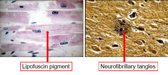
Lipofuscin pigment (cellular debris) and neurofibrillary tangles
In heart & neurons → they don’t divide (non-mitotic)
Coagulation Necrosis
Architecture of tissue is preserved (first few days after death)
Firm tissue
Injury denatures structural proteins & enzymes → blocks proteolysis of dead cells
Leukocytes are recruited to digest dead cells (phagocytosis clears up debris)
Characteristic of infarcts (necrosis caused by ischemia) in all solid organs except brain
Gangrenous Necrosis
Limb that has lost blood supply & has undergone coagulative necrosis involving multiple tissue layers
Changes to liquefactive necrosis when bacterial infection is introduced
Liquefactive Necrosis
Focal bacteria
Microbes stimulate rapid accumulation of inflammatory cells → enzymes of leukocytes liquefy the tissue
Pus
Hypoxic death of cells within CNS
Caseous Necrosis
Cheeselike
Tuberculosis infection
Tissue architecture is obliterated & cellular outlines are indiscernible
Often surrounded by macrophages & inflammatory cells
3 Cardinal body changes following death
Algor mortis
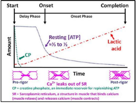
Rigor mortis
Livor mortis
Algor mortis
Decreased body temp
-1oC/hour @ ambient temp (20oC)
Rigor Mortis
Rigidity of body
Several hours after death
Caused by hydrolysis of muscle ATP → without ATP myosin molecules adhere to actin filaments → rigid muscles
Livor Mortis (AKA postmortem hypostasis)
Blood settles in the lower portion of the body → purplish/red discoloration of skin
areas corresponding to where the body was at the time of death may appear dark red/purple with whiteish areas
blood vessels become compressed and prevent hypostasis in those areas
Oxyhaemoglobin forms at the expense of reduced haemoglobin
Post Mortem Events - Autolysis
Spontaneous destruction of tissues by intracellular enzymes
Occurs in body after death
Post Mortem Events - Autolysis Putrefaction
Decomposition of organic matter (like proteins) by microorganisms → foul smell
Post Mortem Events - Postmortem Blood Clots
Clots formed in heart/large vessels after death
4 general types of PHYSICAL injuries
Abrasion
Contusion
Incision
Penetration
7 Head Injury Types
Skull trauma
Concussion
Edema
Cerebral contusion
Epidural hemorrhage
Subdural hemorrhage
Subarachnoid hemorrhage