Vertebrate Histology Lab Exam 1
 Call Kai
 Learn
Learn Practice Test
 Spaced Repetition
 Match
Match Flashcards
 Knowt Play
1/45

There's no tags or description
Looks like no tags are added yet.
Name | Mastery | Learn | Test | Matching | Spaced | Call with Kai | Chat |
|---|
No analytics yet
Send a link to your students to track their progress
46 Terms
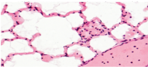
Slide 1
Simple Squamous Epithelium
Alveoli
Squamous Epithelial Cells
Blood Vessel
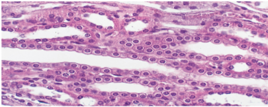
Slide 2
Simple Cuboidal Epithelium
Tubules of kidneys
Lumen of Kidney Tubule
Cuboidal Epithelial Cells
Basement Membrane
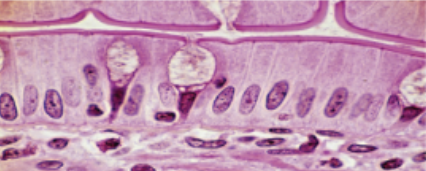
Slide 3
Simple Columnar Epithelium
Duodenum of small intestine
Brush border (microvilli)
Connective Tissue
Nuclei
Goblet Cell
Columnar Cells
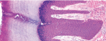
Slide 4
Keratinized Stratified Squamous Epithelium
Epidermis
Keratin Layer
Dead epithelial cells
Living epithelial cells
Basement membrane
Connective Tissue
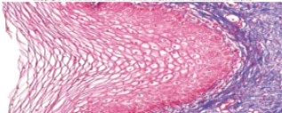
Slide 5
Nonkeratinized Stratified Squamous Epithelium
Near cervix and walls of vagina
Living epithelial cells
Basement membrane
Connective tissue
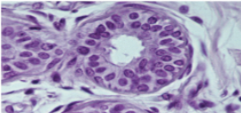
Slide 6
Stratified Cuboidal Epithelium
Sweat Glands
Blood capillary
Connective Tissue
Duct Lumen
Stratified cuboidal epithelial cells
Basement membrane
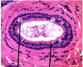
Slide 7
Stratified Columnar Epithelium
Some glands and male urethra
Goblet cells
Columnar cells w/ large nucleus
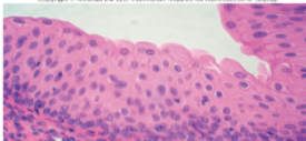
Slide 8
Transitional Epithelium (Urothelium)
Kidneys and ureters
Basement membrane
Connective tissue
Binucleate umbrella cell
Umbrella cells
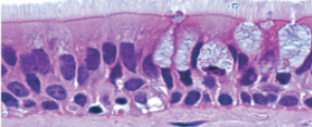
Slide 9
Pseudostratified Columnar Epithelium
Trachea
Cilia
Basement membrane
Basal cells
Goblet cells
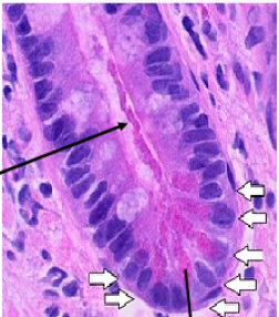
Slide 10
Exocrine Gland Duct System
Sweat Gland
Secretory Cells are cuboidal
Duct lined by epithelium
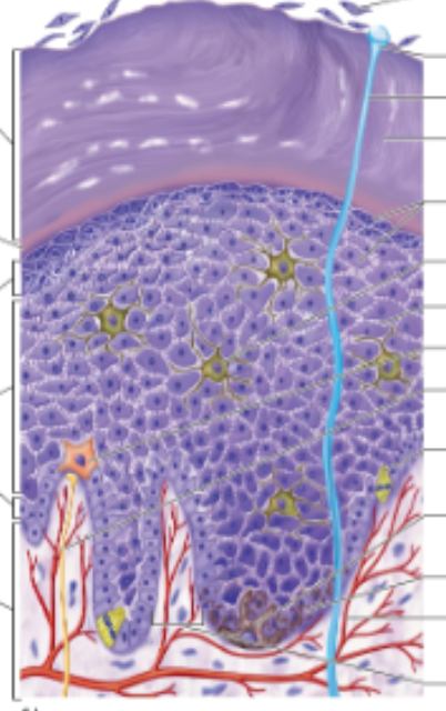
Slide 11
Epidermis
Stratum Corneum
Exfoliating dead cells
Sweat pore
Dead keratinocytes
Stratum Lucidum
Stratum Granulosum
Living Keratinocytes
Stratum Spinosum
Dendritic Cells
Tight Junctions
Tactile Cell
Stratum Basale
Stem cell in mitosis
Melanocyte
Melanized Keratinocytes
Dermis
Dermal blood vessel
Tactile nerve fiber
Dermal papilla
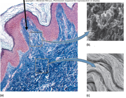
Slide 12
Dermis
Dermal Papilla
Papillary Layer
Reticular Layer
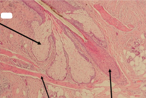
Slide 13
Hair and Hair follicle
Sebaceous Gland
Arrector Pili Muscle
Hair in Hair follicle
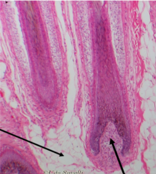
Slide 14
Papilla of the Hair
Subcutaneous Layer
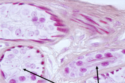
Slide 15
Sweat Glands
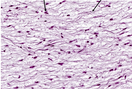
Slide 16
Mesenchyme Tissue
Mesenchyme Cells
Fibers
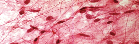
Slide 17
Areolar Connective Tissue
Under mucous membranes
Ground Substance
Elastic fibers
Collagenous Fibers
Fibroblasts
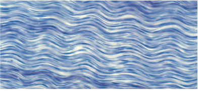
Slide 18
Dense Regular Connective Tissue
Tendons & Ligaments
Collagen Fibers
Ground Substance
Fibroblast Nuclei
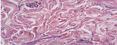
Slide 19
Dense Irregular Connective Tissue
Deep Layers of Skin and heart chambers
Bundles of collagen fibers
Gland ducts
Fibroblast nuclei
Ground substance
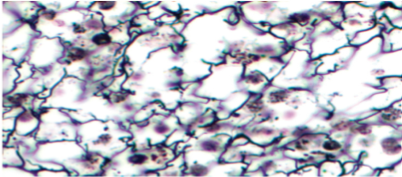
Slide 20
Reticular Tissue
Lymphatic Organs
Leukocytes
Reticular Fibers
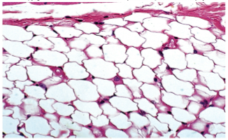
Slide 21
White Adipose Tissue
Subcutaneous
Blood Vessel
Adipocyte nuclei
Lipid in adipocyte
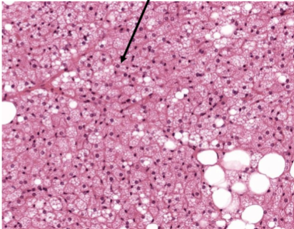
Slide 22
Brown Adipose Tissue
Brown adipocytes
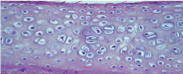
Slide 23
Hyaline Cartilage
Ends of long bones
Matrix
Cell Nest
Perichondrium
Lacunae
Chondrocyte nuclei
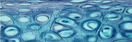
Slide 24
Elastic Cartilage
Ears, nose, epiglottis
Perichondrium
Elastic Fibers
Lacunae
Chondrocytes
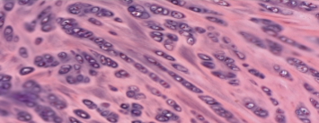
Slide 25
Fibrocartilage
Pubic Symphysis and between vertebrae
Collagen Fibers
Chondrocytes
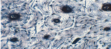
Slide 26
Compact Bone
Covers all bones
Lacunae
Canaliculi
Concentric lamellae of osteon
Central Canal
Osteon
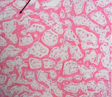
Slide 27
Spongy Bone
Within bones
Trabeculae
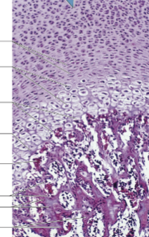
Slide 28
Epiphyseal Plate
In metaphysis
Multiplying chondrocytes
Enlarging Chondrocytes
Breakdown of lacunae
Calcifying cartilage
Bone marrow
Osteoblasts
Osteocytes
Trabeculae of spongy bone
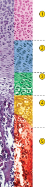
Slide 29
Epiphyseal Plate Zones
Zone of reserve cartilage
resting hyaline cartilage
Zone of cell proliferation
chondrocytes multiplying & lining up in rows of flattened lacunae
Zone of cell hypertrophy
cessation of mitosis; enlargement of chondrocytes & thinning of lacuna walls
Zone of calcification
temporary calcification of cartilage matrix between columns of lacunae
Zone of bone deposition
breakdown of lacuna walls, leaving open channels
death of chondrocytes
bone deposition forms trabeculae of spongy bone
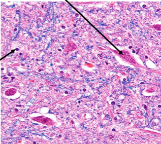
Slide 30
Human nerve tissue
Neuroglial cell
Neuron
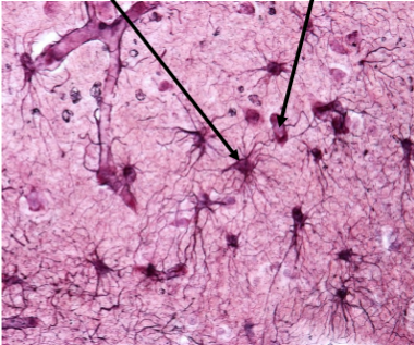
Slide 31
Astrocytes
Oligodendrocytes
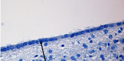
Slide 32
Ependymal Cells
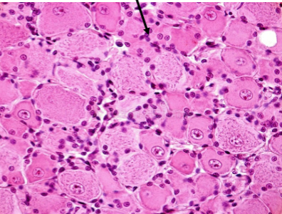
Slide 33
Satellite Cells
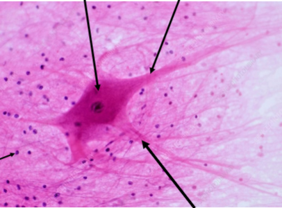
Slide 34
Multipolar Neuron
Cell body with nucleus
Axon
Dendrite
Glial Cell
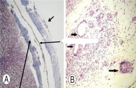
Slide 35
Cranial Meninges
Dura Mater
Pia Mater
Arachnoid Layer
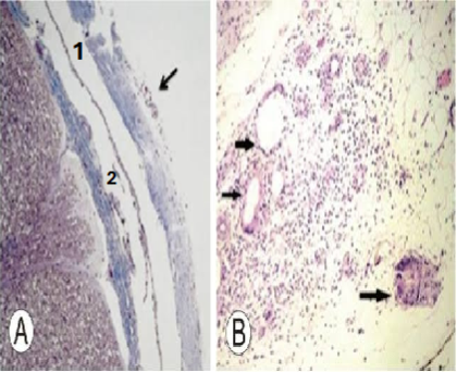
Slide 36
Subdural Space (1)
Subarachnoid Space (2)
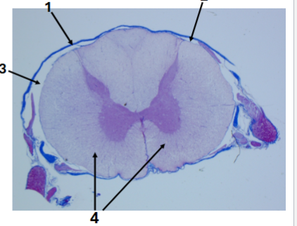
Slide 37
Cross section of human spinal cord
Dura mater (1)
Pia mater (2)
Subdural Space (3)
White Columns (44
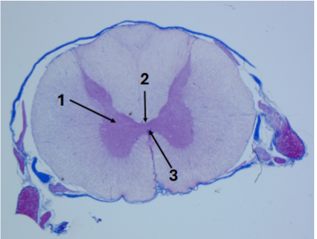
Slide 38
cross section of human spinal cord
Gray matter (1)
Gray commissure (2)
Central Canal (3)
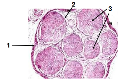
Slide 39
Nerve structure
Epineurium (1)
Perineurium (2)
Fascicles (3)
Slide 40
Myelin Sheath
Nerve Axon
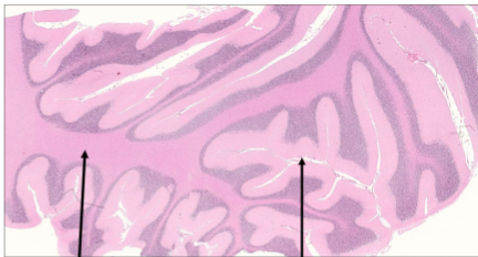
Slide 41
Cerebellum cross section
Gray Matter
Arbor vitae (white matter)
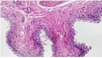
Slide 42
Cerebrum cross section
Gyri (folds)
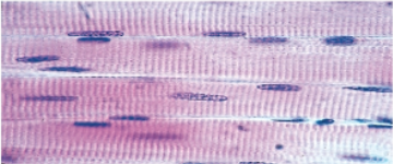
Slide 43
Skeletal Muscle Tissue
Striations
Nuclei
Muscle fiber
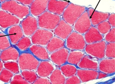
Slide 44
Skeletal muscle tissue structure
Epimysium
Perimysium
Fascicles
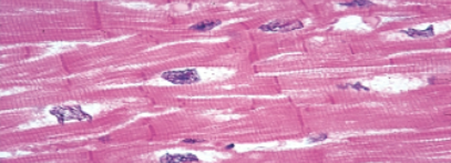
Slide 45
Cardiac Muscle Tissue
Intercalated discs
Striations
Glycogen
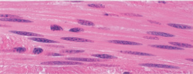
Slide 46
Smooth Muscle Tissue
Nuclei
Muscle Cells