204 Week 6 - Spinal Cord + Nerves
1/9
There's no tags or description
Looks like no tags are added yet.
Name | Mastery | Learn | Test | Matching | Spaced |
|---|
No study sessions yet.
10 Terms
SPINAL CORD
PROTECTED: by Vertebral Columns
LOCATION: Vertebral Canal
FUNCTION: Facilitates communication to/from Periphery + Brain
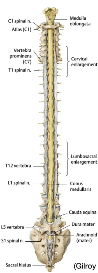
Spinal Cord External Anatomy
Begins: Foramen Magnum AFTER Medulla Oblungata
Finishes: L1-2 in healthy adult (After is the Cauda Equina)
Cervical + Lumbosacral Enlargements
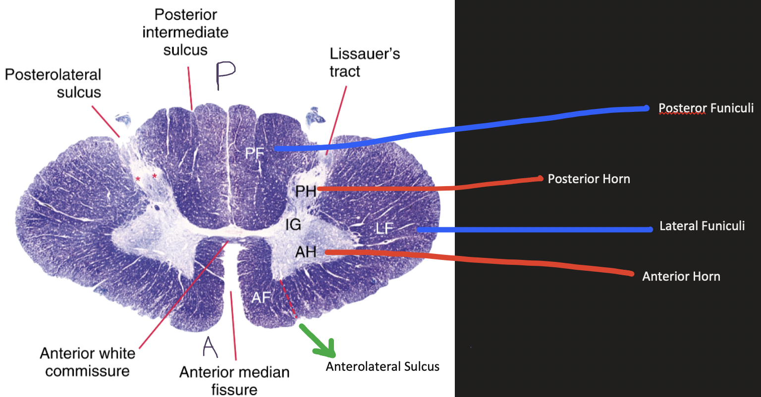
Spinal Cord Internal Anatomy
Anterior Horns: Motor Cell Bodies
Posterior Horns: Sensory Cell Bodies + Interneurons
Lateral Horns: Autonomic Nuclei
Protective Structures of Spinal Cord
1st Layer: Bony
Skull (Brain)
Vertebral Columns (Spinal Cord)
2nd Layer: Connective
3 Meningeal Layers (Pia, Arachnoid, Dura - Mater)
3rd Layer: Fluid
CSF
Provides cushion
Spinal Cord Blood Supply
1 Anterior Spinal Artery (ASA) (2/3rds of Blood Supply)
2 Posterior Spinal Artery (PSA)
White & Grey Matter
Grey Matter:
Motor commands
Contain Cell Bodies (Organelles etc)
White Matter:
Sensory Commands
Outer
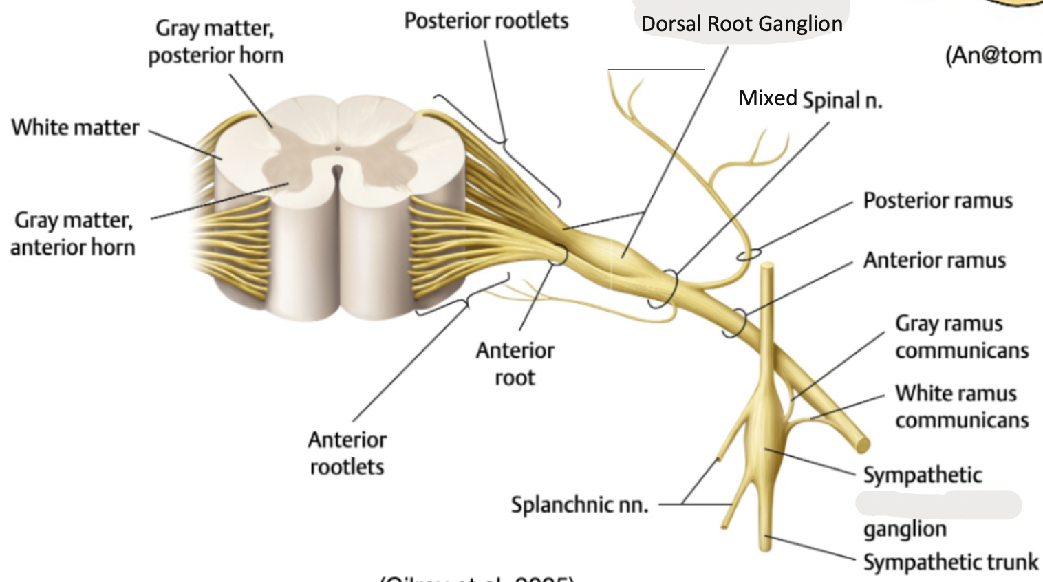
Spinal Nerves Order
Rootlets (M/S) → Roots (M/S) → Mixed Spinal Nerves (M+S) → Rami (Supplies Back/Neck, Trunk + Limbs)
Cauda Equina
Dura Mater Ends: S2