Exam 2
 Call Kai
 Learn
Learn Practice Test
 Spaced Repetition
 Match
Match Flashcards
 Knowt Play
1/74

There's no tags or description
Looks like no tags are added yet.
Name | Mastery | Learn | Test | Matching | Spaced | Call with Kai | Chat |
|---|
No analytics yet
Send a link to your students to track their progress
75 Terms
Give an overview of Catecholamines
The main ones are norepinephrine, epinephrine, and dopamine
Catecholamines are monoamines (biogenic amines), a large group of NTs derived from aromatic amino acids like phenylalanine -> tyrosine, tryptophan, and thyroid hormones
Organic chemistry wise, they have an amino group, 2-carbon chain, and aromatic rings (catechol)
Describe Catecholamine synthesis in general
Begins with the amino acid tyrosine
A multi-step long process including several enzymes
TH is the rate-limiting enzyme in the pathway, meaning it determines the overall rate of DA or NE synthesis
How is dopamine made?
Process: Tyrosine → tyrosine hydroxylase turns it into L-DOPA → aromatic L-amion acid decarboxylase turns that into dopamine
Occurs in: Substantia nigra (body movements), VTA (reward/motivation/pleasure), hypothalamus (DA to contorl body functions)

How is norepinephrine made?
Process: Tyrosine → tyrosine hydroxylase turns it into L-DOPA → aromatic L-amino acid decarboxylase turns that into dopamine → DBH turns it into NE
Occurs in: The brain (locus coerulus in the brainstem) and body (adrenal glands)

How is epinephrine made?
Process: Tyrosine → tyrosine hydroxylase turns it into L-DOPA → aromatic L-amino acid decarboxylase turns that into dopamine → DBH turns it into NE → phenylethanolamine and n-methyltransferase add a CH3 group → EPI
Occurs in: Adrenal medulla (chromaffin cells) and CNS (neurons in the brainstem for use as NT)
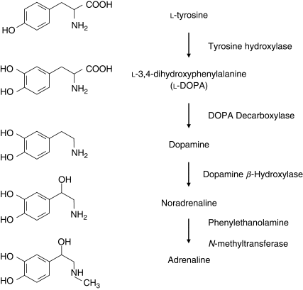
Why do we care about the enzymes used for catecholamine synthesis?
They can be stained to track where certain processes are taking place.
Can also be used to see if someone has a certain disorder.
Neurons that are positive for TH make ___ and are likely ___ neurons
Dopamine; dopaminergic
Neurons that make DBH make ___ and are ___ neurons
Norepinephrine; noradrenergic
Note: Dopamine neurons don’t have DBH because they stop at dopamine
The adrenal medulla secretes ___% EPI and ___ % NE
80%; 20%

How is TH activity regulated?
Increased: When cells are firing (when neurons are firing at a high rate, like during stress, so TH is stimulated and catecholamine synthesis accelerates)
Decreased: Via negative feedback (high catecholamine levels inhibit TH activity)
Why does L-DOPA allow so much DA to be made?
L-DOPA is a way to increase dopamine synthesis by side supplying a precursor to dopamine, which boosts such levels because you skip the TH step (the slowest/rate-limiting one)

How can you block catecholamine synthesis?
AMPT (alpha-methyl-para-tyrosone) locks TH and prevents overall catecholamine synthesis

What is the role of VMAT and what does reserpine do to it?
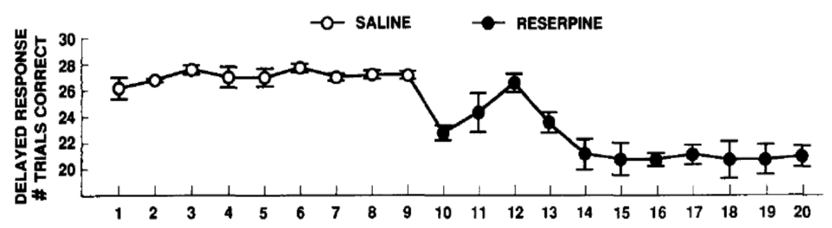
Post synthesis, catecholamines are packaged into vesicles by VMAT (vesicular monoamine transporter) which can be blocked by the drug reserpine so DA and NE aren’t protected in the vesicle and are broken down. VMAT1 is in the adrenal medulla; VMAT2 is in the brain.
Elaborate on reserpine
Resperpine was isolated by Arvid Carlsson from the dried root of Rauvolfa serpentina for the treatment of insanity/calming and fever/snakebites. It depletes catecholamines from peripheral sympathetic nerve endings and produces depression-like events in animals and patients such as fatigue-like symptoms. Helped with Parkinson’s research!
What happens when given amphetamine (AMPH) or methamphetamine (mAMPH)
Given to promote DA. There is increased locomotor activity; at higher doses, this is replaced by stereotyped behaviours (intense sniffing, repetitive head/limb movements, licking, biting) as increasing stimulation of DA receptors in the nucleus accumbens and striatum.
This happens because DA modulates movement – it can drive it, but at very high measures, behaviours become disorganized. Too much DA means you can’t switch from one movement to another one.
Vesicular vs Non-vesicular release with DA
Catecholamines are normally released by exocytosis when a nerve impulse reaches the terminal (called vesicular release) but some drugs cause release independently of cell firing (non-vesicular release) as it tricks the transporter into thinking it is DA and reversing the transporter

How do autoreceptors work with catecholamine release
Catecholamine release is inhibited by autoreceptors which enhance the opening of voltage-gated K+ channels to shorten the duration of APs and reduces Ca++ influx which leads to decreased vesicle exocytosis. This is a Gi-coupled process!

Drugs that stimulate autoreceptors ___ catecholamine release; autoreceptor antagonists ___ rate of release
Decrease; increase
What happened to mice w/o D2 receptors?
Mutant mice w/ no D2 autoreceptors (but normal postsynaptic D2 receptors) were more active than the controls and more sensitive to cocaine due to cocaine being a monoamine transporter blocker
Clonidine vs Yohimbine
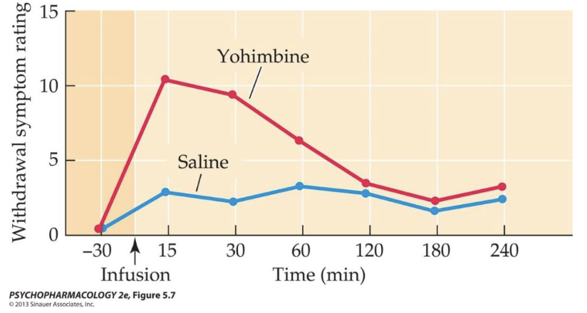
Clonidine: This is an alpha2 agonist; withdrawal from opioid drugs activates the noradrenergic system = withdrawal symptoms, so clonidine can be used for such symptoms
Yohimbine: This is an alpha2-antagonist which increases NE release (and noradrenergic cell firing) which provokes withdrawal symptoms and drug caving in opioid-dependent patients
Inactivation of catecholamines
Metabolites → CSF → bloodstream and eliminated via urine (thus, levels of metabolites are a rough indication of catecholaminergic activity)

What happens to mutant mice w/o DA transporters (DAT knockout)?
There is a lot more dopamine left in the synapses aka less reuptake, and they enter a hyper-dopaminergic state as they cannot recycle dopamine. Likewise, there is receptor downregulation. Might appear like wild mice with stimulants. Cocaine doesn’t have as much as an effect since it is a DA transporter blocker but there is no DA transporters, but they will have an effect still as it will continue to impact NE and SER.

Examples of transporter-blocking drugs
TCAs (tricyclic antidepressants) inhibit re-uptake of NET and 5-HT
Atomoxetine aka Strattera for ADHD blocks NE transporters
Cocaine inhibits reuptake of all monoamines
Drugs that alter catecholamine breakdown
MAO Inhibitors: Used to treat clinical depression via phenelzine (nardil) or tranylcypromine (parnate)
COMT Inhibitors: Entacapone (comtan) and tolacapone (tasmar) enhance the effectiveness of L-DOPA in treating Parkinsons’s disease by preventing breakdown of DOPA
How is the dopaminergic system organized in the brain?
The dopaminergic system originates in several cell groups but mainly in the brainstem (A9/A10)
DA neurons are only 1% of neurons in the brain but have a profound impact on behaviour
4 major pathways: nigrostriatal, mesolimbic, mesocortical, and tuberoinfundibular
Elaborate on the Nigrostriatal pathway
Anatomy: The nigrostriatal pathway is composed of cell bodies in the substantia nigra (A9 cell group) with axons extending to the caudate-putamen (striatum). This pathway is impacted more than anything else (we don’t know why)
Function: The function is muscle tone, posture, and voluntary movements (and abnormal ones) like with how Parkinson’s disease involves the loss of DA neurons in the substantia nigre (80%+) and consequent denervation of the striatum with neurotoxins 6-OHDA and MPTP being used in animals to damage such a pathway as a model for it
What two dopaminergic pathways rise from the VTA? What do they do?
Cell bodies in the ventral tegmental area (VTA; A10 cell group) gives rise to the mesolimbic pathway (in the limbic system) or to the prefrontal cerebral cortex to make the mesocortical
Mesolimbic = brain “reward” pathway (anticipation/detection of rewarding stimulus), compulsive behaviours, perseverance, and is implicated in addiction
Mesocortical = abnormal function in psychoses like schizophrenia

Explain the basics of the tuberoinfundibular pathway
The tuberoinfundibular pathway is associated with the hypothalamus (arcuate nucleus) where DA gets into the median eminence, into portal vessels, then the anterior pituitary to inhibit prolactin
What are the DA receptor families?
D1 and D5 are similar whilst D2 and D3 and D4 are a separate family (D1-like, D2-like) but all 5 DA receptor subtypes are metabotropic as they interact with G proteins and second messengers
What do DA agonists and antagonists show?
DA receptor agonists and antagonists have provided a lot of information about the behavioural functions of DA like behavioural activation (similar but not identical to amph or cocaine). For example, antagonists show suppression of exploratory and locomotor activity as at higher doses, such drug
What is catalepsy?
Neurological condition characterized by muscular rigidity and fixity of posture regardless of external stimuli… PD, Epilepsy, SZ patients on some anti-psychotics (Haloperidol)
Explain how DA knockout mice were impacted
D1: D1 receptor knockout mice exhibit deficits in several kinds of cognitive tasks
D2: D2 knockout mice show impairment in spontaneous movement, coordination, and posture control
Double: Double knockout (D1 and D2) receptor genes leads to fatality in the 2nd/3rd week of life
If a ___ receptor ___, such as haloperidol, is given to rats on a long-term basis, the rats develop ___.
D2; antagonist; behavioural supersensitivity
Explain the basic anatomy of the noradrenergic system
NS: The noradrenergic system has CNS and PNS components
Central: Cell bodies in the brainstem like the locus coeruleus (A6 cell group located in the pons) and their ascending fibers to to multiple structures in the forebrain, the spinal cord, and cerebellum
Peripheral: Part of the sympathetic nervous system

Adrenergic receptors in the noradrenergic system
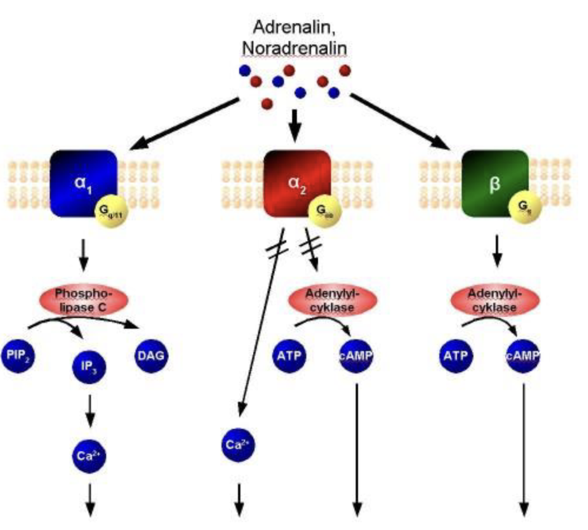
Alpha-2 receptors (Gi) inhibits adenylyl cyclase and reduce synthesis of cAMP which are (typically) autoreceptors
Alpha-1 receptors (Gq, calcium) operate via the phosphinositide second-messenger system
Beta-1 and Beta-1 adrenoreceptors (Gs) stimulate the adenylyl cyclase and will thus enhance synthesis of cAMP
What are the 3 main behaviours of the norargenergic system?
Arousal, cognition, and consolidation (of memories tied to emotional events)
Explain the noradrenergic system and arousal
LC neurons fire more rapidly in awake states rather than sleeping states
Pathways from the LC to the medial septal and medial preoptic areas are involved in wakefulness
Experiment: alpha-1 receptor agonist phenylephrine and/or general beta-receptor agonist isoproterenol were injected into the septal area of mice

Explain the noradrenergic system and cognition
LC projects to the prefrontal cortex which has roles in cognitive functions like attention and working memory but administration of α2-receptor agonists (clonidine) into the PFC enhances working memory
Alpha-1 and Alpha-1 effects help to explain the role of NE in stress-induced impairment of PFC-dependent cognitive functions like working memory
NE affinity: a1 < a2
Under normal conditions, NE facilitates PFC function and cognitive tasks by activating local a2 adrenoreceptors but under stress conditions, the increased NE also activates a1 receptors which can lead to cognitive impairment
Explain the noradrenergic system and consolidation
NE (EPI, and glucocorticoid hormones) modulates the consolidation of emotional memories, especially if they are encoded in the basolateral amygdala
A mouse experiment shows that passive avoidance learning paradigm is commonly used to evaluate memory consolidation

Arnstern reading questions
Question 1: The article describes an "inverted-U" relationship between catecholamine activity and prefrontal cortex (PFC) function. Why might both too little and too much dopamine or norepinephrine impair working memory and executive function?
Question 2: Norepinephrine acts at different adrenergic receptors depending on its concentration. How does the shift from α2A-receptor activation to α1- and β-receptor activation help explain cognitive changes during stress?
Question 3: The authors argue that dopamine and norepinephrine have complementary actions in the PFC. What are these complementary roles, and why is their balance important for cognitive performance?
Question 4: Why might stress impair complex cognition while simultaneously strengthening emotional and habitual forms of behavior?
Question 5: How do the findings on chronic stress alter our understanding of psychiatric disorders and cognitive health?
Question 1: The inverted U relationship is about how too little catecholamine activity means the receptors are under-stimulated, so PFC networks lack baseline activation for processing/holding on to thoughts. However too much activity oversaturates it and shuts down firing which impairs working memory and executive function.
Question 2: When the conditions don't stress out an individual they will have moderate NE levels with a higher binding affinity but under acute stress the NE levels will jump and activate a lower affinity receptor which triggers pathways to open K+ channels and disconnect PFC networks.
Question 3: NE enhances a signal by strengthening the firing of neurons for the task one is doing whilst DA can "reduce" the "noise" via dampening firing of neurons that aren't task-centric and help keep distractions away.
Question 4: The surge of catecholamines during stress pushes the PFC to shut down and disables your ability to use advanced functions like working memory and executive function. However it can also strengthen subcortical structures like the amygdala which is the fear control center and basal ganglia which drives habits.
Question 5: This relates to altered views of disorders to show network impairments being related to loss of top-down PFC control. Also, shows how atrophy and hypertrophy in different areas of the brain are caused by long-term exposure to stress and its pathways.
Where are MAO-A and MAO-B found?
MAO-A is mostly found in catecholamine neurons whilst MAO-B is mostly found in other cells and glial cells/astrocytes
List the steps of Serotonin synthesis
Starts with L-Tryptophan, which is an essential amino acid from high-protein food
The rate limiting step involves TPH (tryptophan hydroxylase) converting L-Tryptophan into L-5-Hydroxytroptophan (5-HTP)
Then AADC (aromatic l-amino acid decarboxylase) turns 5-HTP into 5-HT/serotonin aka 5-Hydroxytryptamine

How diet impacts Serotonin
Increase: Synthesis can be modestly increased by giving subjects large doses of Trp or 5-HTP. Just eating the supplements isn’t enough since the amount getting into the brain depends on the ratio of tryptophan to other amino acids in a diet. High-protein, low-carb meals result in no changes but a low-protein and high-carb diet results in increased L-Trp to avoid competition with the available L-Trp.
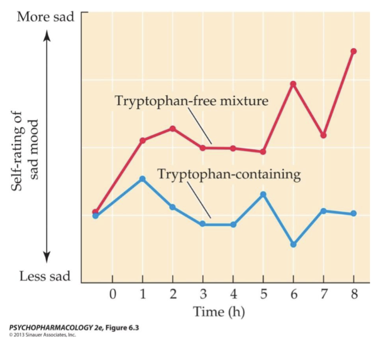
Decrease: Serotonin synthesis can also be partially diminished through diet via the Trp depletion test in which a “cocktail” of AAs w/o tryptophan is given which leads to temporary depletion of brain 5-HT (protein synthesis is stimulated so plasma Trp is diminished and the large neutral amino acids inhibit entry of remaining Trp to the brain)
How can serotonin synthesis be pharmacologically manipulated
Para-chlorophenylalanine (PCPA) blocks 5-HT synthesis by irreversibly inhibiting tryptophan hydroxylase. Used in research - to deplete 5-HT to determine its role in mediating certain behavior.
What drugs impacted serotonin packaging into vesicles and release
Reserpine (VMAT blocker) depletes 5-HT, which is broken down when not protected in vesicles
5-HT release is stimulated by amphetamine-like comp.
Para-chloroamphetamine (PCA) is a releaser, also toxicity, used experimentally to kill 5HT neurons
Fenfluramine was prescribed for appetite suppression in obese patients (x1997 heart disease).
3,4- methylenedioxymeth-amphetamine (MDMA) is a recreational drug.
List the history and original use of ecstasy
MDMA aka ecstasy was first synthesized in 1912 by Merck and was used in psychotherapy in the 1970s but later became a common illicit street drug that can, in repeated high doses, produce serotonin neurotoxicity (you develop hypothermia at high doses)
Serotonin reuptake
After release, 5-HT is rapidly removed from the synaptic cleft via reuptake by the 5-HT transporter aka SERT which is blocked by SSRIs like Prozac/Fluoxetine
Cocaine and MDMA also block SERT but aren’t selective (also block DA)
Serotonin breakdowm
Breakdown of 5-HT is catalyzed by MAO to yield 5-hydroxyindoleacetic acid (5-HIAA)
Serotonin anatomy
Almost all serotonergic neurons in the CNS are found along the midline of the brainstem, associated with the raphe nuclei (dorsal and median; B8 and B9)
Serotonin receptors and their antagonists/agonists
5-HT1A receptors are concentrated in the hippocampus, septal area, amygdala, and dorsal raphe nucleus and are Gi-coupled inhibitory metabotropic receptors, agonists for these receptors can be successful anxiolytics
5-HT2A receptors have large numbers in the cortex and through Gq activate the phosphoinositide second-messenger system which increases Ca++ levels in postsynaptic cells and activates protein kinase C. Agonists include DOI and related drugs being hallucinogenic in humans (hallucinogenic effects of LSD are believed to stem from its ability to stimulate these receptors) whilst antagonists include clozaril and risperdal block 5-HT2A AND D2 receptors (used in schizophrenia treatment with fewer side effects than drugs that only block D2 receptors)
5-HT6 receptors are mainly found in the cortex and are expressed most prominently in the olfactory tubercle, striatum, and hippocampus