Final = Nervous System of Lower Limb
 Call Kai
 Learn
Learn Practice Test
 Spaced Repetition
 Match
Match Flashcards
 Knowt Play
1/68

There's no tags or description
Looks like no tags are added yet.
Name | Mastery | Learn | Test | Matching | Spaced | Call with Kai | Chat |
|---|
No analytics yet
Send a link to your students to track their progress
69 Terms
How is the nervous system structurally divided?
CNS - brain and spinal cord
PNS - cranial nerves, spinal nerves, rami, plexuses, named peripheral nerves, ganglia
Spinal cord
extends from foramen magnum → L1/L2
enlarges at the cervical at the cervical and lumbosacral regions
This is because there are more motor cell bodies (more grey matter) within the ventral horns at these points as this is where the brachial and lumbosacral plexuses are formed supplying the highly muscular upper and lower limb
How many pairs of spinal nerves?
31 pairs:
8 cervical
12 thoracic
5 lumbar
5 sacral
1 coccygeal
How many pairs of cranial nerves?
12 pairs;
2 arise from the cerebrum
10 arise from brainstem
How is the nervous system functionally divided?
Somatic - voluntary
Autonomic - involuntary (parasympathetic vs sympathetic)

Where are cell bodies to efferent axons?
Somatic: ventral horn
Autonomic:
presynaptic cell body in grey matter of CNS
sympathetic: lateral horn
parasympathetic: spinal cord or brain stem
postsynaptic cell body in PNS autonomic ganglia
both multipolar axons
Where are the cell bodies to afferent axons?
Somatic: dorsal root ganglia
Autonomic: dorsal root ganglia or cranial ganglia
both pseudounipolar neurons
Describe formation of spinal nerve - somatic
efferent (motor) - cell body in ventral horn
axons carries signal through ventral root, spinal nerve and either ventral or dorsal horn
afferent (sensory) - cell body in dorsal root ganglia
axons carry signal from receptor down axon through either ventral or dorsal horn, spinal nerve, dorsal root ganglion, dorsal root and dorsal horn
What is the difference between ventral and dorsal rami?
Dorsal ramus → innervates muscle and skin of deep (intrinsic) muscles of back
Ventral ramus → innervates all muscle and skin of the anterolateral trunk, upper and lower limbs, superficial back (muscles only) and most of the neck
also combine to form plexuses which give off named peripheral nerves
Why does the back have decreased sensation?
Back skin has a lower density in of tactile receptors, contributing to a less fine tuned sensory perception. This makes evolutionary sense, as we don’t interact with our environment using our backs.
Intercostal nerves - cutaneous innervation
in the ribcage, the intercostal nerves are direct continuations of the the ventral rami
give off lateral and anterior cutaneous branches to innervate skin of the lateral and anterior trunk
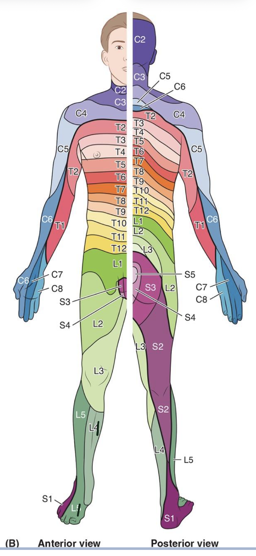
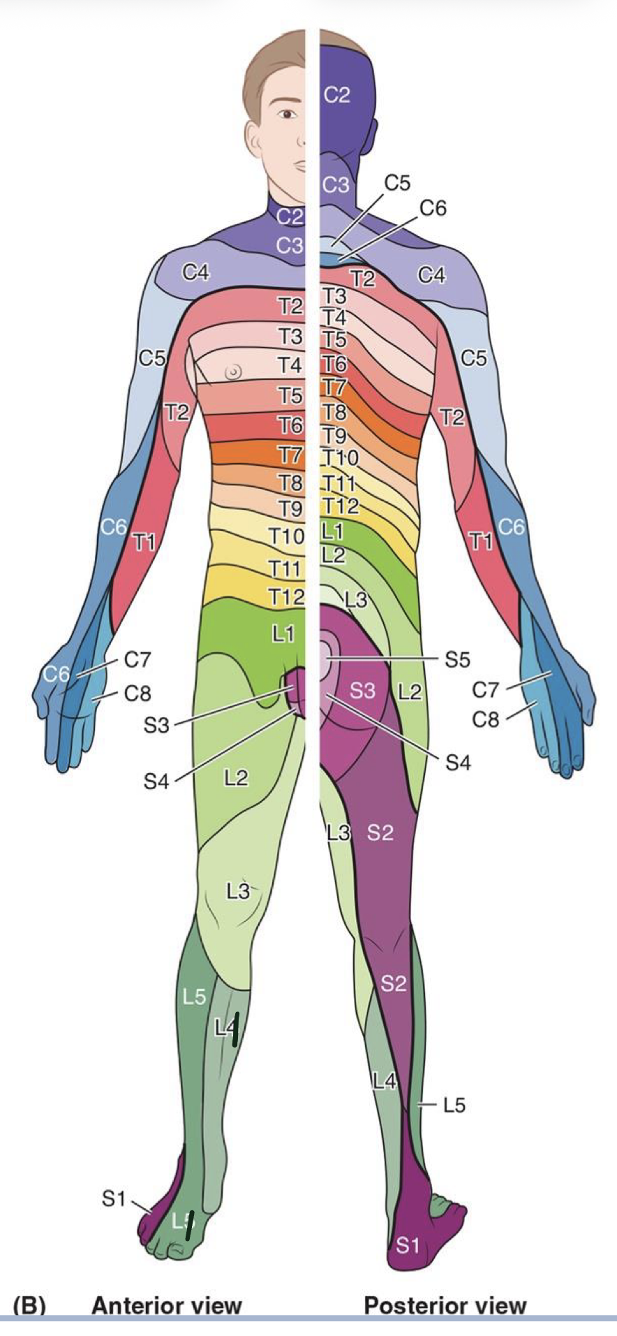
Dermatomes
each spinal nerve with somatic sensory axons (all but C1, typically lacks dorsal root ganglion) provide sensory information for a strip of skin
face cutaneous innervation is done by cranial nerves

L1 dermatome
anterior thigh/inguinal region

L2 dermatome
anterior lateral thigh (superior to knee)
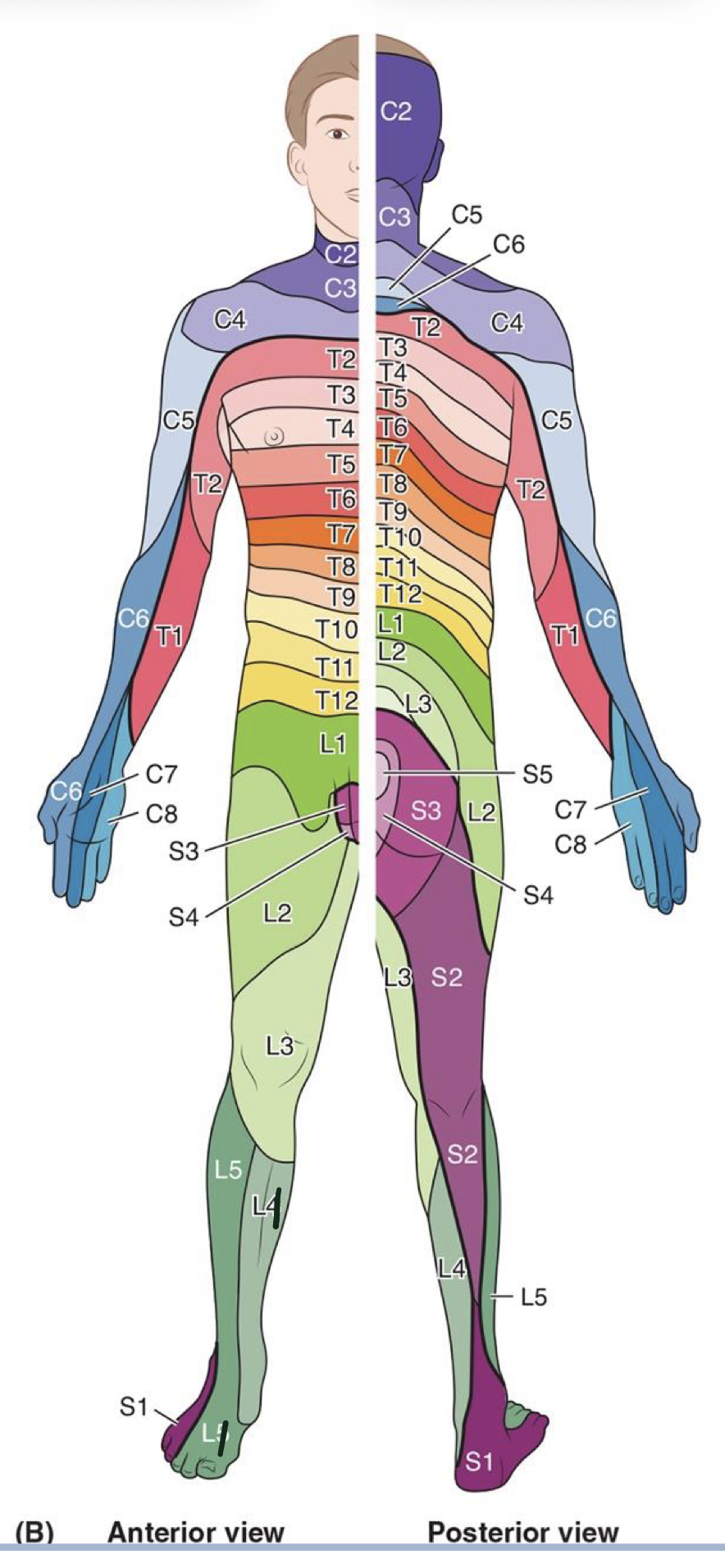
L3 dermatome
anterior medial thigh and knee

L4 dermatome
medial leg and malleolus
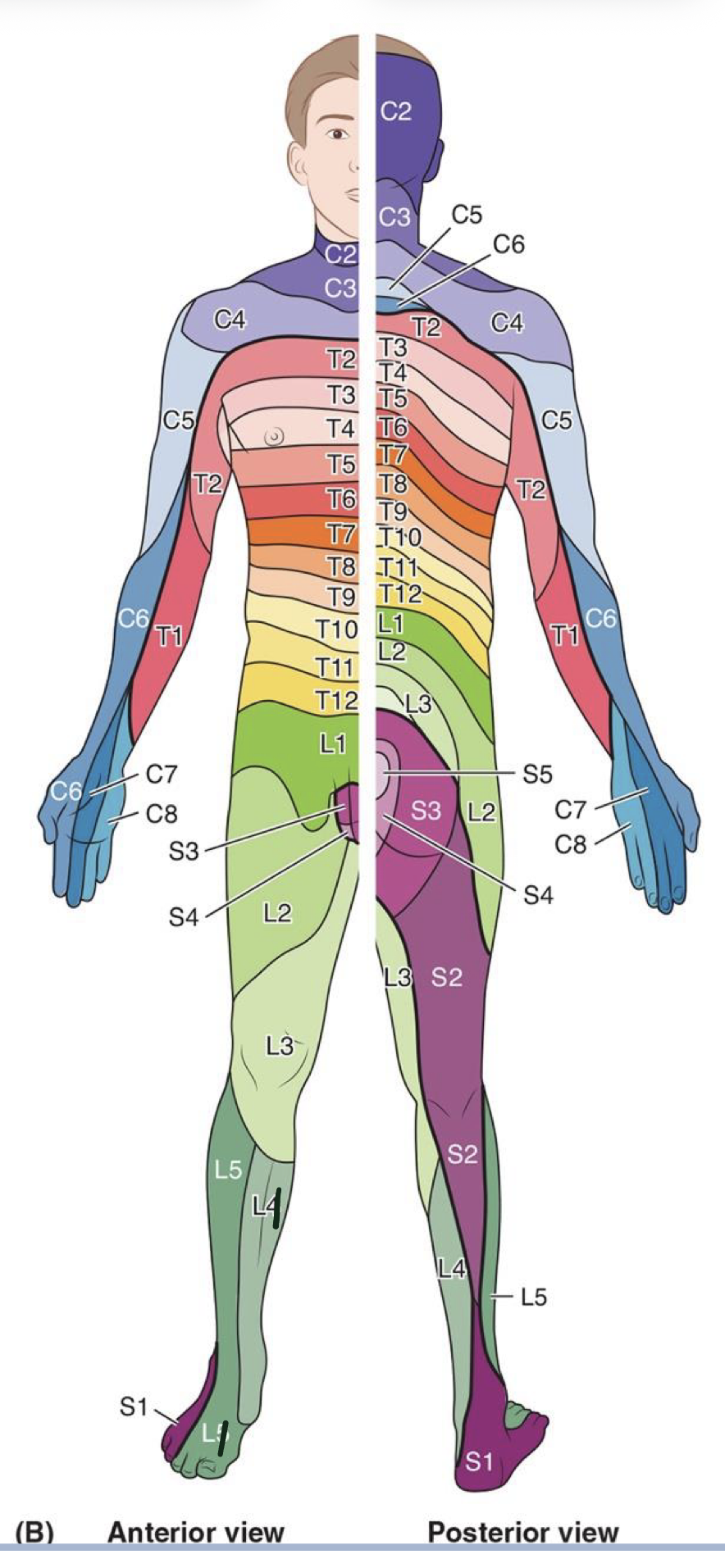
L5 dermatome
anterior lateral leg, medial dorsum of foot, digits 1-3

S1 dermatome
distal posterior lateral leg, ankle and foot, digits 4-5
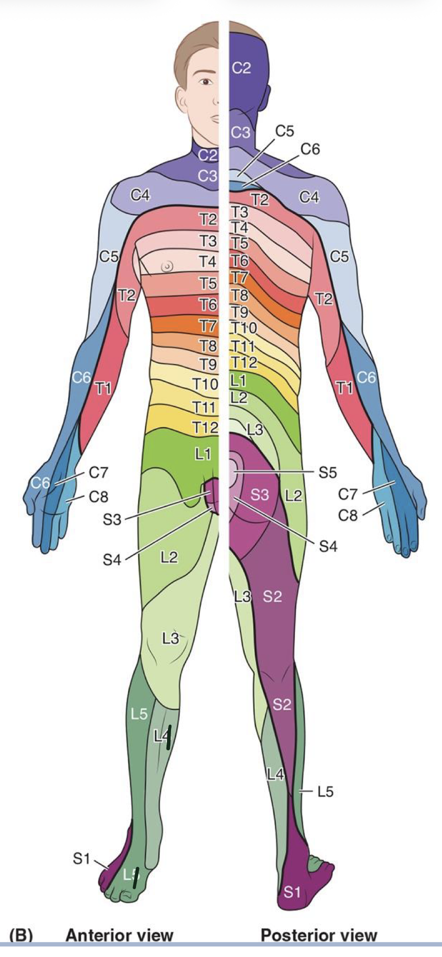
S2 dermatome
posterior midline of the thigh and posterior knee
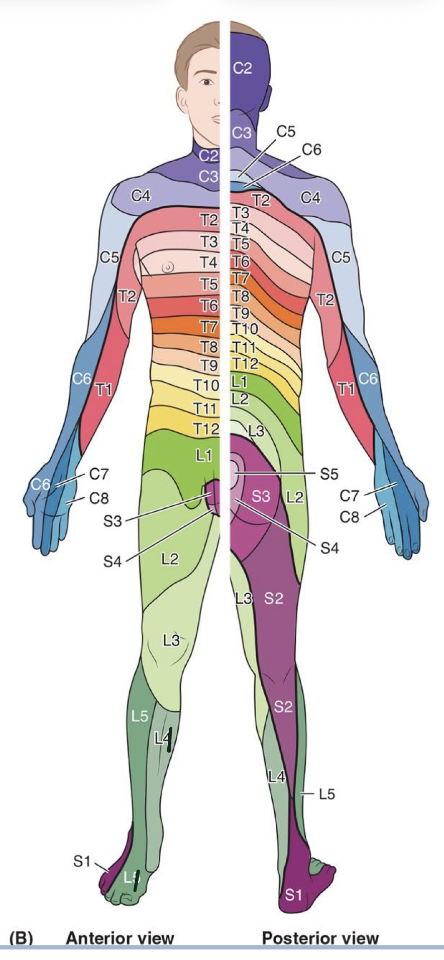
S3 dermatome
outermost ring of gluteal region posteriorly and superior genital area anteriorly (toilet lid)
S4 dermatome
intermediate ring of the gluteal region posteriorly and inferior genital area anteriorly (toilet lid hole)
S5 dermatome
innermost ring of gluteal region at anus/coccygeal area posteriorly

Myotome
the motor axons within each spinal nerve are responsible for innervating specific muscle fibers
Herpes Zoster virus
causes shingles
viral infection that presents following a reactivation of a previous infection of the virus (chickenpox)
virus typically remains dormant within the dorsal root ganglion
the rash is referred to as a “self-limited dermatomal rash” as it spreads long the are of skin associated with the sensory nerve it laid dormant within
How would injury to a spinal nerve compare to injury of a named nerve?
Ventral rami form plexuses from multiple spinal nerve levels that intertwine to give off named nerves
This means if a single spinal nerve was injured, there may be multiple structures weakened but function wont be entirely depleted as other spinal nerves also contribute
Contrarily, if the actual named nerve is injured, this would cut off all spinal nerve contributions, meaning the structures it supplies will lose all signaling.
There are some exceptions with dual innervated muscles, such as pectineous
Herniated (slipped) disc: what is it? what can it cause? symptoms?
between each vertebral body is a disc
if the disc ruptures, the inside material can leak posteriorly
common consequence → compression of the nerve roots for a single spinal nerve (radiculopathy)
if a single spinal nerve is compressed, a patient will present with sensory and motor deficits associated with the same dermatome/myotome
What invervates the hip joint?
femoral n.
obturator n.
superior gluteal n.
nerve to quadratus femoris n.
what inervates the knee joint?
femoral
saphenous
tibial
common fibular
what innervates the ankle joint?
saphenous
tibial
sural
superficial fibular
deep fibular
Hip joint myotomes
hip flexion: L1-L2
hip adduction: L2-L3
hip extension: L5-S1
What structures travel through the greater sciatic foramen?
superior and inferior gluteal avn.
piriformis m.
pudendal nva.
sciatic n.
posterior femoral cutaneous n.
What structures travel through the lesser sciatic notch?
pudendal nva. (reenters pelvis)
obturator internus tendon passes through to attach to femur
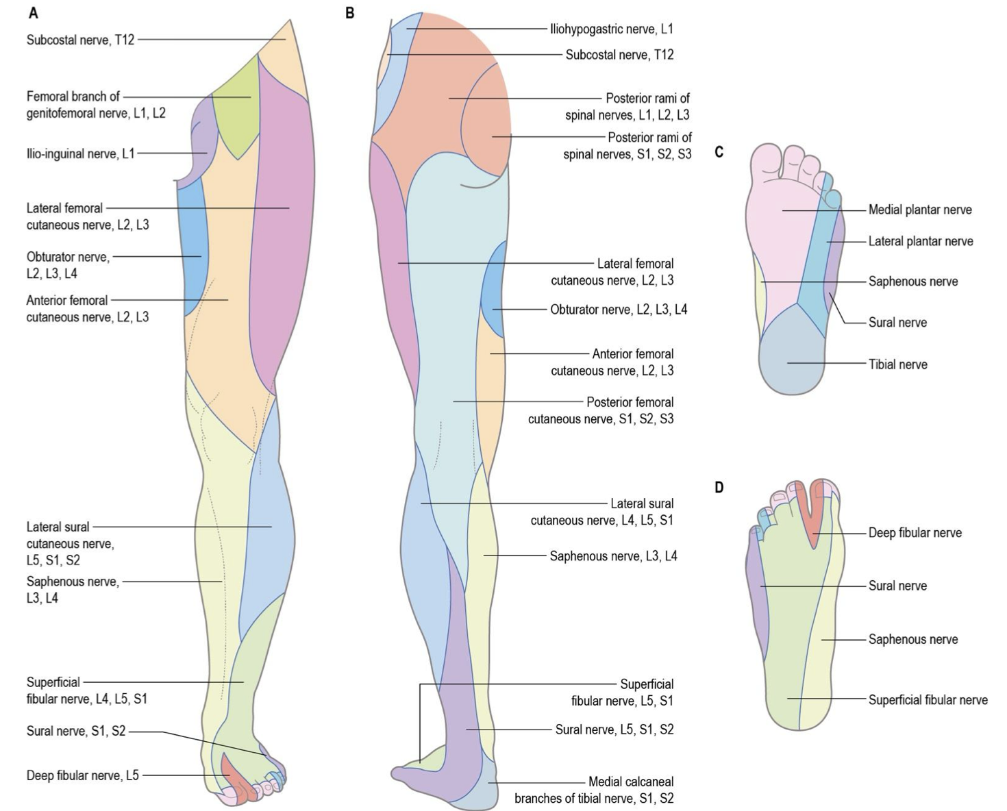
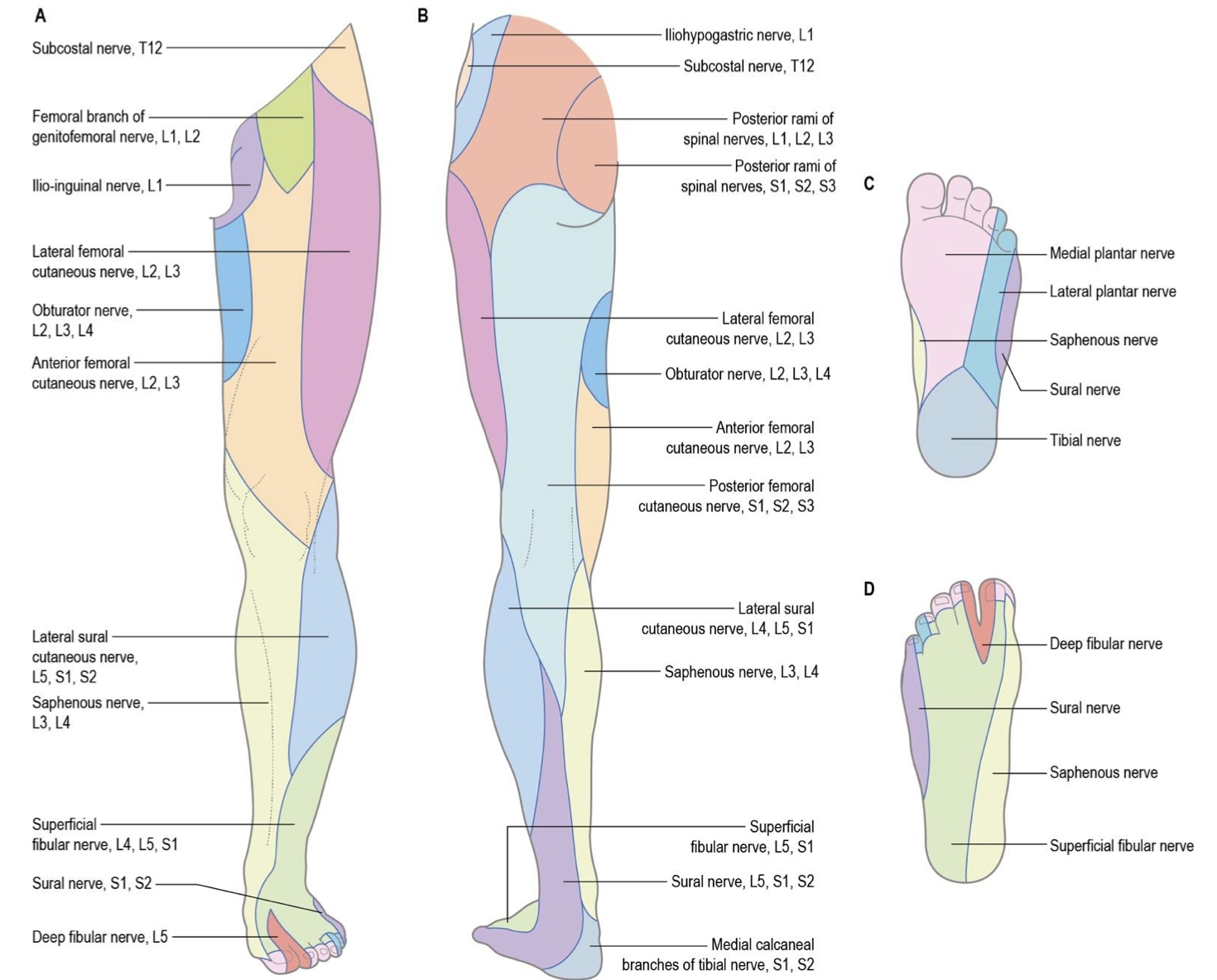
What is cutaneous innervation? how does it compare to dermatomes?
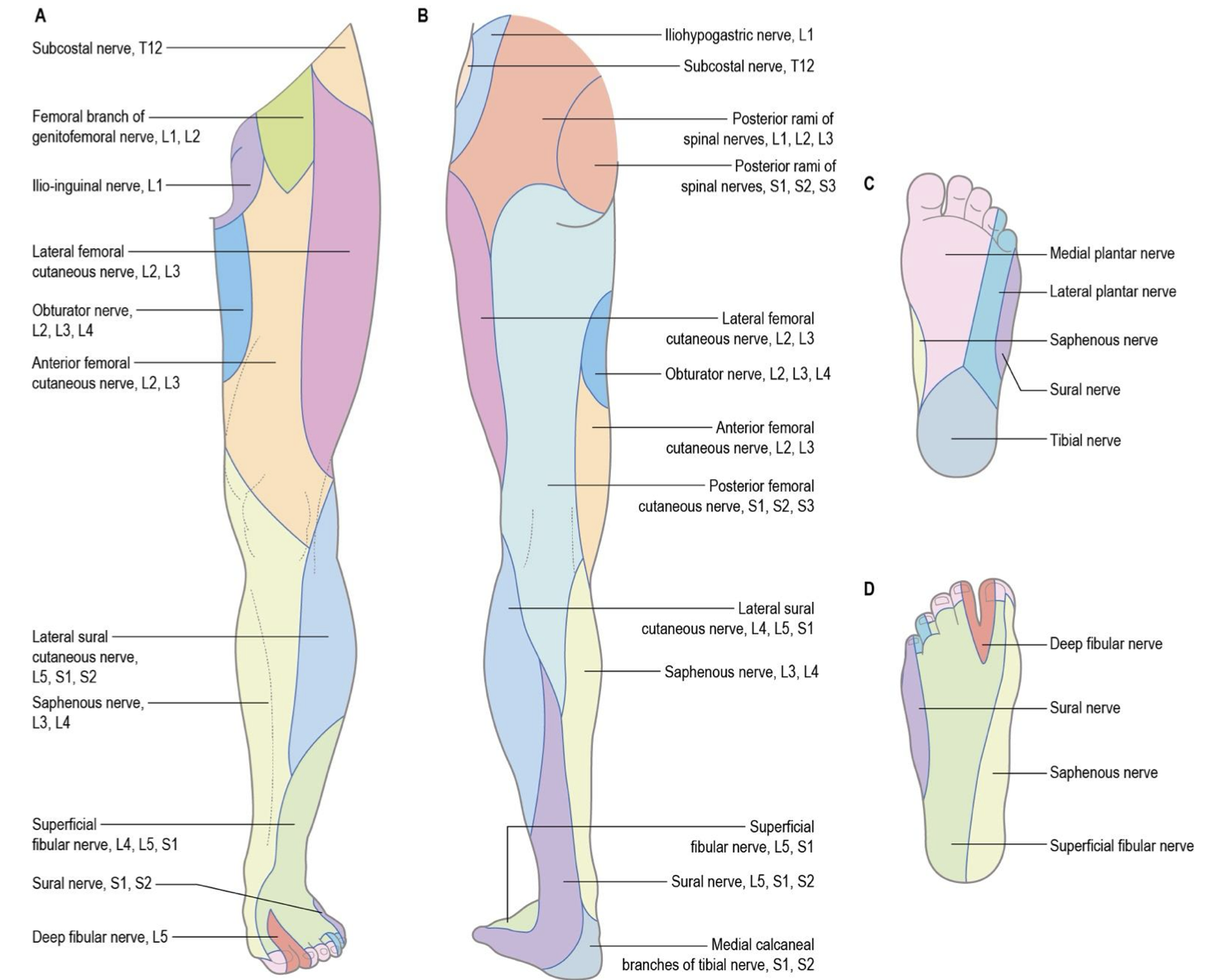
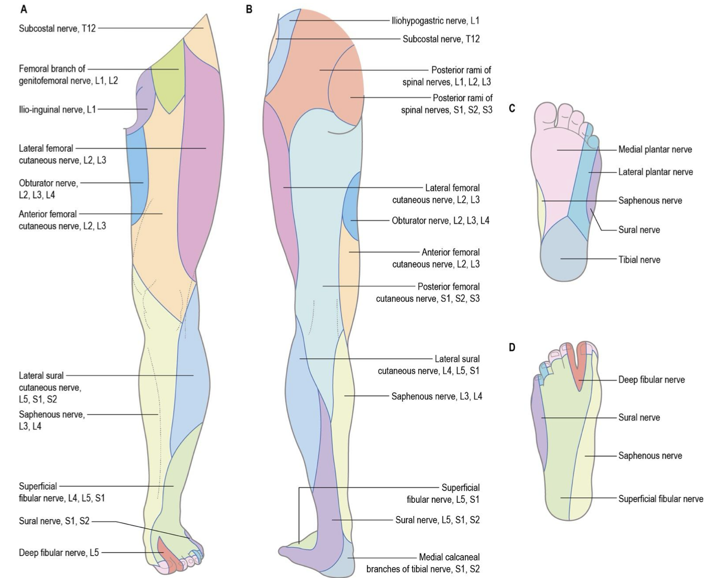
Named nerves almost always provide sensory information to an area of skin, called cutaneous innervation
Named nerves that are formed by a plexus differ from dermatomes because there are typically multiple spinal nerves contributing
dermatomes are the strips of skin that only a single spinal nerve contributes to
cutaneous innervation and dermatome maps are visibly different

Lumbar plexus ventral rami? where does it form?
L1-L4
within psoas major
subcostal n. (levels, path, innervation)
T12 (lumbar)
runs inferior to 12th rib
lateral cutaneous branch innervates
skin of hip region inferior to anterior part of iliac crest and anterior to greater trochanter

iliohypogastric n. (levels, path, innervation)
L1 (lumbar)
emerges from upper lateral border of psoas major and crosses over quadratus lumborum
lateral cutaneous branch innervates
superolateral quadrant of buttocks

Ilioinguinal n. (levels, path, innervation)
L1 (lumbar)
runs on iliac crest to ASIS where inguinal canal starts
innervates
skin over medial femoral triangle

Genitofemoral n. (levels, path, innervation)
L1-L2
runs through center of psoas major, pops out along anterior surface and runs down
gives off genital and femoral branch
innervates
femoral branch → skin over lateral part of femoral triangle

Lateral femoral cutaneous n. (levels, path, innervation)
L2-L3
runs diagnolly across ilaicus and pops up out just below ASIS
innervates
skin of anterior and lateral margin of the thigh
Anterior femoral cutaneous n. (path, innervation)
from anterior division of femoral n.
intermediate anterior thigh, distal medial margin of thigh, proximal lateral knee

Saphenous n. (path, innervation)
from posterior division of femoral n.
medial knee, medial margin of leg, ankle and foot


Femoral n. (levels, path, innervation, injury (motor/sensory)?)
L2-L4
runs lateral to psoas major and exits pelvis deep to inguinal ligament
divides into anterior and posterior divisions
lateral circumflex a. runs in between
innervates
iliacus and pectineus
Injury → motor
Femoral n. → flexion, adduction and medial rotation of hip
Anterior division of Femoral n. → flexion, abduction, and lateral rotation of hip, knee flexion
Posterior division of Femoral n. → flexion of hip, extension of knee
Injury → sensory
Anterior femoral cutaneous n. → anterior and medial thigh
Saphenous. n. → medial knee, leg, ankle and margin of foot
Anterior division of femoral n. (levels, path, innervation)
L2-L4
splits after femoral n. exits pelvis deep to inguinal ligament
mainly sensory, with little motor
innervates
sartorius m.
Injury → motor
flexion, abduction, and lateral rotation of hip, knee flexion
Injury → sensory
Anterior femoral cutaneous n. (medial and intermediate branches) → anterior and medial thigh
Posterior division of femoral n. (levels, path, innervation, injury (motor/sensory)?
L2-L4
splits after femoral n. exits pelvis deep to inguinal ligament
mainly motor, with little sensory
Innervates:
rectus femoris, vastus lateralis, vastuc medialis, vastus intermedius
Injury → motor
extension of knee, flexion of hip
Injury → sensory
Saphenous. n. → medial knee, leg, ankle and margin of foot

Obturator n. (levels, path, innervation, injury (motor/sensory)?
L2-L4
runs medial to psoas major and exist pelvis to medial thigh through obturator canal of obturator membrane
divides into anterior and posterior divisions that sandwhich over adductor brevis m.
Innervate
obturator externus m.
Injury → motor
Obturator n. → lateral rotation and stabilization of hip
Anterior division of obturator n. → adduction, flexion, and medial rotation of hip, knee flexion and rotation when flexed
Posterior divsion of obturator n. → hip adduction and flexion
Injury → sensory
medial thigh
Anterior division of obturator n. (levels, path, innervation, injury (motor/sensory)?
L2-L4
sits anterior to adductor brevis m.
Innervate
adductor longus, adductor brevis, gracilis, pectineus
Injury → motor
Anterior division of obturator n. → adduction, flexion, and medial rotation of hip, knee flexion and rotation when flexed
Injury → sensory
medial thigh
Posterior division of obturator n. (levels, path, innervation, injury (motor/sensory)?
L2-L4
sits posterior to adductor brevis m.
Innervates
adductor portion of adductor magnus m.
Injury → motor
adduction and flexion of hip
Lumbosacral trunk (levels, path, function
L4-L5
medial and deep to psoas major
nerve bridge fromed by L4-L5 connecting lumbar and sacral plexus
Sacral (lumbosacral) plexus ventral rami
L4-S3
Superior gluteal nerve (levels, path, innervation, injury (motor/sensory)?)
L4-S1 (sacral)
exits the pelvis via the greater sciatic foramen superior to piriformis m
travels with the superior gluteal artery and vein
innervates
gluteus medius, gluteus minimus, tensor fascia latae
injury → motor
weakness of medial hip rotation, abduction, stabilization of the hip (pelvic drop), some hip flexion
Inferior gluteal nerve (levels, path, innervation, injury (motor/sensory)?)
L5-S2 (sacral)
exists the pelvis via the greater sciatic foramen inferior to the piriformis m
travels with the inferior gluteal artery and vein
Innervates
Gluteus maximus
injury → motor
weakness in extension of the hip, particularly with rising from sitting/walking up stairs and lateral rotation of the hip
Nerve to piriformis (levels, innervation, injury (motor/sensory)?)
S1-S2 (sacral)
Innervation
piriformis m.
Injury → motor
weakness of lateral rotation of extended hip, abduction of flexed hip, hip stability
Nerve to obturator internus (levels, innervation, injury (motor/sensory)?)
L5-S2 (sacral)
Innervation
obturator internus m
gemellus superior m.
Injury → motor
weakness of lateral rotation of extended hip, abduction of flexed hip, hip stability
Nerve to quadratus femoris (levels, innervation, injury (motor/sensory)?)
L4-S1 (sacral)
Innervation
quadratus femoris m.
gemellus inferior m.
Injury → motor
weakness of lateral rotation of extended hip, abduction of flexed hip, and stabilization of hip
Posterior femoral cutaneous n. (levels, path, innervation, injury (motor/sensory)?)
S1-S3 (sacral)
Exits pelvis via the greater sciatic foramen, inferior to piriformis m.
gives off inferior clunial n.
Innervation
cutaneous innervation to the inferior buttocks (inferior clunial n) and skin along the posterior thigh and popliteal fossa
Injury → cutaneous
above

Pudendal n. (levels, path)
S2-S4 (sacral plexus)
Exits pelvis via the greater sciatic foramen
enters the perineum via the lesser sciatic foramen
Sciatic nerve (levels, path, innervation, injury (motor/sensory)?)
L4-S3 (sacral)
Continuation of sacral plexus, exiting the plevis via the greater sciatic foramen
inferior to piriformis m. between the ischial tuberosity and the greater trochanter
Innervation
none, but composed of two nerves (tibial and common fibular)
Injury → motor
Tibial n. (posterior thigh) → extension of hip, flexion of knee, medial/lateral rotation of flexed knee
Tibial n. (posterior leg) → plantarflexion, toe flexion, inversion
Medial plantar n. (foot) → hallux MTP flexion/abduction, digit 2-5 flexion MTP/PIP, digit 2 (1st lumbrical) MTP flexion and IP extension
Lateral plantar n. (foot) → digit 3-5 adduction, digit 2-4 abduction, digit 2-5 toe flexion MTP/IP, hallux adduction, digit 3–5 (2-4 lumbricals) MTP flexion and IP extension
Superficial fibular n. (lateral leg) → eversion, weak plantarflexion
Deep fibular n. (anterior leg) → dorsiflexion/inversion, hallux extension MTP/IP, digit 2-5 extension MTP/IP.
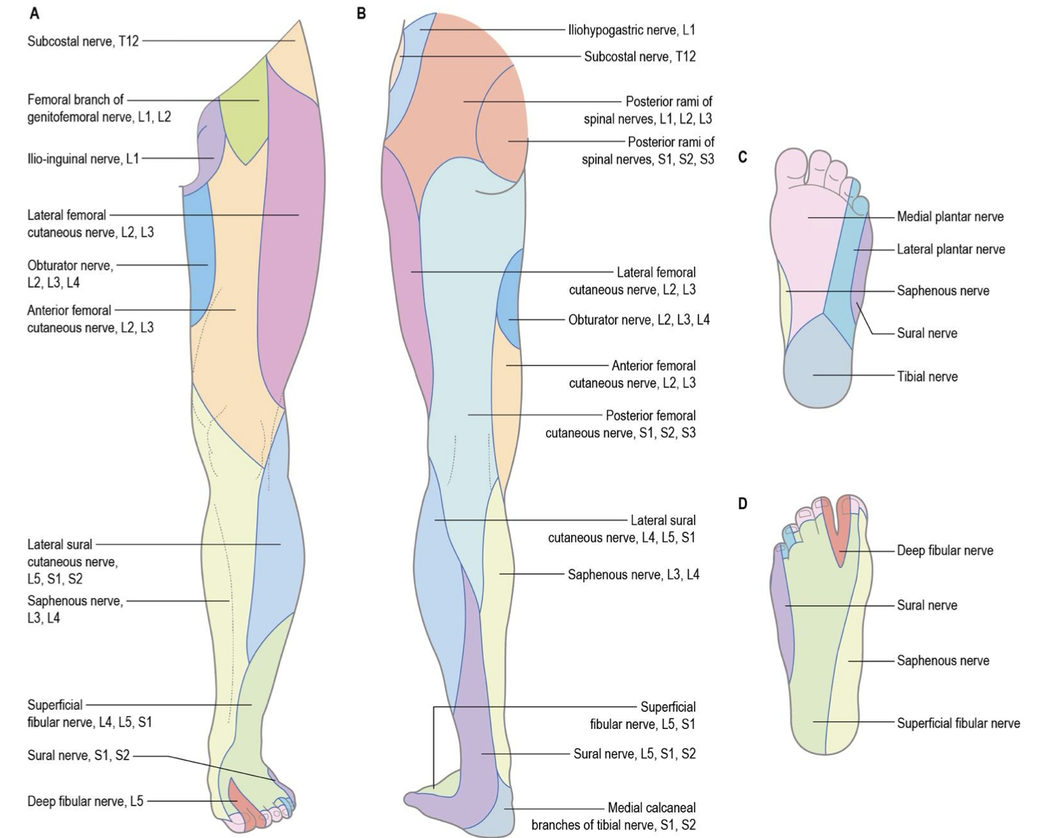
Injury → sensory
Tibial → inferior heel
Medial plantar n. → plantar surface of medial 3.5 digits and nails
Lateral plantar n → plantar surface of lateral 1.5 digits and nails
Superficial fibular n. → distal anterolateral leg and dorsum of foot excluding medial and lateral margins, 1st interweb space and toe nails
Deep fibular n. → 1st web space
Sural n. → lateral foot, lateral malleolus and posterior distal leg
Lateral sural cutaneous n. → proximal anterior/posterior lateral leg
Tibial n. (levels, path, innervation, injury (motor/sensory)?)
L4-S3
starts as part of sciatic n. exiting pelvis through greater sciatic notch, inferior to piriformis
branches off of sciatic n. near the popliteal region, running medially down posterior leg between two heads of gastrocnemius and through tendinous arch of soleus muscle and then wrapping posteriorly around medial malleolus (tarsal tunnel)
gives off medial and lateral plantar n. in plantar foot and medial calcaneal branch either before or within the tunnel
runs with popliteal av.
also gives off medial sural cutaneous n. which joins with lateral sural cutaneous n. to form sural n.
Directly innervates
posterior thigh → semitendinosus, semimembranosus, and biceps femoris long head
posterior leg → gastrocnemius, plantaris, soleus, popliteus, tibialis posterior, flexor digitorum longus, flexor hallucis longus
Injury → motor
Tibial n. (posterior thigh) → extension of hip, flexion of knee, medial/lateral rotation of flexed knee
Tibial n. (posterior leg) → plantarflexion, toe flexion, inversion
Medial plantar n. (foot) → hallux MTP flexion/abduction, digit 2-5 flexion MTP/PIP, digit 2 (1st lumbrical) MTP flexion and IP extension
Lateral plantar n. (foot) → digit 3-5 adduction, digit 2-4 abduction, digit 2-5 toe flexion MTP/IP, hallux adduction, digit 3–5 (2-4 lumbricals) MTP flexion and IP extension
Injury → sensory
Tibial → inferior heel
Medial plantar n. → plantar surface of medial 3.5 digits and nails
Lateral plantar n → plantar surface of lateral 1.5 digits and nails
Sural n. → lateral foot, lateral malleolus and posterior distal leg
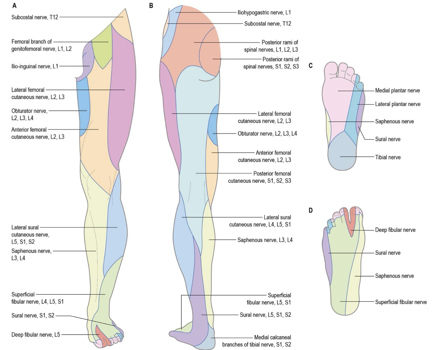
medial calcaneal branch → posterior heel triangle

Medial sural cutaneous n. (path, injury)
branches off of tibial n. in popliteal fossa and combines with sural communicating branch (of lateral sural cutaneous n.) to form sural n.
Injury → sensory
sural n. → lateral foot, lateral malleolus and posterior distal leg

Medial calcaneal branches of tibial n. (path, innervation)
branches off tibial n. either before or within the tunnel
innervates
posterior heel triangle
Medial plantar n. (path, innervation, injury (motor/sensory)?)
branches from tibial nerve after passing deep to flexor retinaculum from tibial n.
enters foot by passing deep to abductor hallucis m.
after motor innervation divides into 3 common plantar digital n. which then divide into proper plantar digital n. for cutaneous innervation
innervates
plantar foot → abductor hallucis, flexor digitorum brevis, 1st lumbrical and flexor hallucis brevis
Injury → motor
hallux MTP flexion/abduction, digit 2-5 flexion MTP/PIP, digit 2 (1st lumbrical) MTP flexion and IP extension
Injury → sensory
plantar surface of medial 3.5 digits and nails

Lateral plantar n. (path, innervation, injury (motor/sensory)?)
branches from tibial nerve after passing deep to flexor retinaculum from tibial n.
enters foot by passing laterally between the flexor digitorum brevis and quadratus plantae m.
after motor innervation divides into proper plantar and common plantar digital n, which will divide into proper plantar digital n.
Innervates
foot → abductor digiti minimi, quadratus plantae, lumbricals 2-4, adductor hallucis, flexor digitorum brevis and plantar/dorsal interossei
Injury → motor
digit 3-5 adduction, digit 2-4 abduction, digit 2-5 toe flexion MTP/IP, hallux adduction, digit 3–5 (2-4 lumbricals) MTP flexion and IP extension
Injury → sensory
plantar surface of lateral 1.5 digits and nails

Common fibular n. (levels, path, innervation, injury (motor/sensory)?)
L4-S2
branches off of sciatic nerve near the popliteal region, running laterally over fibular neck
divides into deep (anterior leg) and superficial fibular (lateral leg) n.
lateral sural cutaneous n branches in popliteal fossa giving off sural communicating branch that joints medial sural cutaneous n. to form sural n.
Innervates
biceps femoris short head
Injury → motor
common fibular n. → flexion and lateral rotation of the knee
deep fibular n. → dorsiflexion/inversion, hallux extension MTP/IP, digit 2-5 extension MTP/IP.
superficial fibular → eversion of ankle, weak plantarflexion
Injury → sensory
deep fibular n. → 1st web space
superficial fibular n. → distal anterolateral leg and dorsum of foot excluding medial and lateral margins, 1st interweb space and toe nails
lateral sural cutaneous n. → proximal anterior/posterior lateral leg
sural n. → lateral foot, lateral malleolus and posterior distal leg
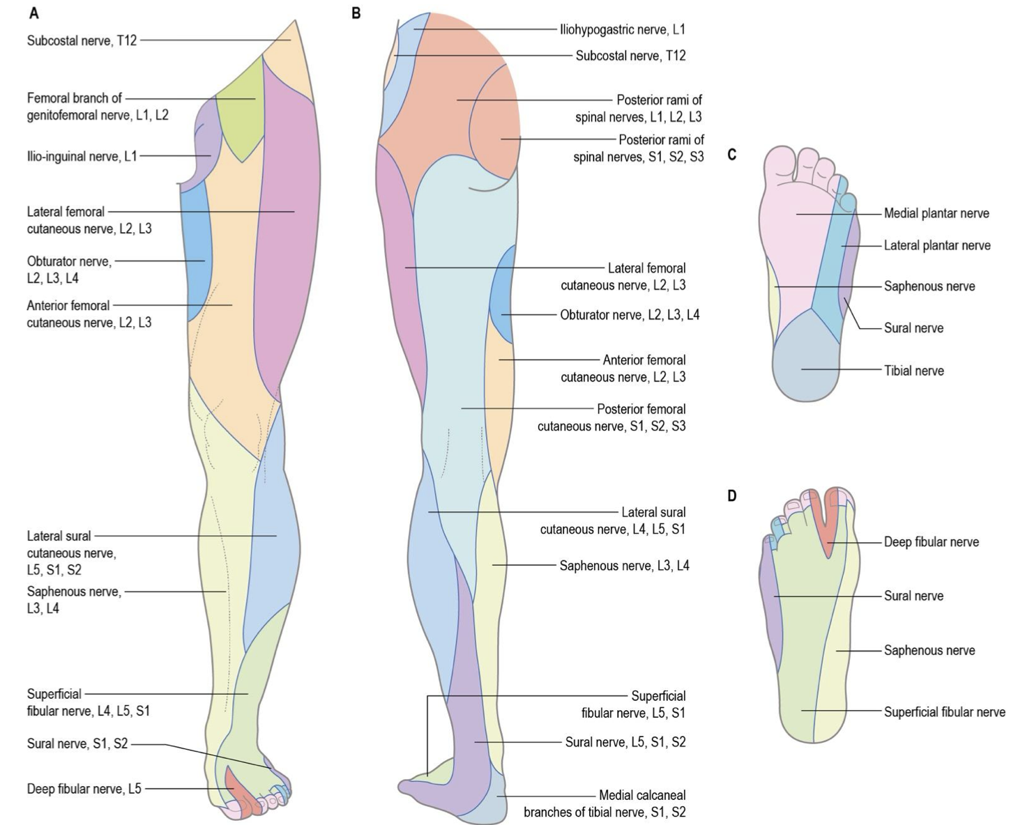
Lateral sural cutaneous (path, innervation)
branches off of common fibular n. and gives off sural communicating branch that joints medial sural cutaneous n. to form sural n.
innervates
lateral sural cutaneous n. → proximal anterior/posterior lateral leg
sural n. → lateral foot, lateral malleolus and posterior distal leg

Superficial fibular n. (levels, path, innervation, injury (motor/sensory)?)
L4-S2
branches off common fibular nerve at fibular neck and dives into lateral leg compartment b/w fibularis longus and brevis
become subcutaneous halfway down the leg
Innervates
fibularis longus and fibularis brevis
Injury → motor
eversion of ankle, weak plantarflexion
Injury → sensory
distal anterolateral leg and dorsum of foot excluding medial and lateral margins, 1st interweb space and toe nails

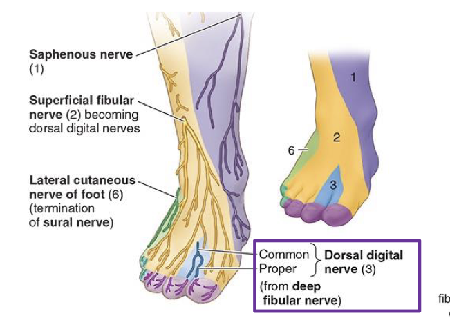
Deep fibular n. (levels, path, innervation, injury (motor/sensory)?)
L4-S1
branches off common fibular nerve at fibular neck and dives into anterior leg
runs along anterior surface of interosseous membrane, sitting lateral to tibialis anterior belly/tendon and running with anterior tibial a
passes deep to extensor retinacula running between extensor digitorum and hallucis brevis (anterior tibial becomes dorsalis pedis)
after motor innervation, it transitions to common dorsal digital n. which will divide into proper dorsal digital n. in the first interdigital space
Innervation
anterior leg → tibialis anterior, extensor hallucis longus, extensor hallucis brevis, fibularis tertius
dorsal foot → extensor hallucis brevis, extensor digitorum brevis
Injury → motor
dorsiflexion/inversion, hallux MTP and IP extension, digit MTP and IP 2-5 extension
Injury → sensory
1st web space
Superior cluneal n. (levels, innervation)
dorsal rami of L1-L3 (no plexus)
innervates
skin overlying superior and central parts of buttocks
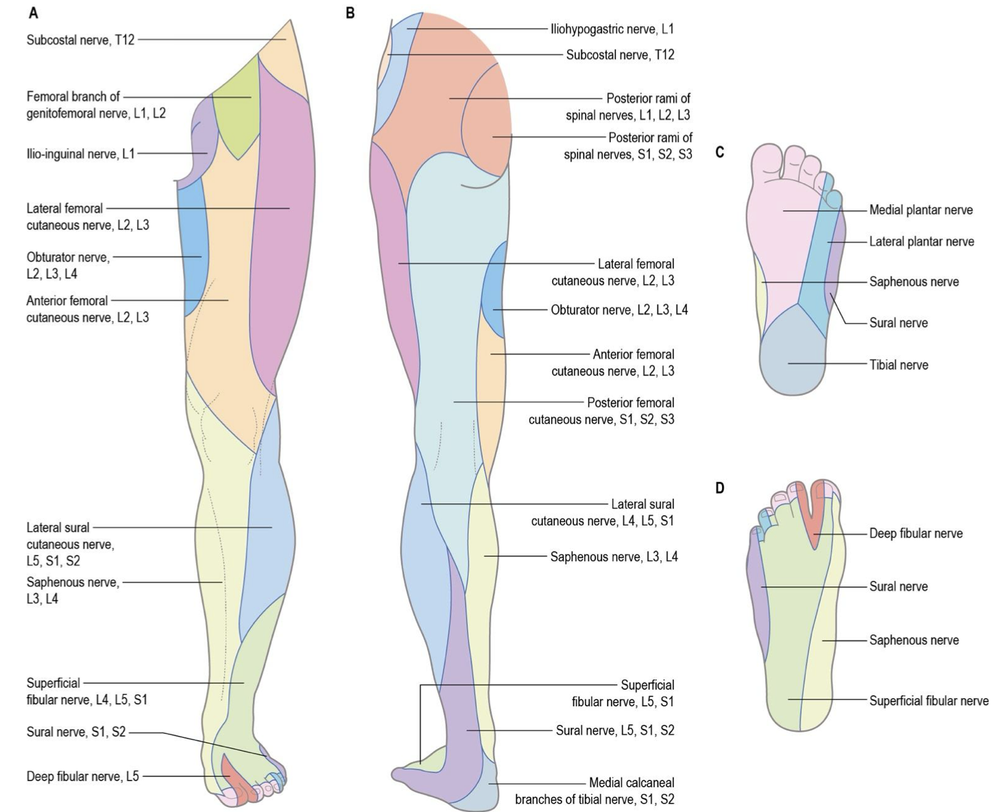
Middle cluneal n. (levels, innervation)
dorsal rami of S1-3
innervates
skin of medial buttock and intergluteal cleft

Sciatic nerve variations
can travel below, split around or through piriformis