Adv Med Surg: Exam 1
 Call Kai
 Learn
Learn Practice Test
 Spaced Repetition
 Match
Match Flashcards
 Knowt Play
1/315

Earn XP
Description and Tags
dysrhythmia, cv disease - mi, sympathomimetic, preload and afterload
Name | Mastery | Learn | Test | Matching | Spaced | Call with Kai | Chat |
|---|
No analytics yet
Send a link to your students to track their progress
316 Terms
What are pacemaker cells?
automatic cells designed to create/start a node (capable of generating an electrical impulse)
What are muscle cells?
cells that contract and relax
What is the order of the cardiac conduction system?
SA Node—> AV Node—> Bundle of His—> R and L Bundle Branches—> Purkinje Fibers
If there is a change in cardiac rhythm…
assess the patient
What do tachys do that can be bad/detrimental?
increase myocardial o2 demand, decrease ventricular filling time, decrease coronary artery perfusion time
What do bradys do that can be bad/detrimental?
decrease cardiac output, can cause syncope, chest pain
Cardiac Output=
Cardiac Output = Heart Rate x Stroke Volume
What is stroke volume
amount of blood pumped out with every beat of the heart
How to get HR from a 6 second EKG strip
count QRS complexs and multiple by 10
P wave
atrial depolarization

What is the star represent
It is a time delay so the atria have time to pump and fill the ventricles
Normal length of PR interval
0.12-0.20 seconds
What does the QRS Complex represent
ventricular depolarization
Normal QRS Complex length
0.06-0.10 seconds

What does the ST Segment represent?
early repolarization
if the ST segment is above/below the isoelectric line, it could be a sign of
cardiac ischemia/infarction
What does the T wave represent
ventricular repolarization
What does the QT wave represent
ventricular depolarization and repolarization
If QT interval becomes prolonged, pt could be at risk for what rhythm?
torsades de pointes (lethal ventricular dysrhythmia)
Calculating a regular rhythm heart rate
number of small blocks between each complex and follow to the corresponding HR. only use rate table for regular rhythm
calculating HR for irregular rhythm
count number of QRS complex’s in a 3 second strip and multiply by 20. count the number of QRS complexes in a 6 second strip and multiply by 10
Normal Sinus Rhythm
electrical impulse starts in SA node, regular rate and rhythm (60-100), impulse travels through normal conduction pathway
Atrial conduction=
1 P wave before each QRS, consistant in shape
Sinus Tachycardia
everything is normal, but rate is above 100 BPM. Happens when SA Node creates an impuse at a faster than normal rate
how to treat sinus tach
treat underlying cause!!! monitor (most lethal dysrhythmia).. also can be put on beta blocker
sinus brady
occurs when SA Node creates an impulse at slower than normal rate. rate less than 60
tx for sinus brady
monitor, treat underlying cause, atropine if caused by vagal mechanism
causes of sinus brady
decreased metabolic needs, athletic training, hypothyroidism, vagal stimulation, meds (beta blockers, calcium channel blockers), idiopathic SA Node dysfunction, increased ICP, MI
vagal stimulation
enhanced pressure in heart (having BM, vomiting, bearing down)
causes of sinus tach
stress, blood loss, anemia, shock, hypovolemia, heart failure, pain, fever, exercize, anxiety, hypoxia….
What are atrial dysrhythmias
a fib, a flutter, premature atrial contractions (PAC)
Premature Atrial Contraction (PAC)
premature, ectopic electrical impulse discharges in atrium before next normal impulse of SA Node
Ectopic beat
a pacemaker site outside of the SA node (not where is it supposed to be)..complex comes in early
PAC criteria
rhythm: regular except where premature beat occurs
rate: rate of underlying rhythm
atrial conduction: P wave before the ectopic beat is different from P wave of underlying rhythm
PAC causes
caffine, alc, HF, anxiety
True or false, PACs are common in normal, healthy hearts
true
tx for PAC
dont really treat, just get rid of cause
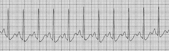
what is this
sinus tach
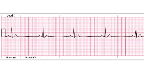
what is this
normal sinus rhythm
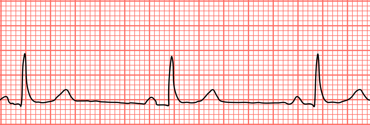
what is this
sinus brady

what is this
sinus with PAC
look for _____ when looking at PAC in a rhythm strip
pointy t wave, p wave hiding on top of t wave
Atrial Flutter
atria are depolarized at rates of 250-400x/min, multi p’s for every QRS
A flutter cause
reentry circut
what happens in A flutter
not all the atrial impulses are conducted into the ventricle, causing a therapeutic block at the AV node
t/f: the adult heart can tolerate a flutter
false, we need to slow it down
How to tell A flutter on a strip
“p” waves produce a saw-tooth pattern called flutter waves
causes of a flutter
underlying cardiac disease, long standing HTN, CAD, hypoxia, HF…
what can occor due to lack of atrial contraction during a flutter
blood stasis and thromboembolisms
tx for A Flutter ****
control heart rate, convert to normal sinus rhythm, (amiodarone, cardioversion) vagal maneuvers, anticoagulation
examples of vagal maneuvers for A Flutter
carotid sinus massage, cough
what meds can help control HR for A Flutter
diltiazem, beta blockers, digitalis

what is this
A Flutter
A Fib
uncoordinated atrial electrical activation from multiple areas in the atria. Results in a rapid, disorganized, quivering, fibrillating atria
A Fib charateristics
no p waves!!
EKG baseline is irregular and undulating
irregularly irregular
clinical significance of A Fib
loss of atrial systolic volume “atrial kick”
loss of AV synchrony
reduced cardiac output (blood clots)
increased potential for thromboembolism
causes of A Fib
HTN long term, MI, HF, pulmonary disease…
t/f: A fib rarely occurs in those with no functional heart disease
true
A Fib tx
convert to NSR, decrease ventreicular raate, prevent clots, rate control meds: (calcium channel blockers, beta blockers, digoxin) conversion meds: amiodarone, dofetilide, flecanide
antiplatelet agent
aspirin
antithrombotic meds
anticoagulant: warfarin
factor Xa inhibitors: dabigatran, rivaroxaban, apixaban
A Fib tx
cardioversion if unstable
t/f: some pts remain chronically in A Fib
true
what is cardioversion
synchronized defibrillation

what is this
A Fib
ventricular dysrhythmias
premature ventricular contraction, ventricular tachycardia, ventricular fibrillation, PEA
premature ventricular contraction (PVC)
premature ectopic impulse that originates in the ventricle. conducted through the ventricles before the next normal sinus impulse
can PVCs occur in healthy ppl
yes
characteristics of PVC
ventricular complex that comes in early
QRS is wide, shape is bizzare and there are ST and T wave changes
QRS and T tend to go in opposite directions
causes of PVC
caffine, nicotine, alc, cardiac ischemia, hypoxia…
tx for PVC
if infrequent- none needed
remove causing agent
amiodarone, lidocaine
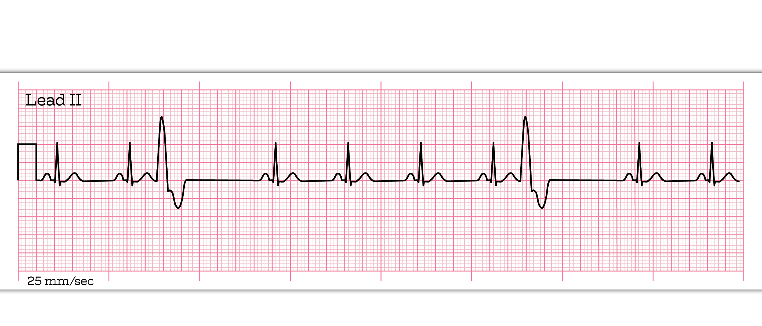
what is this
sinus with PVC
V tach
3 or more PVCs in a row with a rate exceeding 100bpm
t/f: v tach is an emergency situation and can be a life-threatening dysrhythmias
true
characteristics of v tach
ventricular rate greater thhan 100, rhythm is usually regular, QRS complexes r wide, no P waves present
causes of V Tach
MI, ischemia, hypoxia, electrolyte imbalence, CAD, acidosis, HF…
treatment for v tach with a pulse (stable)
antiarrhythmic drugs, amiodarone
treatment for v tach with a pulse (unstable but conscious)
sedate, cardioversion, antiarrhythmic drugs
Cardioversion
uses quick, low-energy shocks to restore a regular heart rhythm
treatment for v tach without a pulse
defibrillate
T/F: you can get someone out of this rhythm with just CPR
false, early defibrillation is VERY important

what is this
v tach
torsades de pointes
type of polymorphic VT that is preceded by a long QT interval
T/F: torsades de pointes is a life threatening dysrhythmia with no P waves
true
causes of torsades
long QT interval, electrolyte imbalance, coronary vasospasm and meds
tx for torades
IV magnesium, treat the cause, defibrillate if pt is pulseless

what is this
torsades de pointes
V Fib
rapid, disorganized ventricular rhythm that causes ineffective quivering and pumping of the ventricles
characteristics of V Fib
no atrial activity seen, ventricular rhythm is extremely irregular and chaotic with no pattern. QRS shape is irregular
most common cause of cardiac arrest
V Fib
tx of V Fib
CPR, defibrillation, epi, amiodarone, vasopressin, lidocaine, sodium bicarb

what is this?
V Fib
Asystole
cardiac arrest, cardiac standstill
Asystole charaterisitcs
no electrical activity on EKG, no cardiac output
tx for Asystole
CPR, epi
T/F: Asystole has a poor prognosis
true
What is pulseless electrical activity
the monitor shows an identifiable electrical rhythm but no pulse is detected
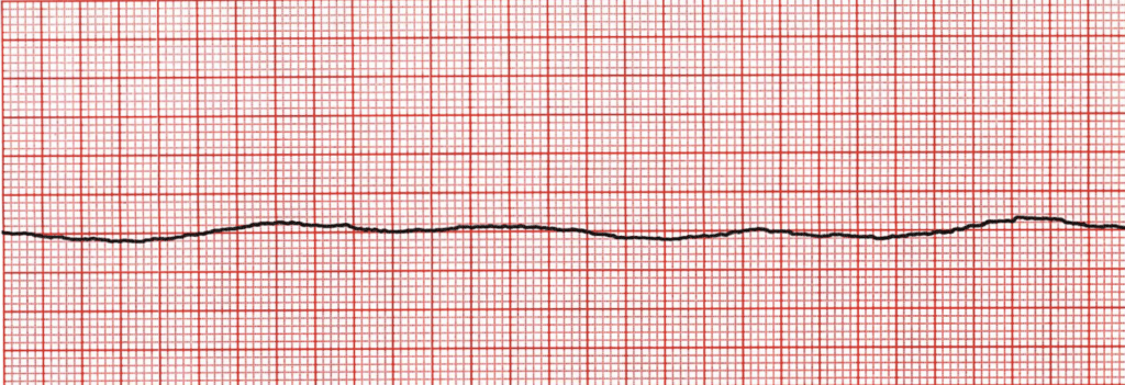
what is this?
asystole

what is this (its pulseless)
PEA
1st degree AV heart block
occurs when all the atrial impulses are conducted through the AV node into the ventricles at a slower rate than normal