shoulder girdle
1/76
There's no tags or description
Looks like no tags are added yet.
Name | Mastery | Learn | Test | Matching | Spaced | Call with Kai |
|---|
No analytics yet
Send a link to your students to track their progress
77 Terms
The bones that connect the upper limb to the trunk are collectively termed the:
shoulder girdle.
The acromial extremity of the clavicle articulates with the:
acromion process of the scapula.
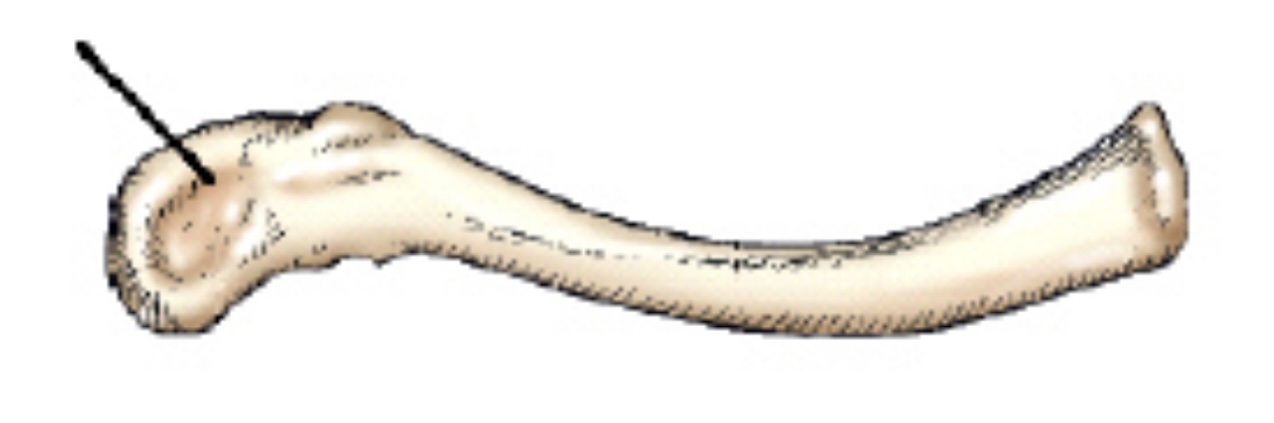
The part identified on the clavicle in this figure is the:
acromial extremity.
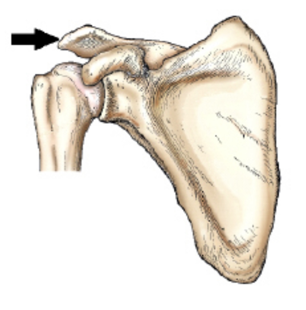
The part identified by the arrow in this figure is the:
Acromion
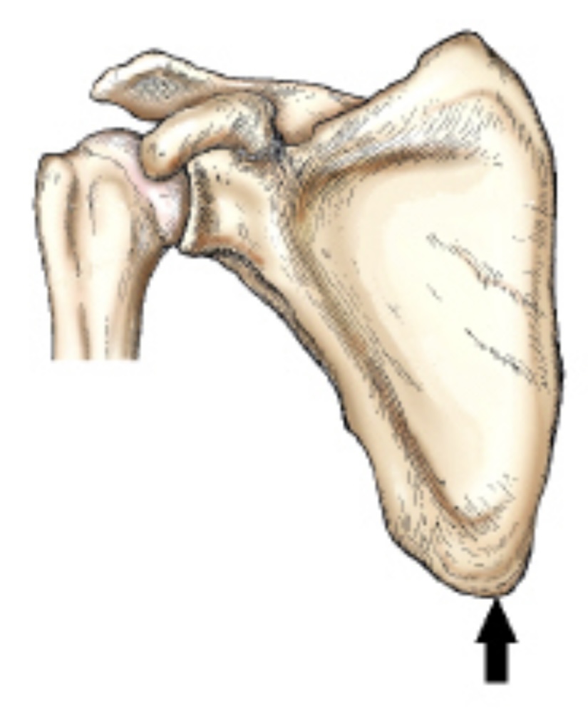
The part identified by the arrow in this figure is the:
inferior angle.
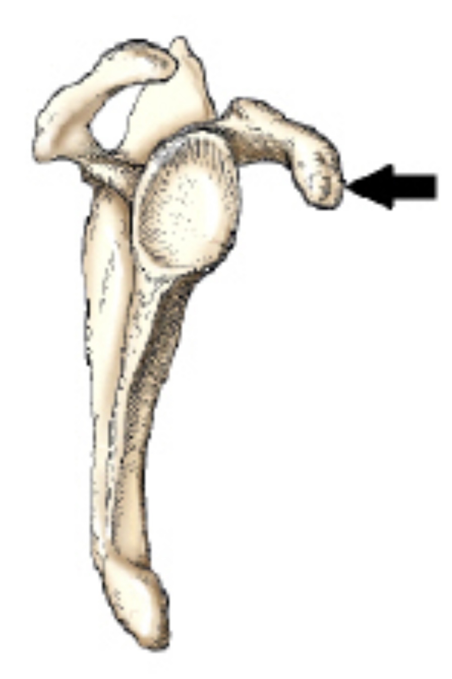
The bone part identified by the arrow in this figure is the:
coracoid process.
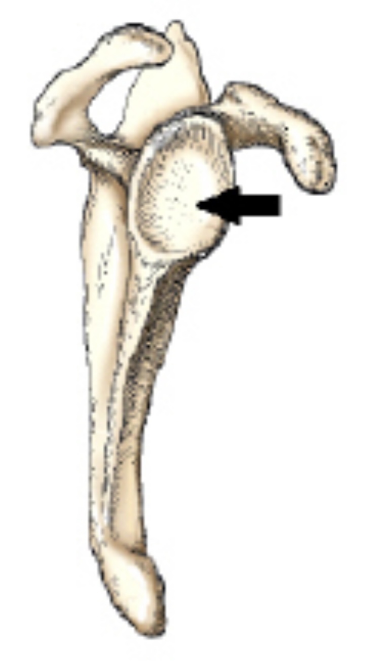
The bony area identified by the arrow in this figure is the:
Glenoid cavity
The area of the proximal humerus located directly below the tubercles, which is the site of many fractures, is called the:
Surgical neck
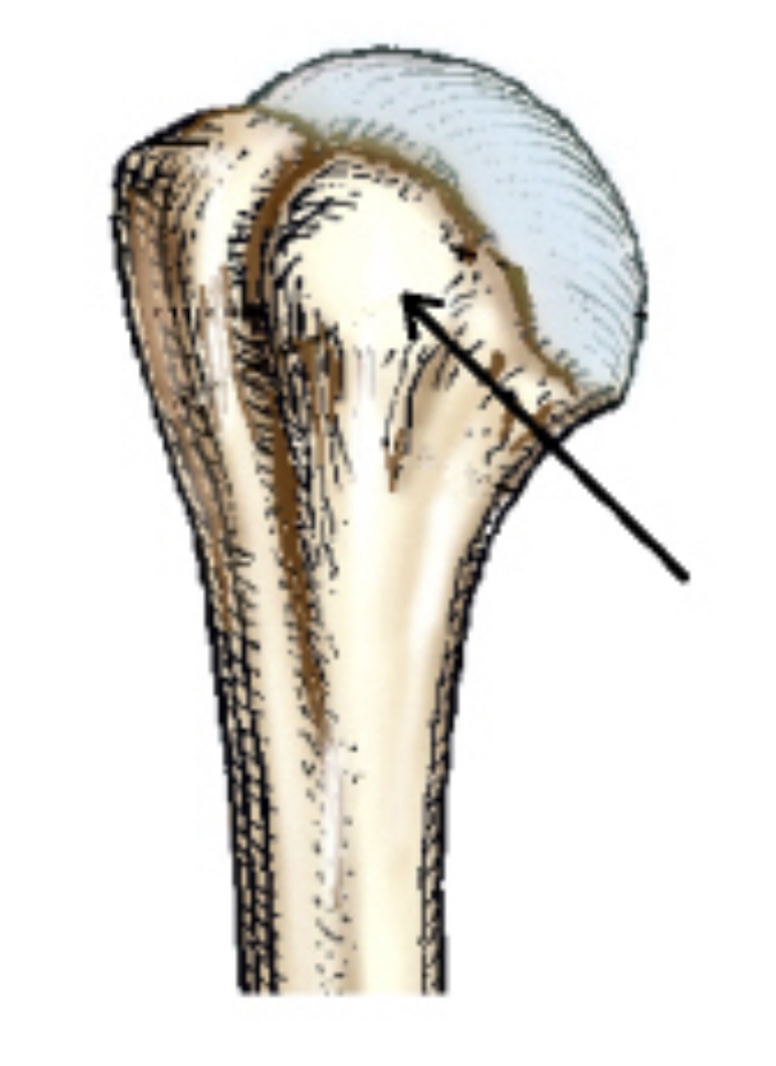
The small, rounded, elevated process identified by the arrow in this figure is the:
lesser tubercle.

The depression identified in this figure is called the:
intertubercular groove.
The large, rounded, elevated process prominently located on the lateral surface of the proximal humerus is the:
greater tubercle.
The small, synovial fluid-filled sacs, which relieve pressure and reduce friction in joint tissues, are called:
bursae.
What joint is formed by the articulation between the glenoid cavity and head of the humerus?
Scapulohumeral
The scapulohumeral articulation is classified as a
____ joint, _____ type.
synovial; ball and socket
All of the following positions of the humerus are commonly used when performing an AP projection of the shoulder, except:
neutral rotation.
superior rotation.
external rotation.
internal rotation.
superior rotation.
To demonstrate the greater tubercle of the humerus on an AP projection of the shoulder, the epicondyles must be:
parallel with the plane of the IR.
What is the recommended exposure field dimensions for the AP projection of the shoulder?
10 × 12 inches (24 × 30 cm)
The respiration phase for an AP projection of the shoulder should be:
Suspended
For an AP projection of the shoulder, the central ray should be directed:
Perpendicular to the IR
For an AP projection of the shoulder, the central ray should enter:
1 inch (2.5 cm) inferior to the coracoid process.
What is prominently shown in profile on an AP projection of the shoulder with the humerus in external rotation?
Greater tubercle
The greater tubercle will be partially superimposed over the humeral head on which projection and position?
AP, neutral rotation
For an AP projection of the shoulder with the arm in a neutral position, how should the humeral epicondyles be positioned in regards to the IR plane?
45 degrees
If the patient places the palm of the hand against the thigh, the humerus will be in:
neutral position.
For an AP projection of the shoulder with the humerus in internal rotation, how should the humeral epicondyles be positioned in relation to the IR plane?
Perpendicular
What structure is prominently shown in profile on an AP projection of the shoulder, internal rotation position?
Lesser tubercle
If the patient places the back of the hand against the hip, the humerus will be in:
internal rotation.
When the arm cannot be rotated or abducted due to injury, which of these can be used to perform a lateral projection of the shoulder?
Tangential projection, Settegast method
Transthoracic lateral projection, Lawrence method
PA axial projection, Holmblad method
AP projection, Pearson method
Transthoracic lateral projection, Lawrence method
Where should the center of the IR be positioned for a lateral projection of the shoulder?
Surgical neck
If the patient can be positioned properly, the central-ray angle for the transthoracic lateral projection (Lawrence) of the shoulder is:
0 degrees
If the patient cannot elevate the unaffected shoulder for a transthoracic lateral projection (Lawrence) of the shoulder, what central ray orientation is needed?
10 to 15 degrees cephalad
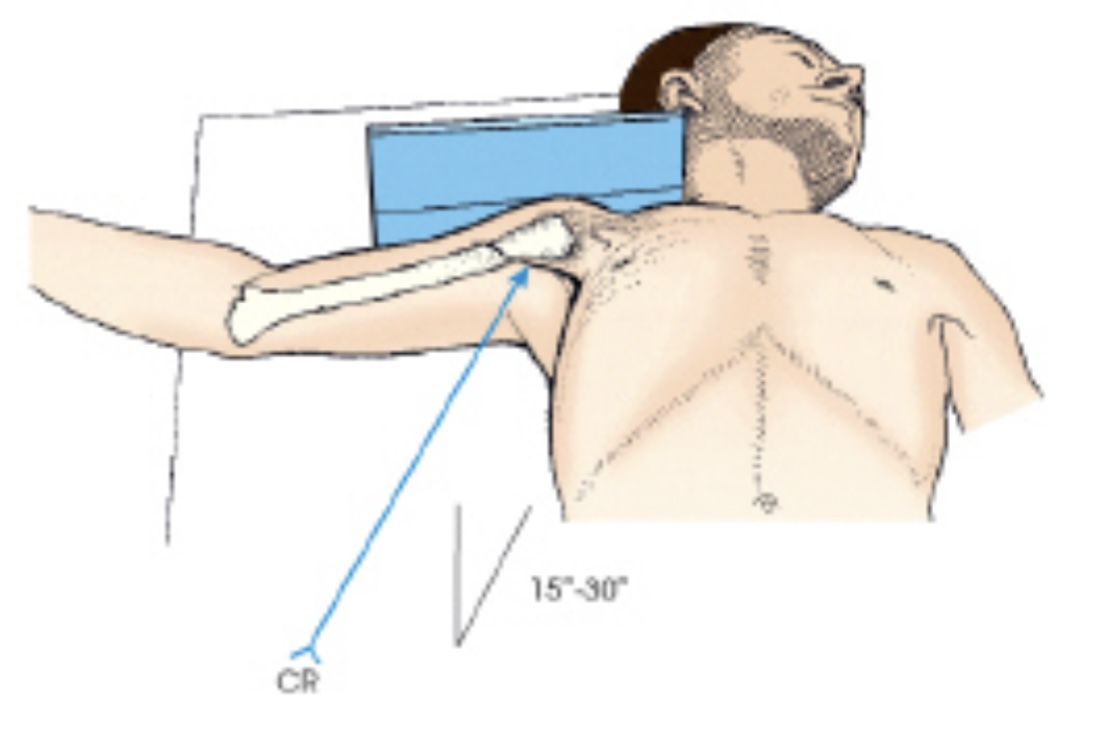
The projection of the shoulder demonstrated in this figure is the:
inferosuperior axial (Lawrence).
How far should the arm be abducted for an inferosuperior projection (Lawrence) of the shoulder joint?
90 degrees
How is the central ray directed for an inferosuperior axial projection (Lawrence) of the shoulder joint?
15 to 30 degrees horizontally
Which of the following are clearly demonstrated on the inferosuperior axial projection (Lawrence) of the shoulder joint?
Proximal humerus
Scapulohumeral joint
Acromioclavicular articulation
1,2, and 3
The PA oblique projection of the shoulder joint (scapular Y) is performed in what positions?
RAO or LAO
PA oblique projection of the shoulder (scapular Y) is performed to evaluate:
Dislocations
For the PA oblique projection (scapular Y) of the shoulder, the body is rotated so that the midcoronal plane is how many degrees from the IR?
45 to 60
What is the central ray angle for the PA oblique projection (scapular Y) of the shoulder joint?
0 degrees
What anatomy is superimposed over the junction of the Y on the PA oblique (scapular Y) projection on a patient with a normal shoulder joint?
humeral head
Which structures is projected in lateral profile on a PA oblique (scapular Y) projection?
Scapula
Which projection clearly demonstrates the glenoid cavity?
AP oblique (Grashey)
How many degrees is the body rotated for the AP oblique projection (Grashey method) of the shoulder joint?
35 to 45 degrees toward the affected side
The Pearson method is an AP projection of the:
acromioclavicular articulation.
How many pounds of weight should be affixed to each wrist for the AP projection of the AC joint?
5 to 8
Two exposures are commonly made of the AC joints-one without weights and one with weights. How are the weights applied?
Affixed to the wrists
How should the central ray be angled for the AP projection (Pearson method) of the AC joints?
0 degrees
The AP projection of the AC joints places the joints at an increased OID. What is the recommended SID to compensate for this distance?
72 inches
In an image of an AP axial projection of the clavicle, the clavicle should be demonstrated with:
most of the clavicle projected above the ribs.
only the lateral end superimposing the coracoid process.
only the medial end superimposing the first or second ribs.
1 and 3 only
All of these projections can be used to demonstrate the clavicle, except.
AP
AP axial
PA
Lateral
Lateral
The central-ray angle for an AP axial projection of the clavicle when performed on a patient in the supine position is _ degrees.
15 to 30
To elevate the clavicle above the ribs and scapula for the AP axial projection, the phase of respiration should be:
full inspiration.
For an AP projection of the scapula, the recommended exposure field is:
10 × 12 inches (24 × 30 cm) lengthwise.
How is the upper extremity positioned for an AP scapula?
Abducted 90 degrees, with elbow flexed
For the lateral projection of the scapula, the body is placed in which position?
45- to 60-degree anterior oblique
For delineation of the acromion and coracoid processes of the scapula in the lateral projection, how is the arm positioned?
Flex the elbow and place the hand on the posterior thorax.
When the patient is positioned properly for a lateral projection of the scapula, the body of the scapula will be ___ the plane of the IR.
perpendicular to
The central-ray angulation for a lateral scapula is:
0 degrees
The clavicle is classified as a(n) ___ bone.
Long
The scapula is classified as a(n) __ bone.
Flat
The rounded head of the humerus fits into an oval depression on the lateral aspect of the scapula called the:
glenoid cavity.
Which position of the hand will place the humerus in external rotation?
supination.
Which position of the hand will place the humerus in neutral position?
Palm against the thigh
Which position of the hand will place the humerus in internal rotation?
Back of the hand against the thigh
For delineation of the body of the scapula for the lateral projection, how is the arm positioned?
Extend the arm upward and rest the opposite shoulder

The projection in the image below was obtained with the arm positioned in:
external rotation.

Letter A in the image below labels the:
clavicle.

Letter B in the image below labels the:
acromion.
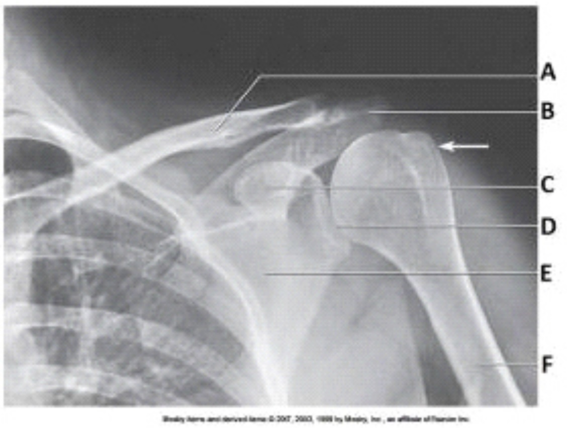
Letter C in the image below labels the:
coracoid process.
The white arrow in the image below points to the:
greater tubercle.

The glenoid of the scapula in the image below is labeled as letter:
D.
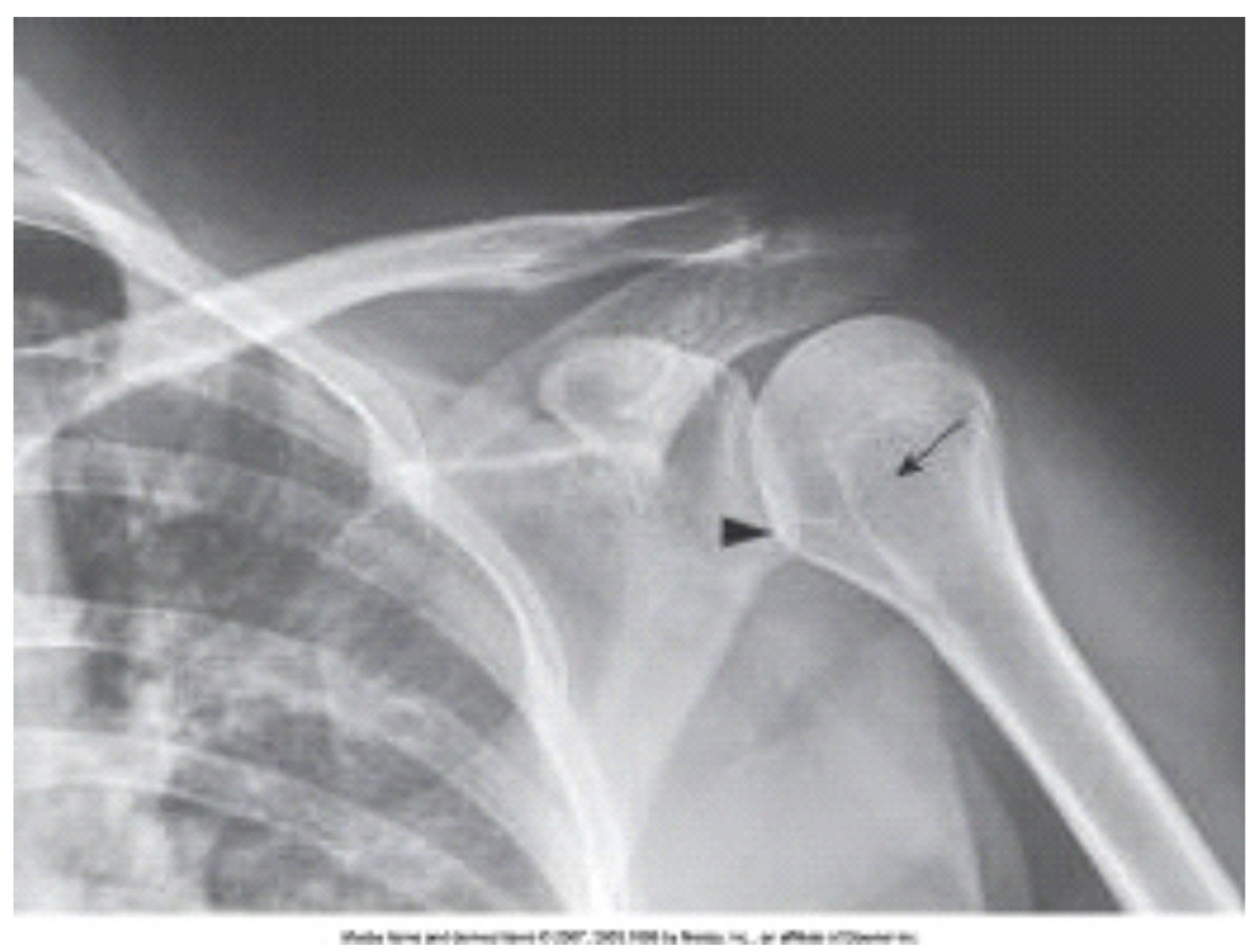
What projection, position, and anatomy of interest in the image below is demonstrated?
AP projection of the shoulder in internal rotation

The anatomy labeled in the image below by the arrowhead is the:
lesser tubercle.

What projection (method) is demonstrated in the image below?
Inferosuperior axial (Lawrence)

What anatomy is labeled as letter A in the image below?
Lesser tubercle
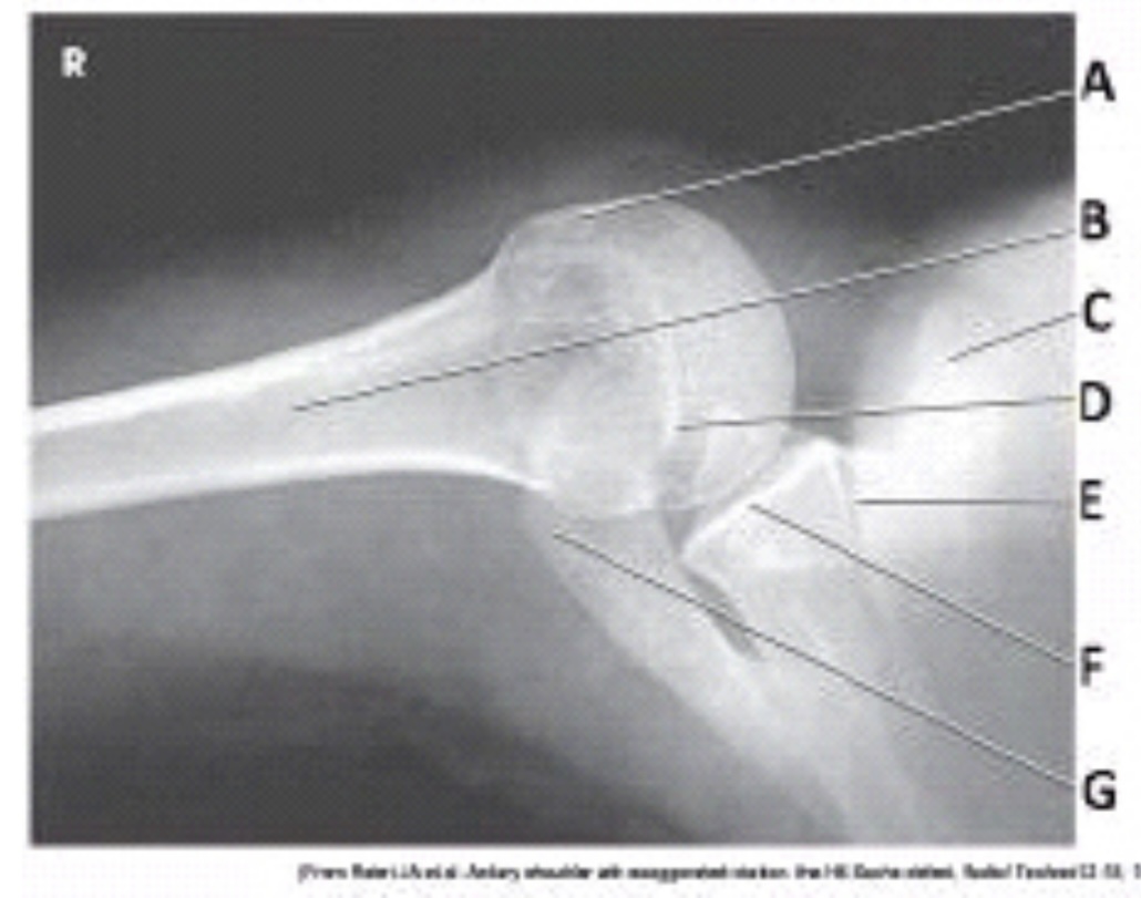
What anatomy is labeled as letter C in the image below?
Coracoid process

What anatomy is labeled as letter D in the image below?
Acromion