Small Animal Anatomy Exam 2
 Call Kai
 Learn
Learn Practice Test
 Spaced Repetition
 Match
Match Flashcards
 Knowt Play
1/360

There's no tags or description
Looks like no tags are added yet.
Name | Mastery | Learn | Test | Matching | Spaced | Call with Kai | Chat |
|---|
No analytics yet
Send a link to your students to track their progress
361 Terms
Suprahamate process of the scapula (cat)
A normal, palpable caudally-projecting process near the distal third of the scapular spine.

Humerus foramina difference (cat vs. dog)
Cats lack a supratrochlear foramen but possess a distinct, slit-like supracondylar foramen on the medial side of the humerus.

Structures passing through the supracondylar foramen (cat)
Brachial artery and median nerve.
Typical number of mammary glands in the queen
4 pairs (8 total), though asymmetric variations can occur.
Insertion of the superficial pectoral muscle (cat)
Distal humerus and proximal ulna (more distal than in dogs).
Insertion of the brachiocephalicus muscle (cat)
Proximal ulna (inserts further distally than in the dog).
Characteristics of the feline clavicle
Predictably ossifies into a small, palpable bone spicule within the brachiocephalicus muscle.
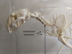
cat clavicle
It is radiographically visible and should not be mistaken for a foreign body.

Brachioradialis muscle clinical relevance (cat)
Well-developed and lateral to the cephalic vein; can be mistaken for the vein during venipuncture.
Dorsal ligaments function in cats
Asymmetrical, well-developed ligaments that passively keep distal interphalangeal joints hyperextended to sheath claws.
Muscle required to expose claws in cats
Active contraction of the deep digital flexor muscle.
Sartorius muscle structure (cat vs. dog)
Undivided in cats, whereas in dogs it is divided into cranial and caudal parts.
Sacrotuberous ligament
cat does not have
Soleus m.
Cats posses this caudal crus muscle, unlike dogs who do not posses this muscle
Nuchal ligament
Cat does not posses
Position of Heart
In all domestic species (besides cat) heart rests between 3rd and 6th intercostal spaces. In the cat, heart resides a bit more caudally, between the 4th and 7th ribs.
What are the 3 classifications of joints?
Fibrous, cartilaginous, synovial
What are fibrous joints made of?
Collagenous connective tissue
Are fibrous joints joints of stability or mobility?
Stability
What are the 3 subtypes of fibrous joints?
Suture, gomphosis, and syndesmosis
What is a suture and an example?
Two pieces of bone filled with collagenous connective tissue
Ex: skull
What is a gomphosis and an example?
Joint between roots of tooth and the bony socket
Ex: tooth
What is a syndesmosis and an example?
side to side fibrous joint
Ex: joint between metacarpal bones
What are cartilaginous joints made of?
Cartilage
Are cartilaginous joints joints of mobility or stability?
Stability
Cartilaginous joints are ______ united by ______
Directly, cartilage
What are the subtypes of cartilaginous joints?
Symphysis and synchondrosis
What is a symphysis and an example?
Hybrid tissue between collagenous connective tissue and hyaline cartilage
Ex: pelvic symphysis
Symphysis is united by _________
fibrocartilage
What is a synchondrosis and an example?
cartilaginous joints made up of hyaline cartilage
Ex: Sacroiliac joint
Synchondrosis is united by _________
Hyaline cartilage
Do synovial joints directly unite?
No, indirectly connected via joint capsule and ligaments
What are the bone ends covered by in synovial joints?
Articular cartilage/hyaline cartilage
What are the components of a synovial membrane?
Joint capsule, fibrous membrane, synovial membrane, synovial fluid
What is unique about cartilage?
It is avascular
What is the function of articular cartilage?
Reduces friction
What two structures of the synovial joint are continuous
Fibrous membrane (around joint) and periosteum (around bone)
What is the function of the periosteum?
Covers the bone like a sleeve
Is the periosteum connective tissue?
Yes
The periosteum allows for _________ connection between the bones
Indirect
What is the joint cavity vs the joint capsule
The synovial joint is completely enclosed by a sleeve like structure called the joint capsule and the space enclosed within it is the joint cavity
What are the 2 layers of a joint capsule
fibrous membrane (outer) and synovial membrane (inner)
What is the fibrous membrane made of?
Collagenous connective tissue
What is the function of the synovial membrane?
Secrete and rebsorbs the synovial fluid
What lines the synovial membrane?
Epithelium (both secreting and absorbing)
What is the function of the synovial fluid?
Lubricates the articular cartilage, nourishes articular cartilage
Is synovial fluid vascular or avascular?
Vascular- nourishes the synovial membrane and hyaline cartilage
What is interesting about the synovial fluid?
It is constantly being turned over, supplying fresh fluid/rich oxygen & nutrients/removing waste
If hyaline cartilage was damaged, what would a joint look like radiographically?
The joints would not have as big of a "gap"
What is the definition of a ligament?
ct band that connects bone to bone, regional thickening of fibrous membrane
The lateral and collateral ligaments are actually just regional thickenings of the _________ __________
Fibrous membrane
What two joints do not have collateral ligaments?
Shoulder and hip joint
If IDing the collateral ligaments on the manus or pes, rather than saying medial or lateral, you would say _____ or ______
Axial or abaxial
What are extracapsular ligaments?
Ligaments outside of the joint cavity
Example of extracapsular ligament
collateral ligaments
Which of the carpal bone complex joints have the most movement
Antebrachiocarpal joint
Which of the carpal bone complex joints communicate?
Middle carpal joint and carpometacarpal joint
UMN or LMN is responsible for contracting the muscles?
LMN
What are the accessory synovial structures
Bursa, tendon sheaths, and sesamoid bones
What is a bursa
Synovial fluid filled sac between a tendon and a bony prominence
What is the function of a bursa
Reduces friction
What does the wall of a bursa look like?
A synovial joint capsule- has a fibrous membrane and synovial membrane!
What is a tendon sheath?
An elongated bursa that wraps around an entire tendon
Where can you find tendon sheaths?
Bony grooves, beneath retinacula, annular ligaments
What are the two major functions of sesamoid bones
Protects tendon by decreasing friction, helps redirect force of a tendon
Where can you find sesamoid bones?
Embedded in muscle tendons
Are sesamoid bones synovial joints?
Yes! They form a synovial joint between the tendon and bony prominence
What muscle is the proximal sesamoid bone embedded in?
Interosseus m
Quadriceps femoris origin
ilium - rectus femoris; Proximal femur -other heads
Quadriceps femoris insertion
Tibial tuberosity
Quadriceps femoris action
Flex hip joint (only rectus femoris) and extend stifle joint
Iliopsoas origin
ilium - iliacus; lumbar vertebrae - psoas major
Iliopsoas Insertion
Lesser trochanter of femur
Iliopsoas action
Flex coxal joint (principal action) Rotate limb laterally
Craniolateral muscles of Crus
crainial tibial, long digitial extensor, lateral digital extensor, peroneus [fibularis] longus. Principal action - flex tarsal joints and extend digital joints
Cranial tibial origin
lateral aspect of proximal tibia
Cranial tibial insertion
plantaroproximal metatarsal bones 1 and 2
cranial tibial action
flex tarsal joints
long digital extensor origin
extensor fossa of femur
long digital extensor insertion
extensor processes of the distal phalanges
long digital extensor action
extend digital joints, flex tarsal joints. Minimally extend stifle joint
Lateral digital extensor of pelvic limb origin
proximal fibula
insertion of lateral digital extensor m.
extensor process of distal phalanges digits 3-5
Lateral digital extensor action
extend lateral digital joints and flex the tarsal joints
Fibularis longus origin
lateral condyle of tibia and proximal fibula
Fibularis longus insertion
Plantaroproximal metatarsal bones
Fibularis longus action
flex tarsal joint
Caudal muscles of crus
Gastrocnemius, soleus (only in cats), superficial digital flexor, deep digital flexor, popliteus. Principal action is to extend tarsal joints and flex digital joints
gastrocnemius origin
Caudodistal femur
Gastrocnemius insertion
tuber calcanei
Gastrocnemius action
extend tarsal joints. Flex stifle joint minimally
Soleus origin
head of fibula
Soleus insertion
tendon of lateral head of gastrocnemius
soleus action
assist the gastrocnemius in extension of tarsal joints
superficial digital flexor m. of pelvic limb origin
lateral supracondylar tuberosity of femur
Superficial digital flexor m. of pelvic limb insertion
tuber calcanei and plantaroproximal aspects of middle phalanges 2-5
superficial digital flexor action
extend tarsal joints and flex digital joints, except distal interphalangeal joints. Minimally flex genual joint
deep digital flexor origin
caudal aspects of tibia and fibula
deep digital flexor insertion
distal phalanges, plantar aspect
deep digital flexor action
flex digital joints and extend tarsal joints