Osteology of the Skull- Development of the Skull Sex Differences
 Call Kai
 Learn
Learn Practice Test
 Spaced Repetition
 Match
Match Flashcards
 Knowt Play
1/50

There's no tags or description
Looks like no tags are added yet.
Name | Mastery | Learn | Test | Matching | Spaced | Call with Kai | Chat |
|---|
No analytics yet
Send a link to your students to track their progress
51 Terms
Bone also serves as a reservoir of __________ and _________
calcium, phosphate
Hydroxyapatite is
calcium phosphate
Bone tissue is also called
osseous tissue
the ECM (extracellular matrix) is a _________ and _______ ________
mineral (hydroxyapatite), collagen fibers
bone is one type of connective tissue: _____ and ____________ _______
cells, extracellular matrix
intramembranous ossification
direct ossification of the mesenchyme during the fetal period
what bones come from intramembranous ossification?
flat bones of the skull, body of the mandible, clavicle
endochondral ossification
cartilage precursor serves as a model for bone formation
what bones come from endochondral ossification?
bones of extremities, vertebrae, cranial base (sphenoid, part of temporal, part of occipital)
intramembranous ossification process (4)
around week 8, some mesenchymal cells migrate to form ossification centers
cells differentiate into embryonic osteoblasts; once encased in bone, osteoblasts become osteocytes
bone grown in linear extensions (spicules), angiogenesis occurs
fusion of spicules, appositional growth
spicules are also called
linear extensions
endochonral ossification process part 1
mesenchymal cells differentiate into chondroblasts which produce cartilage matrix creating hyaline cartilage model of the shape of future bone
bone cuff develops in mid region of cartilage; what was perichondrium converts into periosteum producing osteoblasts
chondrocytes in the center of cartilage enlarge and cartilage matrix calcifies
chondrocytes die, producing cavity
endochondral ossification process part 2
blood vessels grow around cartilage and bony collar, some penetrate through to cavity, taking osteprogenitor cells into the matrix forming primary ossifcation center
bone formation spreads along shaft toward both ends, remodeling continues creating marrow cavity, growth occurs by interstitial and appositional growth
interstitial growth is growth of the _______
length
appositional growth is growth of the ______
diameter
endochondral ossification processs part 3
secondary ossification centers develop in the epiphyses and eventually, only cartilage remaining in articular cartilage on the surface and a thin plate (epiphyseal plate) between the bone of epiphyses and diaphysis
flat bones of the skull are _____ in fetuses
paired
3 main cell types in bone
osteoblasts (building bones, deposit calcium and phosphate to make mineral component)
osteoclasts (dissolve bone, secrete acid)
osteocytes (maintaining cells)
2 types of bone
compact and spongy (cancellous)
fontanelles
flexible fibrous joints
the delayed fusion of sutures allows for
postnatal brain growth
sutures of the skull (6)
internasal, frontal (metopic), sagittal, coronal, squamosal, lambdoidal
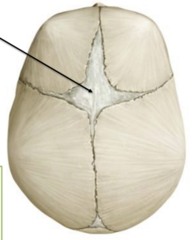
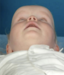
anterior fontanelle
largest, diamond shaped
junction of sagittal, frontal coronal sutures
closure is at around 18 months
future sight of bregma
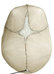
posterior fontanelle
triangular, junction of sagittal and lambdoidal sutures
closure begins at 3 months
future sight of lambda
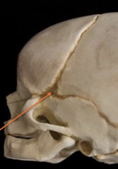
sphenoidal fontanelle
closes at around 6 months
becomes pterion
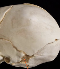
mastoid fontanelle
closure at around 12 months
becomes asterion
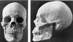
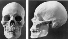
general shape of male skull
large, blocky
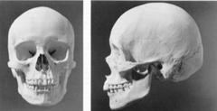
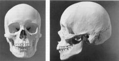
general shape of female skull
smaller, tapered
supraorbital margin of male skull
pronounced, thicker
supraorbital margin of female skull
flatter
zygomatic arch of male skull
pronounced, sharper
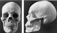
mandible of male skull
square, acute angle
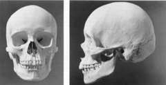
mandible of female skull
rounded
AP diameter of male skull
elongated
frontal bone on male skull
greater slope
frontal bone of female skull
more vertical
surface of male skull
rough and bumpy
surface of female skull
smooth
protuberance on male skull
pronounced
in an x-ray, a child's bone is not going to show up very well because
there is more cartilage present than bone, which does not show up on an x-ray well
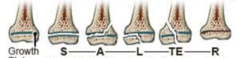
Salter-Harris Fractures
S-traight across
A-bove
L-lower
TE-Through Everything
c-R-ush
in fetuses, the flat bones are paired with fontanelles to allow for _________ during childbirth
distortion
when does the coronal suture close?
30-40 years
When does the sagittal suture close?
around 6 months
when does the frontal suture close?
around 6 months
when does the internasal suture close?
around 20-30 years
when does the squamosal suture close?
around 30-40 years
when does the lambdoidal suture close?
around 30-40 years
which 3 sutures take 30-40 years to close?
coronal, squamosal, lambdoidal
craniosyntosis
premature fusing of the skull bones
scaphocephaly, plagiocephaly, oxycephaly
the anterior fontanelle is in the middle of the _________ and _________ bones
frontal, parietal