Light Microscope
1/10
There's no tags or description
Looks like no tags are added yet.
Name | Mastery | Learn | Test | Matching | Spaced |
|---|
No study sessions yet.
11 Terms
Dark Field Microscope
What
What part is different
Why
How does it create an image
Maximum magnification
Resolution
Advantages
Ideal for
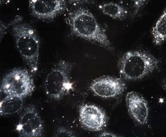
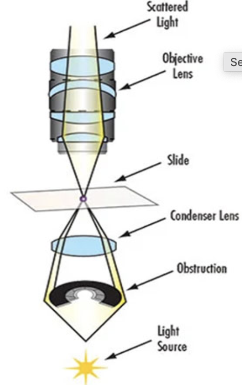
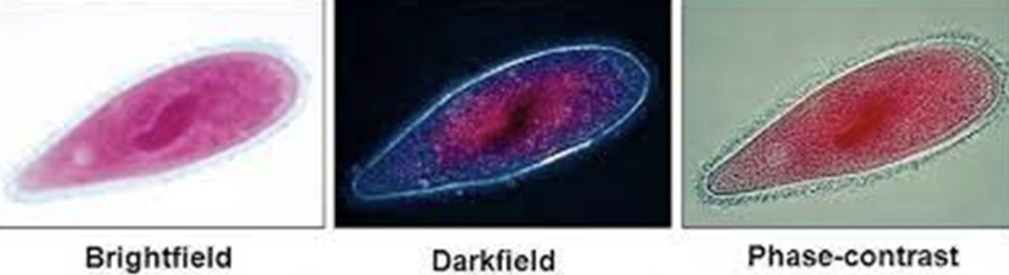
What: Specimen is brightly illuminated against dark background
Part is different: Condensor
Why: Prevents the parallel and oblique rays from entering which makes the microscopic field dark
Creates an image by: In presence of specimen has a different refractive index
Resulting in the oblique rays to scatter by reflection and refraction and enter the objective making the specimen brightly illuminated
Maximum magnification: 1500x
Resolution: 0.1 - 0.2µ
Advantages: Makes it easy to obtain the correct focal plane at low magnification
Ideal for: Small, low contrast specimens
What are the 5 uses of dark field microscope
Morphology and motility of microorganisms
Initial examination of cell suspensions
Initial survey and observation at low powers of pond water samples
Examination of lightly prepared slides
Determination of motility in cultures
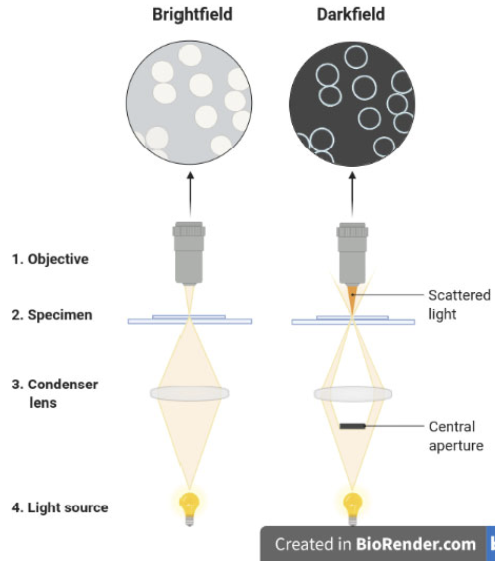
Brightfield vs Darkfield Microscopy
Darkfield has scattered light and central aperture

Dissecting / Inverted Microscope
Designed for
Uses what kind of light rays
Magnification power
Produces what kind of image
How
How many light sources
Designed for: Low magnification observation of a specimen for dissection and parasitology
Uses: Reflected light rays from specimen surface
Instead of transmitted light rays
Magnification power: Ranges from 5-80x
Produces: 3D image
How: Because it contains 2 objective lens and eyepiece
This creates 2 separate optical paths for each eye
How many light sources: 2
1 from upper portion of specimen
Reflected in eyepiece
Other from below portion of sample
For illumination through thinner samples
What is the difference between reflected light rays and transmitted light rays
Sample type
Light path
Application
Illumination method
Transmitted Light Rays | Reflected Light Rays | |
Sample Type | Opaque | Transparent or semi-transparent |
Light Path | Reflects off the surface | Passes through the sample |
Application | Surface features | Internal structures |
Illumination Method | Episcopic (reflected) illumination | Diascopic (transmitted) illumination |
Phase Contrast Microscope
What
How
Colour of background
Advantages
Disadvantages
What: Used to observe transparent and unstained specimens
How: By using the fact that structures have different refractive indexes causing them to bend light differently creating a contrast
A phase plate slows down the highly refracted light rays putting them “out of phase” creating a contrast between the cell and background
Color of background: Dark
Advantages:
Alive specimens can be used
Can observe motility and stimuli responses
No staining needed
Disadvantages:
More expensive than bright field
Must be properly aligned
UV Microscope
What length of wavelengths lead to better resolution
UV rays cannot pass through glass, so what lenses are used
UV are invisible so how is an image displayed
What other type of microscopes uses these principles
Better resolution: Shorter wavelengths
Lenses used: Quartz lenses
Image displayed by: Photographic plates or special filters
Other microscopes: Florescence
What is the phenomenon fluorescence?
When certain chemicals absorb light and reemit part of the radiant energy as light of longer wavelength
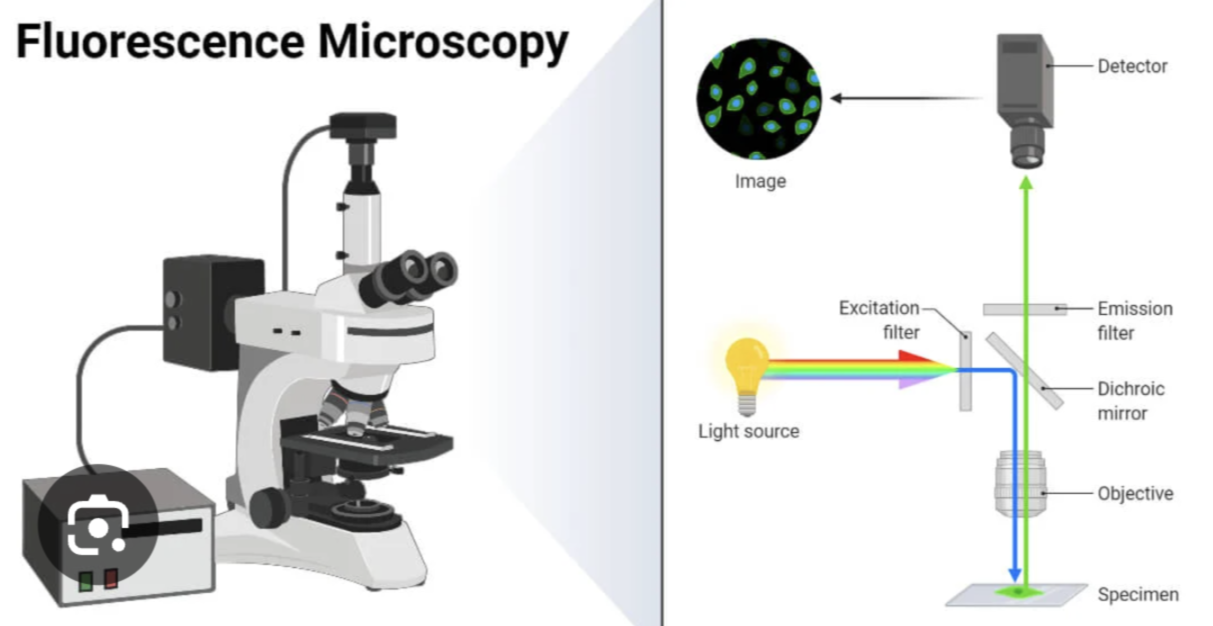
How does fluorescence microscope work?
Light source
Excitation filter
Directing light to specimen
Specimen staining
Emission of fluorescent light
Barrier filter
Observation
UV light option
1.Light source: High intensity mercury lamp which emits white light (containing all colors of spectrum)
2.Excitation filter: White light passes through the exciter filter, removing all colors except blue light
3.Directing light to specimen: Blue light is reflected downward onto specimen by dichroic mirror
4.Specimen staining: Stained with flourscent dye
Parts of specimen retain dye causing it to
Absorb blue light
Emit green light
5.Emission of fluorescent light: Stained areas that emit green light travels back through microscope
6. Barrier filter: Emitted light passes through barrier filter which blocks unwanted blue wavelengths, only allowing the reemitted green light to reach observer
7. Observation: Sees the green fluorescence against a dark background.
Unstained parts = remain invisible, providing high contrast.
8. UV light option: Sometimes UV light is used instead of visible light to excite fluorescent molecules
When is fluorescence microscope used?
In immunology to observe reactions of antigens and antibodies
Confocal Laser Scanning Microscope
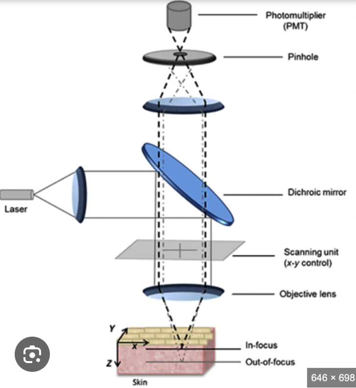
What is the light source and illumination
Works in what mode
Meaning
Spreading laser beam:
Reflection by dichroic mirror:
Fluorescence and pinhole aperture:
Function of pinhole aperture:
Light source and illumination: Laser
Mode: Epi-illumination
Meaning: Laser comes from above and shines directly onto sample
Spreading laser beam: Laser beam is widened using special lens to fill back part of the objective lens
Allows light to focus as a small point onto sample
Reflection by dichroic mirror: A dichroic mirror is a mirror that reflects certain wavelengths but lets other pass through
It directs the laser light to the sample while allowing fluorescent light to emit by the sample to pass through detector
Fluorescence and pinhole aperture:
Sample absorbs laser light and re-emits it as fluorescence
This passes back through objective lens
Goes to detector through small pinhole aperture
Function of pinhole aperture: Ensures the only light from focused spot on sample is collected, eliminating out of focus blur (photo bleaching) that is normally generated by fluoresce microscope
