Hematological System: Structure, Function, and Alterations
1/72
There's no tags or description
Looks like no tags are added yet.
Name | Mastery | Learn | Test | Matching | Spaced | Call with Kai |
|---|
No analytics yet
Send a link to your students to track their progress
73 Terms
Three primary functions of blood
Distribution, regulation, and protection.
Average blood volume in adults
4 to 5 liters in women; 5 to 6 liters in men.
Lifespan of a red blood cell (RBC)
Approximately 120 days.
Lifespan of platelets
Approximately 9 to 10 days.
Difference between plasma and serum
Plasma contains clotting factors; serum does not.
Hematocrit (HCT)
The percentage of total blood volume occupied by erythrocytes.
Normal hematocrit values
Male: 40.7% to 50.3%; Female: 36.1% to 44.3%.
Normal pH range of blood
7.35 to 7.45.
Most abundant plasma protein
Albumin, which contributes most to colloid osmotic pressure.
Transferrin
The plasma protein that transports iron through the circulation.
Ferritin
The primary intracellular storage protein for iron.
Anaerobic ATP generation in RBCs
Prevents erythrocytes from consuming the oxygen they transport.
Hemoglobin (Hb) structure
Four globin chains (two alpha, two beta), each bound to a heme group.
Heme composition
An iron atom (ferrous Fe²⁺) and a porphyrin ring.
Carbonic anhydrase
Enzyme in RBCs that converts carbon dioxide into bicarbonate ions.
Primary site of hematopoiesis in adults
Red bone marrow of the axial skeleton and girdles.
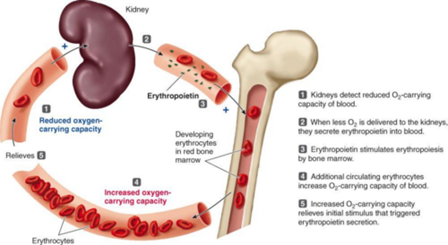
Erythropoietin (EPO)
Hormone released by the kidneys during hypoxia to stimulate RBC production.
Bilirubin conjugation
Occurs in the liver with glucuronic acid to make it water-soluble.
Diapedesis
The movement of white blood cells out of capillaries into tissue spaces.
Megakaryocytes
Giant bone marrow cells whose fragments form platelets.
Platelet inhibition factors
Nitric oxide (NO) and prostacyclin (PGI₂).
Three phases of hemostasis
Vascular spasms, platelet plug formation, and coagulation.
Vitamin K-dependent clotting factors
Factors II, VII, IX, and X.
Fibrinolysis
The breakdown of a clot by plasmin digesting fibrin strands.
Erythroblastosis fetalis cause
Rh incompatibility where maternal antibodies attack Rh-positive fetal red blood cells.
Fetal hemoglobin (HbF) oxygen affinity
Significantly higher than adult hemoglobin (HbA).
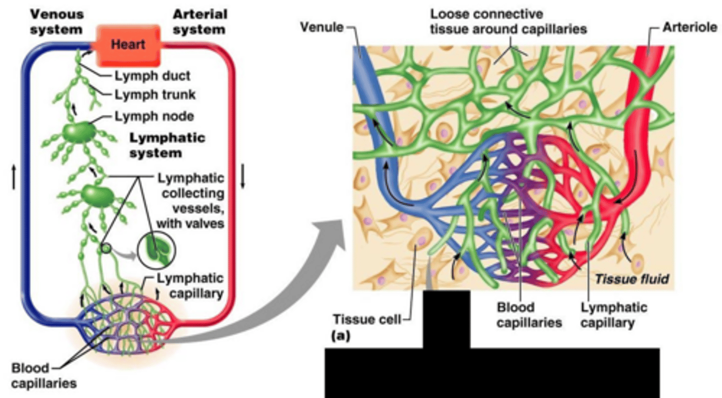
Lymphatic system function
Returns interstitial fluid and leaked plasma proteins to the blood.
Mean Corpuscular Volume (MCV)
Measures the average size of red blood cells.
Prothrombin Time (PT)
Measures the extrinsic pathway of coagulation.
Activated Partial Thromboplastin Time (aPTT)
Measures the intrinsic and common pathways of coagulation.
Normocytic-normochromic anemia causes
Acute blood loss, aplastic anemia, or chronic kidney disease.
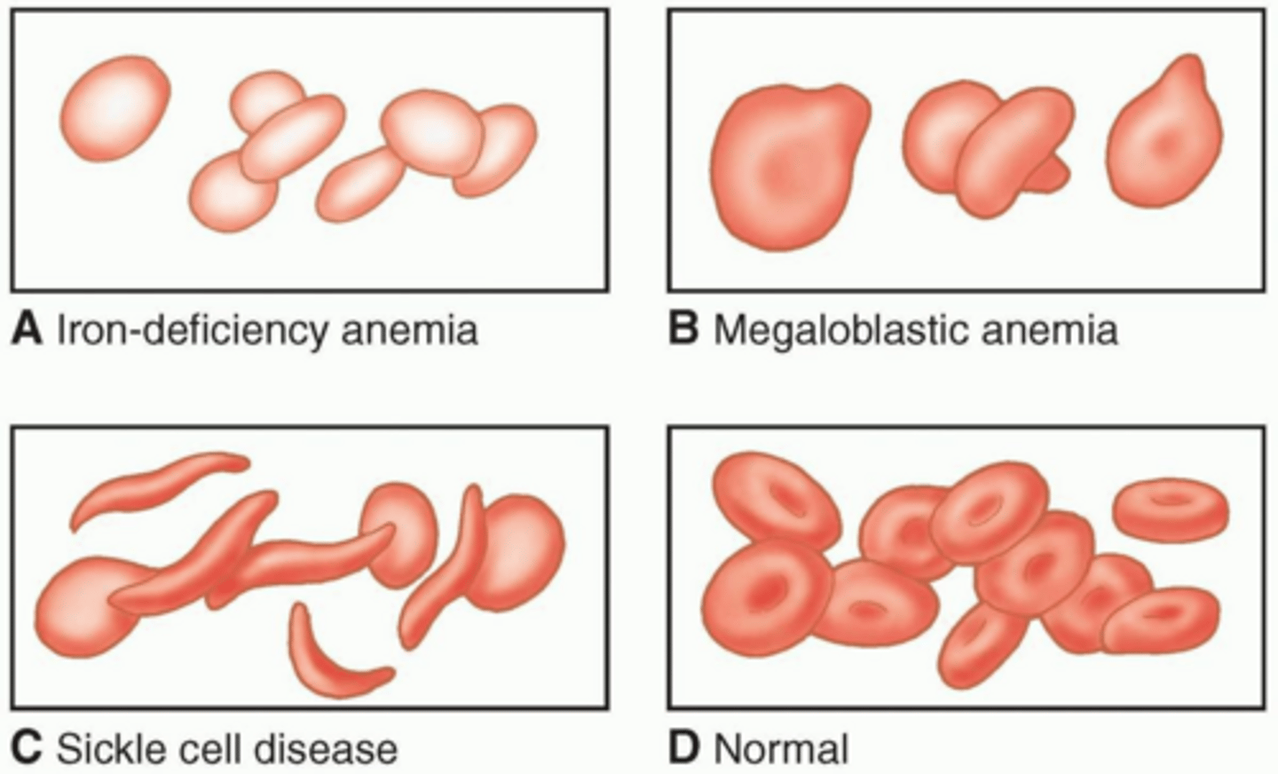
Megaloblastic (macrocytic) anemia cause
Vitamin B12 or folic acid deficiency impairing DNA synthesis.
Pernicious anemia
Anemia caused by autoimmune destruction of parietal cells, depleting intrinsic factor.
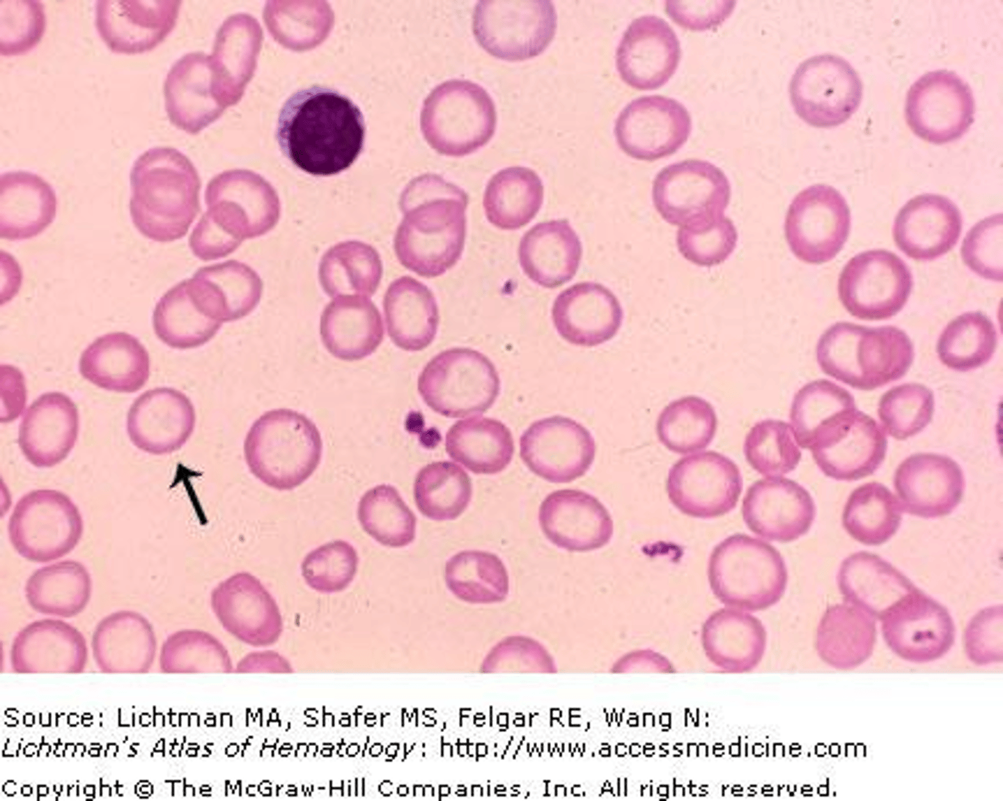
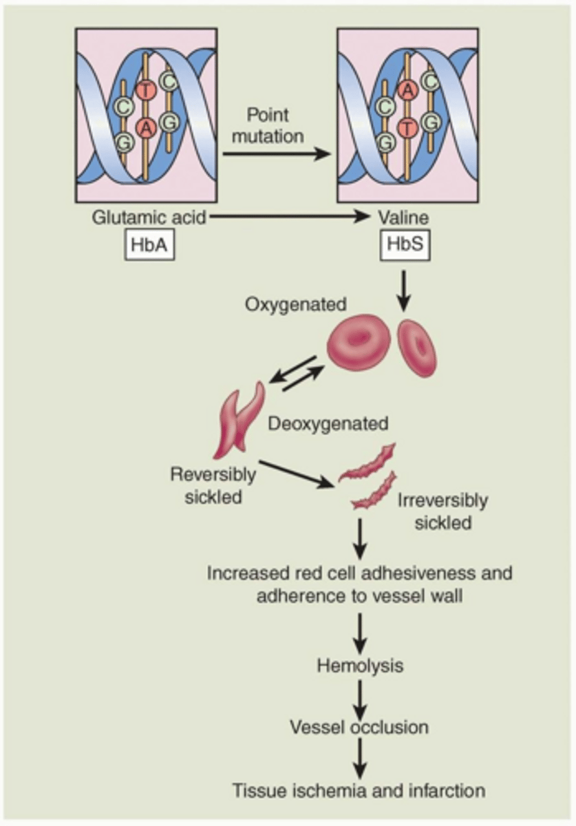
Sickle cell anemia mutation
A single amino acid substitution in the beta-globin chain.
Sickle cell RBC lifespan
Reduced to 20 days or less.
Thalassemia major (Cooley's anemia)
Homozygous genetic defect causing severely reduced alpha or beta chain synthesis.
G6PD deficiency triggers
Infections, diabetic ketoacidosis, and oxidative drugs like aspirin or Bactrim.
Iron deficiency anemia presentation
Microcytic, hypochromic red blood cells with low serum ferritin.
Pancytopenia
Simultaneous decrease in red blood cells, white blood cells, and platelets.
Polycythemia vera
Neoplastic increase in RBCs, WBCs, and platelets, increasing blood viscosity.
Acute vs. chronic leukemia cells
Acute involves poorly differentiated blast cells; chronic involves well-differentiated cells.
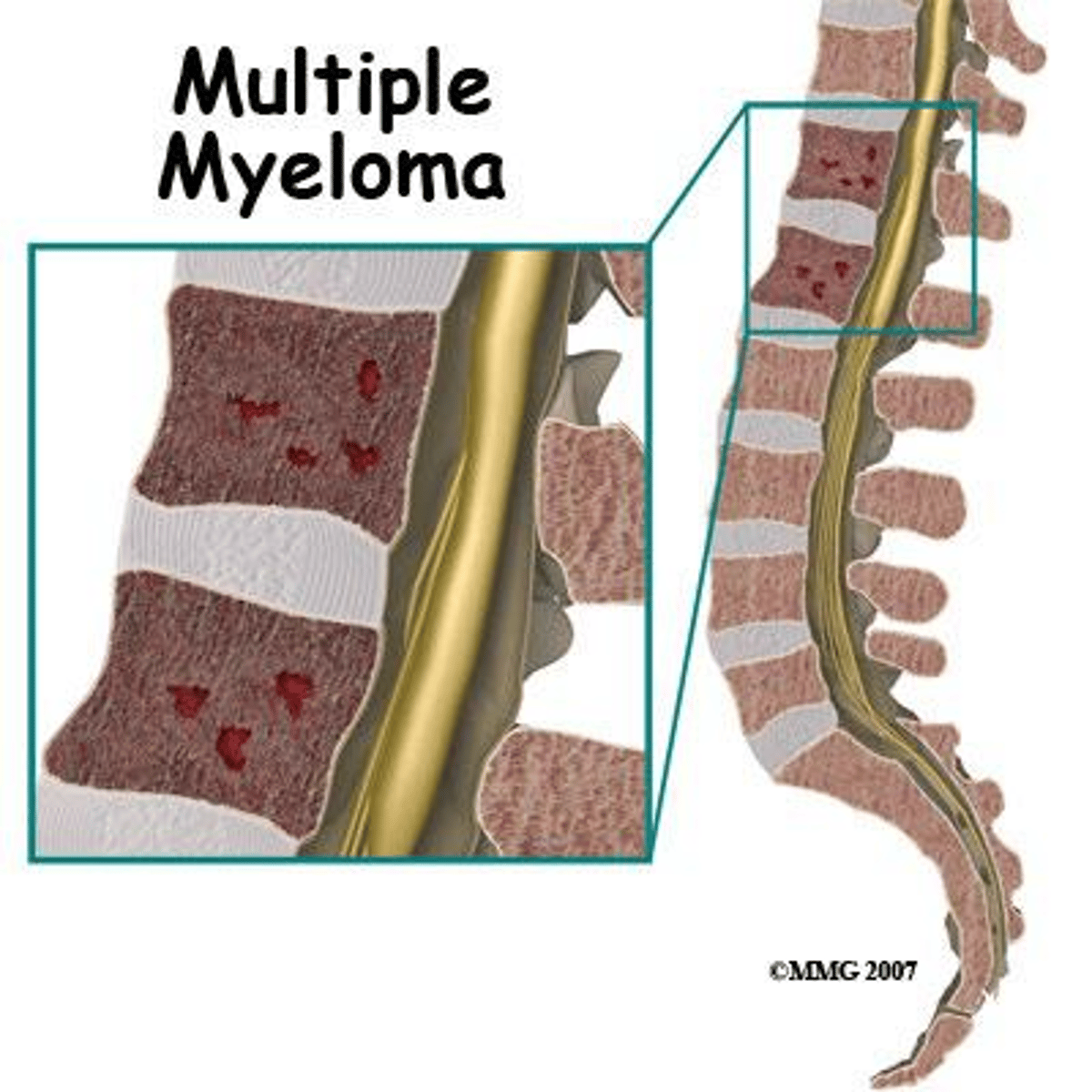
Multiple myeloma hallmarks
Osteolytic bone lesions, elevated calcium, and Bence Jones proteins.
Reed-Sternberg cell
The giant, multinucleated B-cell hallmark of Hodgkin Lymphoma.
Virchow's Triad
Endothelial injury, abnormal blood flow, and hypercoagulability.
Thrombocytopenia threshold
A platelet count below 100,000/µl.
Thrombotic Thrombocytopenic Purpura (TTP) cause
ADAMTS13 enzyme deficiency leading to accumulated von Willebrand factor.
Hemophilia A deficiency
Inherited deficiency of clotting Factor VIII.
Hemophilia B deficiency
Inherited deficiency of clotting Factor IX.
Disseminated Intravascular Coagulation (DIC)
Widespread systemic clotting that consumes coagulation factors, causing severe bleeding.
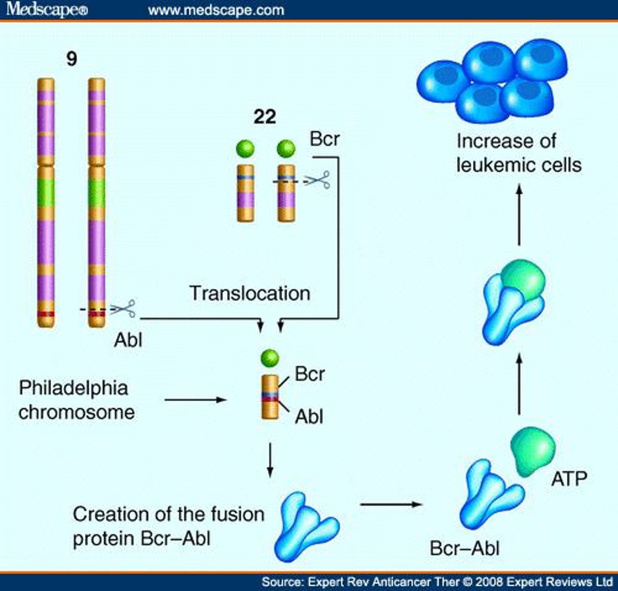
Philadelphia chromosome association
Hallmark of CML, resulting from reciprocal translocation of chromosomes 9 and 22.
Three major aggregation sites of lymph nodes
Inguinal (groin), axillary (armpit), and cervical (neck) regions.
Band cells
Immature neutrophils measured to assess the rate of neutrophil production.
Immune thrombocytopenic purpura (ITP)
Autoimmune condition where the body produces antibodies against its own platelets.
Order of white blood cell abundance
Neutrophils, lymphocytes, monocytes, eosinophils, then basophils.
CD20 surface marker
Target on non-Hodgkin lymphoma cells for monoclonal antibody therapy.
Carbon monoxide lethal mechanism
Competes with oxygen for the ferrous ion in hemoglobin with higher affinity.
Sickle cell disease molecular mutation
Glutamic acid is replaced by valine at position 6 of the beta-globin chain.
Clinical significance of elevated neutrophils
Indicates an active bacterial infection.
Clinical significance of elevated eosinophils
Indicates a parasitic worm infection or allergic response.
Primary substances released by basophil granules
Histamine (a vasodilator) and heparin (an anticoagulant).
Precursor cells of tissue macrophages
Monocytes that have left the bloodstream.
Chloride shift mechanism in RBCs
Chloride ions enter the cell as bicarbonate ions exit.
Location where megakaryocytes release platelets
Bone marrow sinusoids, as blood flow shears off cell fragments.
Structural difference of fetal hemoglobin (HbF) compared to HbA
Contains two gamma chains instead of two beta chains.
Relationship between hematocrit and body fat
Inversely proportional; more adipose tissue leads to lower hematocrit.
Bilirubin transport in blood
Binds to albumin for transport to the liver.
Stimulators of leukopoiesis
Interleukins and colony-stimulating factors (CSFs).
Monocyte nuclear morphology
Kidney-shaped nucleus resembling a McDonald's arch.
Aspirin effect on platelets
Inhibits COX-1, reducing thromboxane A2 synthesis and platelet activation.
Trigger for extrinsic clotting pathway
Tissue factor (Factor 3) released by damaged perivascular tissue.
Natural anticoagulants in blood
Heparin, antithrombin 3, protein C, and protein S.
RhoGAM mechanism of action
Coats fetal RhD antigens, preventing maternal immune recognition and sensitization.
Percentage of population that is Rh-positive
Approximately 85%.