Unit 5- Sonographic Evaluation(GS, YS, Fetal Pole)
1/32
There's no tags or description
Looks like no tags are added yet.
Name | Mastery | Learn | Test | Matching | Spaced | Call with Kai |
|---|
No analytics yet
Send a link to your students to track their progress
33 Terms
When is the typical visualization of embryonic development?
During 5th week
What is sonographic appearance of the embryonic development during the 5th week of embryonic development?
1-2 mm sac with echogenic ring and sonolucent center
Anechoic center is chorionic cavity
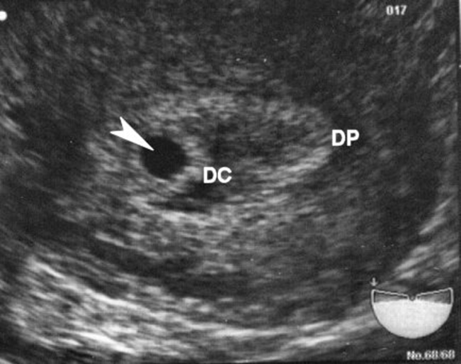
What are these images showing?
Decidual Reaction
What are the different amniotic/chorionic cavities?
Decidua Basalis
Decidua Capsularis
Decidua Parietalis
Decidua Vera
Decidua Capsularis
Decidua Parietalis
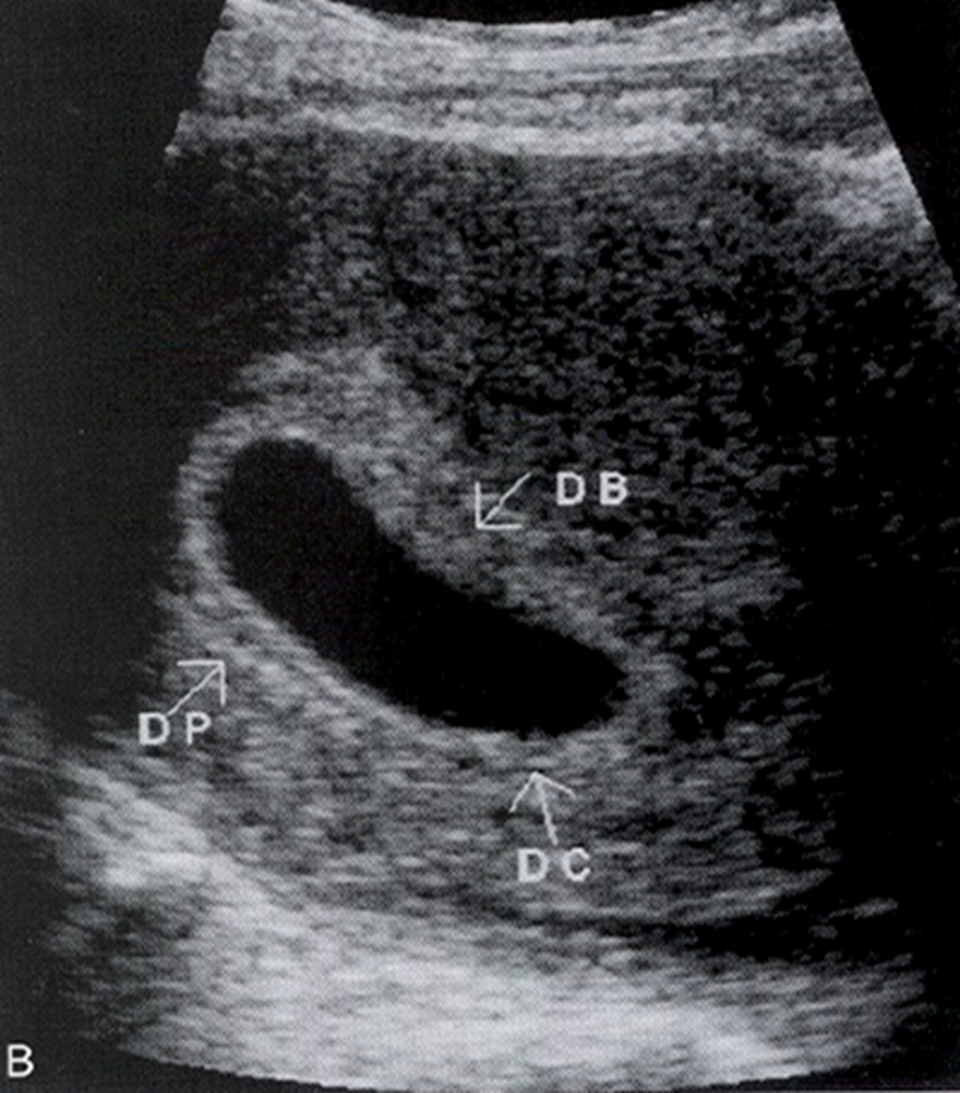
What is this image showing?
Decidua Capsularis
Decidua Parietalis
When is the best visualization of the amniotic membrane transvaginal?
Visualization after 5.5 weeks
When visualizing amniotic and chorionic cavities later, what do the cavities do?
Amniotic cavity expands, chorionic cavity decreases in size with eventual chorioamnionic fusion occurring at approx. 16-17 weeks.
Where does the embryo/fetus grow?
Grows inside the amniotic cavity
When should the amnion/chorion separation fuse?
Around 16-17 weeks
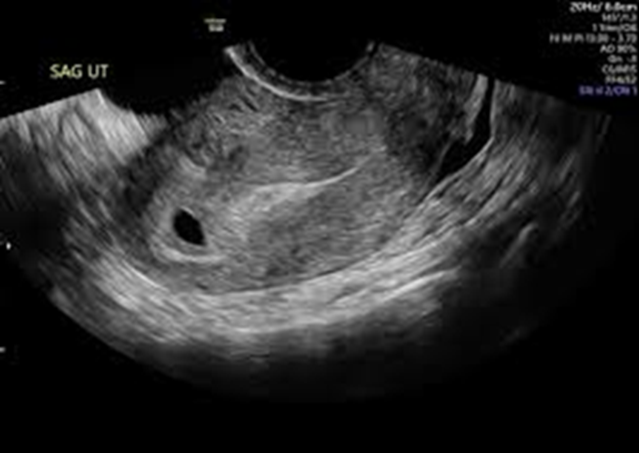
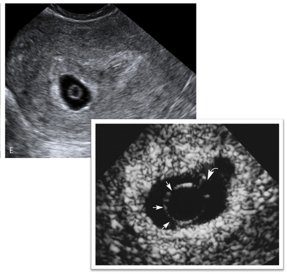
What is this image showing?
Double Decidual sac sign
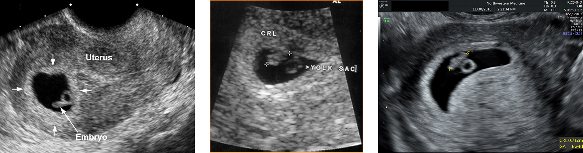
What is the sonographic appearance of the gestational sac?
Round or oval shape
Fundal position in the uterus
Central portion of uterus
Smooth contour
> 3mm decidua wall
If the MSD is > 10 mm (5-5.5 wks) what is it considered?
Yolk sac
If the MSD is > 16 mm ( 6-6.5 wks) what is it considered?
Embryo
How much does the gestational sac grow?
1 mm/day in early pregnancy
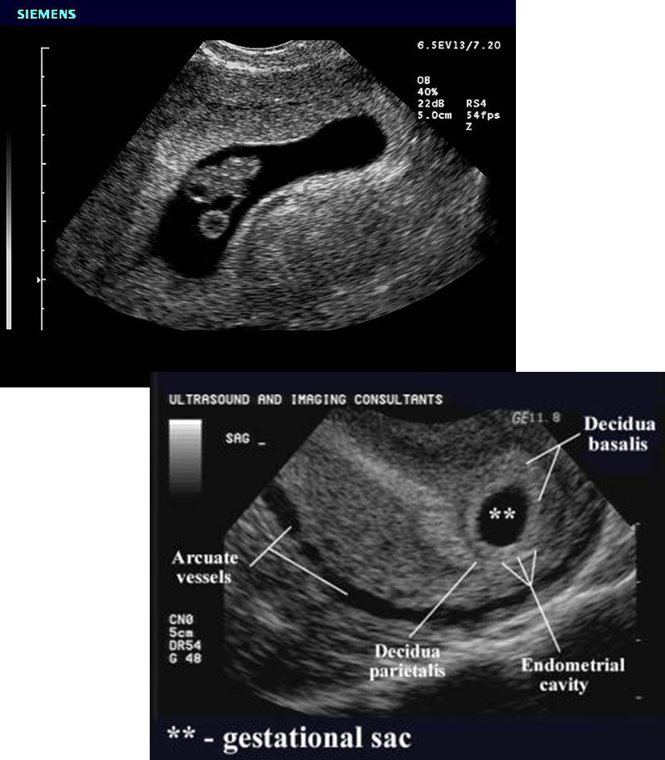
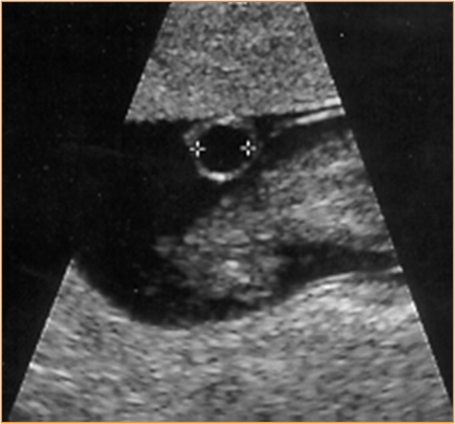
What is this image showing?
Gestational Sac
What are these images showing?
Yolk Sac
When is the mean yolk sac diameter identified transabdominally?
20 mm or greater
When is the mean yolk sac diameter identified transvaginaly?
8 mm or greater
If the yolk sac is not visualized, what is it suspicious for?
Suspicious of abnormal pregnancy
If the yolk sac is >5.6 mm what is it associated with?
Associated with poor outcome from weeks 5-10
When does the yolk sac disappear?
10-12 weeks
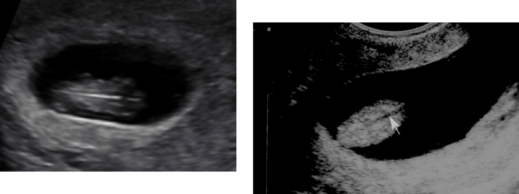
What are these images showing?
Yolk sac
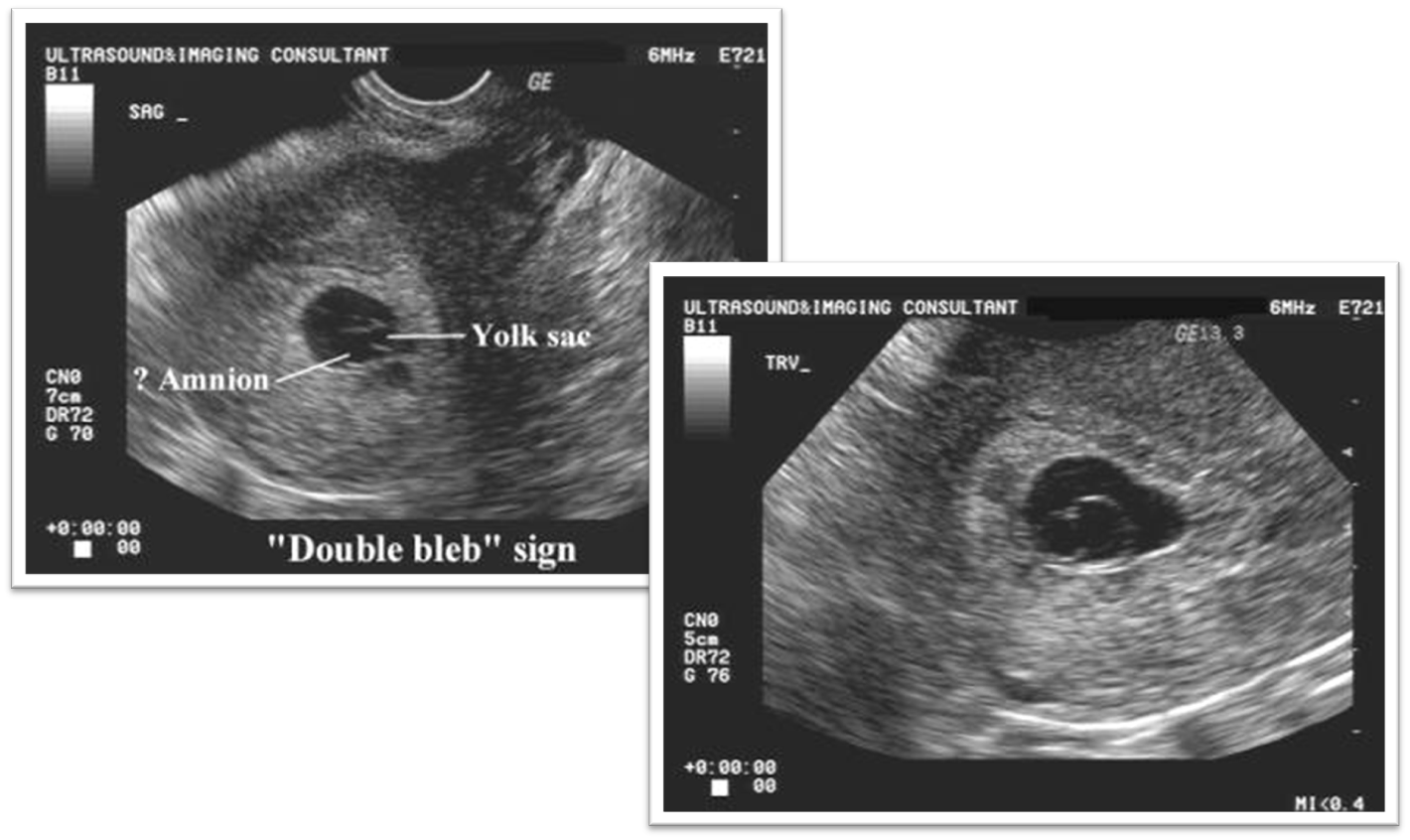
What are these images showing?
Double Bleb Sign
The yolk sac is between ____________ and amniotic cavity contains __________.
Membranes; Embryo
What are these images showing?
Yolk Sac
The embryo conceptus should be seen when the embryo MSD measures __________ TA?
25 mm or greater
The embryo conceptus should be seen when the embryo MSD measures __________ TV.
16 mm or greater
What is a sac highly suspicious for when it measures greater than 25mm without contents
Anembryonic gestation
Cardiac activity should be identified TA at what week and measurement?
7 weeks
9mm
Cardiac activity should be identified TV at what week and measurement?
5-6 weeks
2-5 mm embryo
7mm new absolute criteria
What should be identified on a 7-8 week embryo
Skeletal, muscle, heart and limb buds forming
Fetal head and trunk can be differentiated
Intermittent movement noted
Recognition of the spine
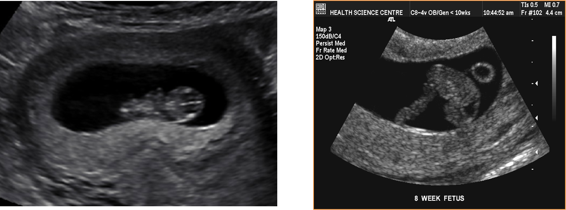
What are these images showing?
Embryo at 8 weeks
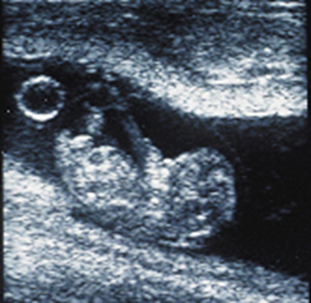
What are these images showing?
Embryo at 8 weeks