NEUROMOTOR SYSTEM
 Call Kai
 Learn
Learn Practice Test
 Spaced Repetition
 Match
Match Flashcards
 Knowt Play
1/77

There's no tags or description
Looks like no tags are added yet.
Name | Mastery | Learn | Test | Matching | Spaced | Call with Kai | Chat |
|---|
No analytics yet
Send a link to your students to track their progress
78 Terms
UPPER MOTOR NEURONS
cortical motor neurons within M1
motor neurons within the brainstem (extrapyramidal neurons)
LOWER MOTOR NEURONS
neurons in the anterior horn of the spinal cord
neurons within the cranial nerve nuclei within the brainstem
FOUR SYSTEM FOR MOTOR CONTROL
Descending Systems
Upper motor neurons
Motor cortex
Brainstem centres
Basal Ganglia
initiation of wanted movement
suppression of unwanted movement
Cerebellum
coordination of ongoing movement
Brainstem circuits and Spinal cord
lower motor neurons
local circuit neurons
motor neuron pools
SKELETAL MUSCLE ESSENTIAL FUNCTIONS
Movement
postural maintenance and joint stability
heat generation (active contraction & shivering)
venous return (veins rely on valves + SkM pumping)
lymphatic drainage
reproduction, digestion, excretion
NEUROMUSCULAR JUNCTION
The specialised connection between the motor nerve ending and skeletal muscle.
biggest chemical synapse in body
excitable
can undergo reinnervation if damaged
MUSCLE ACTION POTENTIAL STEPS
AP arrives at nerve terminal
Triggers opening of voltage gated calcium channels
Calcium influx
ACh released by exocytosis at active zones
ACh crosses cleft and binds to its ligand gated receptor
Ion channels open
Na+ in / K+ out
Depolarisation of endplate
Local current opens voltage gated Na+ channels nearby
Na+ entry causes resting potential to rise from -70 to -60mV
Triggers muscle AP
ACh destroyed by acetylcholinesterase
ACETYLCHOLINESTERASE
Enzyme in synaptic cleft
degrades ACh
recycles choline back to nerve terminal
Keeps ACh concentration controlled so receptors aren’t desensitised
SAFETY FACTOR
The neuromuscular junction has a high safety factor to ensure endplate potentials never fail to trigger an action potential.
This is done by there always being enough AChR’s to bring the endplate potential to threshold.
CLUSTERING ACHR’S
Motor nerves release agrin which binds to its muscle receptor LRP4-MuSK, which drives phosphorylation and recruitment of Dok7, which via Rapsyn, clusters AChR’s into a dense patch below the nerve terminal.
this pathway drives muscle gene expression
mutations in LRP4, MuSK, Dok7 or Rapsyn reduce AChR density and cause congenital myasthenia gravis.
MOTOR NEURON DISEASE
Total failure of neuromuscular synapses leading to muscle paralysis and death due to respiratory muscles failing to contract.
MOTOR NEURON DISEASE CAUSED BY
Loss of neuromuscular connections
Death of alpha motor neurons
nucleocytoplasmic transport defects
altered RNA metabolism
mitochondrial dysfunction
neuroinflammation
failure of neuromuscular signalling to muscle
MOTOR NEURON DISEASE DRUG TREATMENTS
RILUZOLE
anti-excitotoxic
inhibits glutamate release, lowers motor neuron hyper-excitability
EMA
metabolic
restores efficient glycolysis in muscle
EDARAVONE
antioxidant
lowers reactive oxygen species
PMX205
anti-inflammatory
lowers CNS innate immune activation
MYASTHENIA GRAVIS
Partial failure of neuromuscular synapses caused by defects in muscles' postsynaptic region.
Loss of AChR’s → decrease in muscle AP’s → muscle weakness
MYASTHENIA GRAVIS FORMS
Autoimmune form:
antibodies directed against AChR’s
Congenital form:
mutations in LRP4, MuSK, Dok7 and Rapsyn
MYASTHENIA GRAVIS TREATMENT
acetylcholinesterase inhibitors
immunotherapy of antibodies
immunosuppressives (corticosteroids)
MuSK-IgG4 treatment
LAMBERT EATON SYNDROME
Partial failure of neuromuscular synapses caused by defect in the motor nerve terminal.
poor release of neurotransmitter → muscle weakness
Treatment:
K+ channel blocker to increase ACh release
DUCHENNE MUSCULAR DYSTROPHY
Caused by an inherited genetic defect in the dystrophin gene.
PRE-DIAGNOSIS:
born without visual symptoms
ACUTE PHASE:
inflammation
muscle necrosis
muscle weakness
CHRONIC PHASE:
wheel-chair dependent
muscle wasting
cardiac myopathy
diaphragm weakening
DYSTROPHIN
Transduces contractile force across the plasma membrane protecting it from tearing.
Loss of dystrophin = Ca2+ influx + activation of proteases + membrane damage = creatine kinase release in blood as a biomarker of muscle damage.
DUCHENNE MUSCULAR DYSTROPHY TREATMENT
ANTI-INFLAMMATORY DRUGS
corticosteroids (bad long term)
prostaglandin pathway modulators
GENE THERAPY
AVV-mediated
RATE MODULATION
Increased frequency of muscle APs = increased muscle fibre tension
maximal contraction = tetanus
this increases SINGLE muscle fibre tension
MOTOR UNIT RECRUITMENT
Activation of additional motor units to increase tension in whole muscle
1 motor unit = 1 motor neuron + all muscle fibres it innervates
HENNEMAN’S SIZE PRINCIPLE
Neurons with smaller cell bodies are activated first and larger ones activated last.
Slow twitch = myosin 1, small motor neurons
Fast fatigue-resistant = myosin2A, medium motor neurons
Fast fatigued = myosin2B, large motor neurons
COMPOUND MUSCLE ACTION POTENTIALS
The summed electrical signal from all motor units activated within a muscle.
INPUT CONTROL TO MOTOR NEURON OUTPUT
AFFERENT (SENSORY) INPUT
reflex arcs
PRIMARY MOTOR CORTEX
direct, heirarchical, pyramidal (corticospinal)
MULTI-NEURONAL MOTOR SYSTEM
indirect, parallel, extrapyramidal (brainstem, basal ganglia, cerebellum)
PROPRIOCEPTION
Every muscle contraction is accompanied by sensory feedback
allows brain to compute limb position
intended vs actual movement
PROPRIOCEPTORS
MUSCLE SPINDLE
within muscle belly
relative changes in muscle length, movement, velocity
GOLGI-TENDON ORGANS
muscle tendons/connective tissue attachments
muscle tension and force
JOINT RECEPTORS I - IV
joint capsule
capsule tension, ligament tension, pain
MUSCLE SPINDLES
𝛄 = neuromuscular synapses at end of intrafusal muscle fibres (efferent)
sensory connections at centre of intrafusal muscle fibres (afferent)
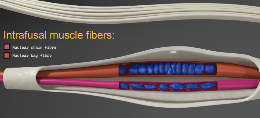
INTRAFUSAL FIBRE TYPES
Nuclear chain fibres
sensitive to muscle length
Nuclear bag fibres
sensitive to velocity of length change
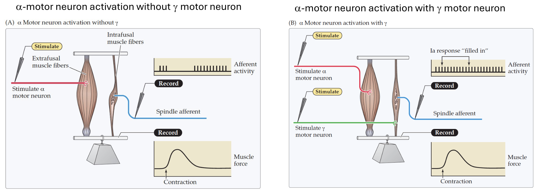
𝛄 MOTOR NEURON IMPORTANCE
⍺ motor neuron activation with 𝛄 motor neurons keep intrafusal fibres taut during contraction, preserving spindle sensitivity throughout the movement.
SPINAL CORD ORGANISATION
Motor neuron cell bodies are organised into motor columns:
Lateral Motor Column:
innervates limb appendicular muscles
distal muscles for fine movement
Medial Motor Column:
innervates axial (trunk) muscles
posture and whole body orienting
INTERNEURONS
Connection between afferent inputs and motor neurons
ipsilateral and contralateral across segment levels
coordinate activity of muscle groups and their afferents
POST-SYNAPTIC GATING
Monosynpatic reflex
inhibition of neurons ability to fire
constantly active at low level
Ex. reciprocal inhibition (knee-jerk reflex)
CONVERGENCE REFLEX
multiple inputs onto one motor neuron
enhance stretch reflex
DIVERGENCE REFLEX
one sensory input distributed to many motor neurons
ex. walking
BRAIN STEM INPUTS
Proprioception (muscle spindles, golgi-tendon organs, joint receptors)
Vestibular apparatus (head position/balance)
Eyes (visual context for posture)
BRAIN STEM OUTPUTS
Modify motor neuron output for:
balance
stabilisation
alignment of head/body with gravity
protective responses
Achieved via two mechanisms:
Rapid, stereotyped feedback (reflex)
Pre-programmed feedforward (anticipatory) control
Don’t need higher order processing from motor cortex to modify motor neuron output
BRAINSTEM MOTOR CENTRES
Vestibular Nuclei
recieve vestibular afferents with regards to head position relative to gravity
Reticular Formation
connections from premotor regions & coordinate lower motor neuron pools
Superior Colliculus
Integrates eyes, vestibular and proprioceptive input
Red Nucleus
Plays a role in motor learning
VESTIBULAR NUCLEI
LATERAL
maintaining stance
proximal muscles of limbs
MEDIAL AND SUPERIOR
coordination of eyes with head movements
INFERIOR AND DORSAL
integrates afferents and cerebellum to higher centres
FEEDBACK
Rapid, stereotyped, reflex like
fixed space-time organisation
triggered AFTER disturbance is detected
FEED-FORWARD
pre-programmed based on experience
unlike simple reflexes
scale of response is refined with repitition
FEEDBACK VS FEEDFORWARD EXAMPLE
Standing on platform that is suddenly pulled backwards and fall.
FEEDBACK
Proprioceptors closest to distrubance (ankle, foot)
vision and vestibular
combine to pull body back upright
FEEDFORWARD
if repeated brain anticipates the disturbance
amplitude of correction is smaller
don’t fall as far due to quickened reflex
VOLUNTARY MOVEMENT
MOTOR CORTEX
‘I chose to move’
‘how should i move’
BASAL GANGLIA
‘what action should i perform’
CEREBELLUM
‘is the movement unfolding as expected’
MOTOR CORTEX
Goal → Plan → Command → Movement
Consists of:
Primary motor cortex
Premotor cortex
Supplementary motor area
Prefrontal cortex
Posterior parietal cortex
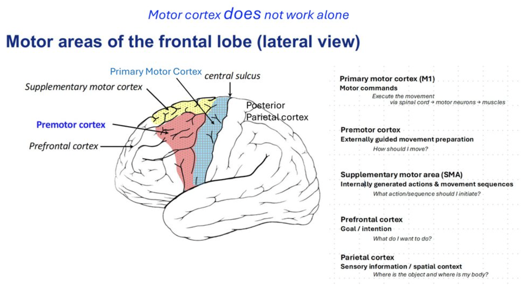
PRIMARY MOTOR CORTEX (M1)
motor commands
executes movements via spinal cord → motor neurons → muscles
PREMOTOR CORTEX
guided movement preperation
how should i move?
SUPPLEMENTARY MOTOR AREA
internally guided action sequences
what action/sequence should i initiate?
PREFRONTAL CORTEX
goal / intention
what do i want to do?
POSTERIOR PARIETAL CORTEX
spatial / sensory context
where is the object and my body?
CORTICOSPINAL NEURONS
Upper motor neurons
layer V of M1
largest, longest projecting neurons in nervous system
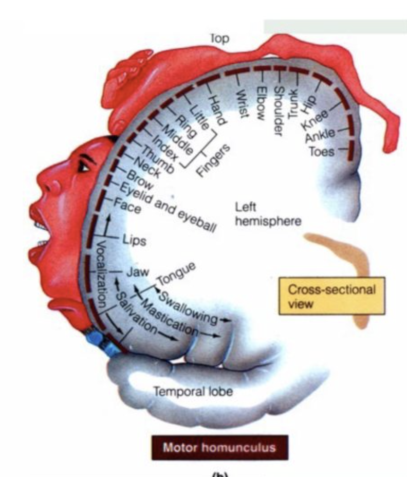
Somatotopic organisation:
more at muscles requiring fine complex control
less at muscles that are less refined
disproportionate representation = motor homunculus
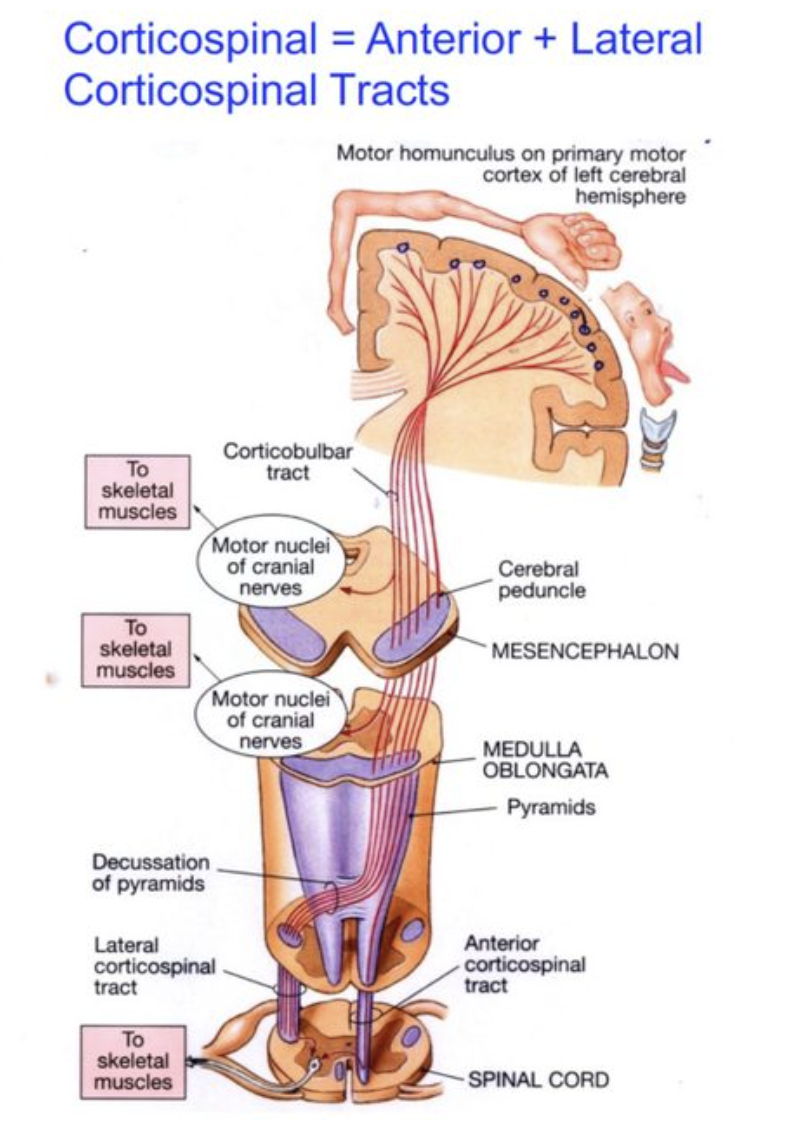
CORTICOSPINAL TRACT
descending corticospinal neurons
anterior and lateral divisions
cross at medulla pyramids
causes each hemisphere to control opposite side of body
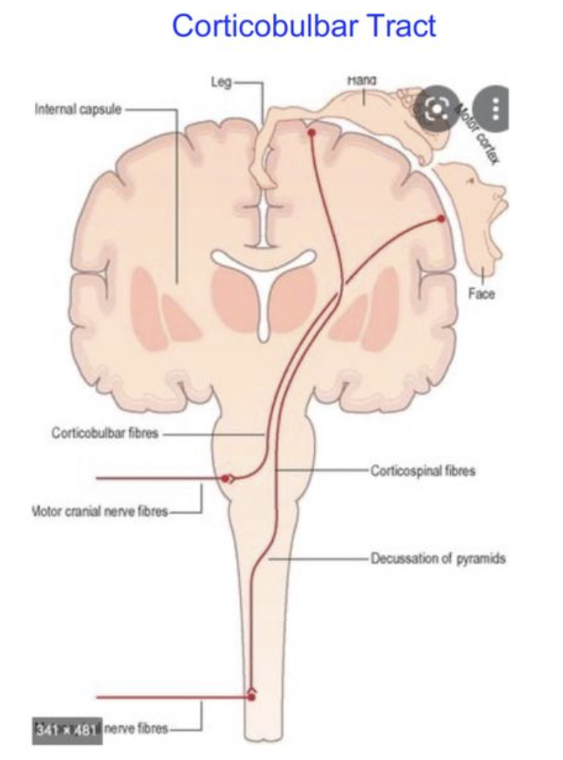
CORTICOBULBAR TRACTS
fibres destined for cranial nerve motor nuclei (face, neck, head)
cross earlier in brainstem
DIRECT CONNECTIONS
Lateral corticospinal neurons
direct excitatory connections to motor neurons
for fine movement control
INDIRECT CONNECTIONS
spinal interneurons
can excite or inhibit different motor neuron pools
dual direct/indirect wiring can produce spasticity
SUPPLEMENTARY MOTOR CORTEX AND REHEARSAL
Performing finger flexion activates:
Somatomotor/somatosensory areas
Performing finger movement sequence activates…
Somatomotor/somatosensory areas + supplementary motor cortex
Mentally rehearsing finger movement sequence activates…
just supplementary motor cortex
Therefore, SMA plans actions independently regardless of their physical execution.
CEREBELLUM - BASAL GANGLIA LOOP
Cortex → basal ganglia → thalamus → cortex
action selection
ex. initiating walking
CEREBELLUM - CEREBELLAR LOOP
Cortex → cerebellum → thalamus → cortex
prediction / correction
ex. correcting balance while walking
CEREBELLUM SUB-DIVISIONS
Vestibulocerebellum
Spinocerebellum
Cerebrocerebellum
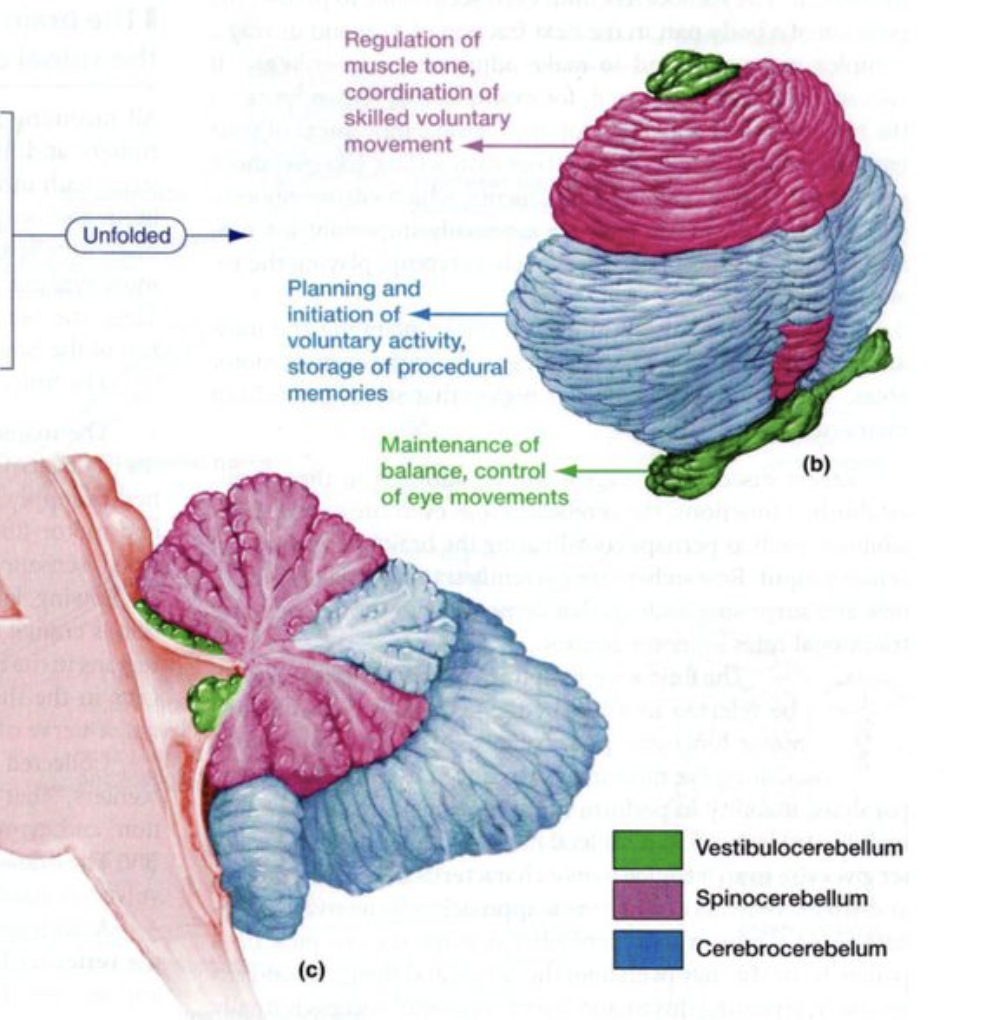
VESTIBULOCEREBELLUM
input = vestibular apparatus
output = lateral vestibular nucleus
function = balance, eye movement coordination
SPINOCEREBELLUM
input = proprioceptors
output = spinal cord
function = muscle tone, correct ongoing movement, coordination of voluntary movement
CEREBROCEREBELLUM
input = cerebral cortex
output = premotor cortex
function = planning + initiation of voluntary movement, storage of procedural memory
MOVEMENT IMPROVEMENT
Internal prediction = what should happen
Sensory feedback = what actually happened
Difference = motor error = improve future movement
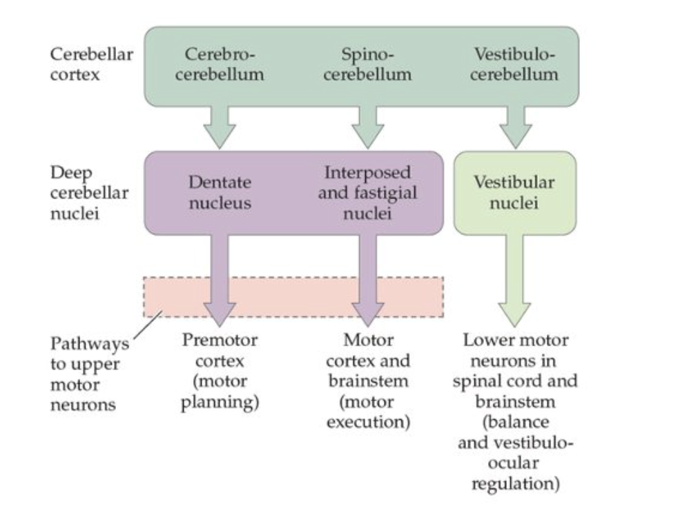
DEEP CEREBELLAR NUCLEI
Dentate nucleus
Interposed & fastigial nuclei
vestibular nuclei
Excitatory inputs from mossy and climbing fibres
inhibitory inputs from Purkinje cells
EXAMPLE COMPARATOR CIRCUIT
Motor command send to spinal cord → ⍺ motor neurons → muscle
Copy of motor command sent to cerebellum
Proprioceptors report back sensory consequences of actual movement
Cerebellum compares copy of command and sensory feedback
Any mismatch is computed and an updated motor command is sent out

ATAXIA
Damage of cerebellum reflects faulty integration of proprioceptive information
irregular reach
undershoot/overshoot movements due to impaired predicting vs sensing
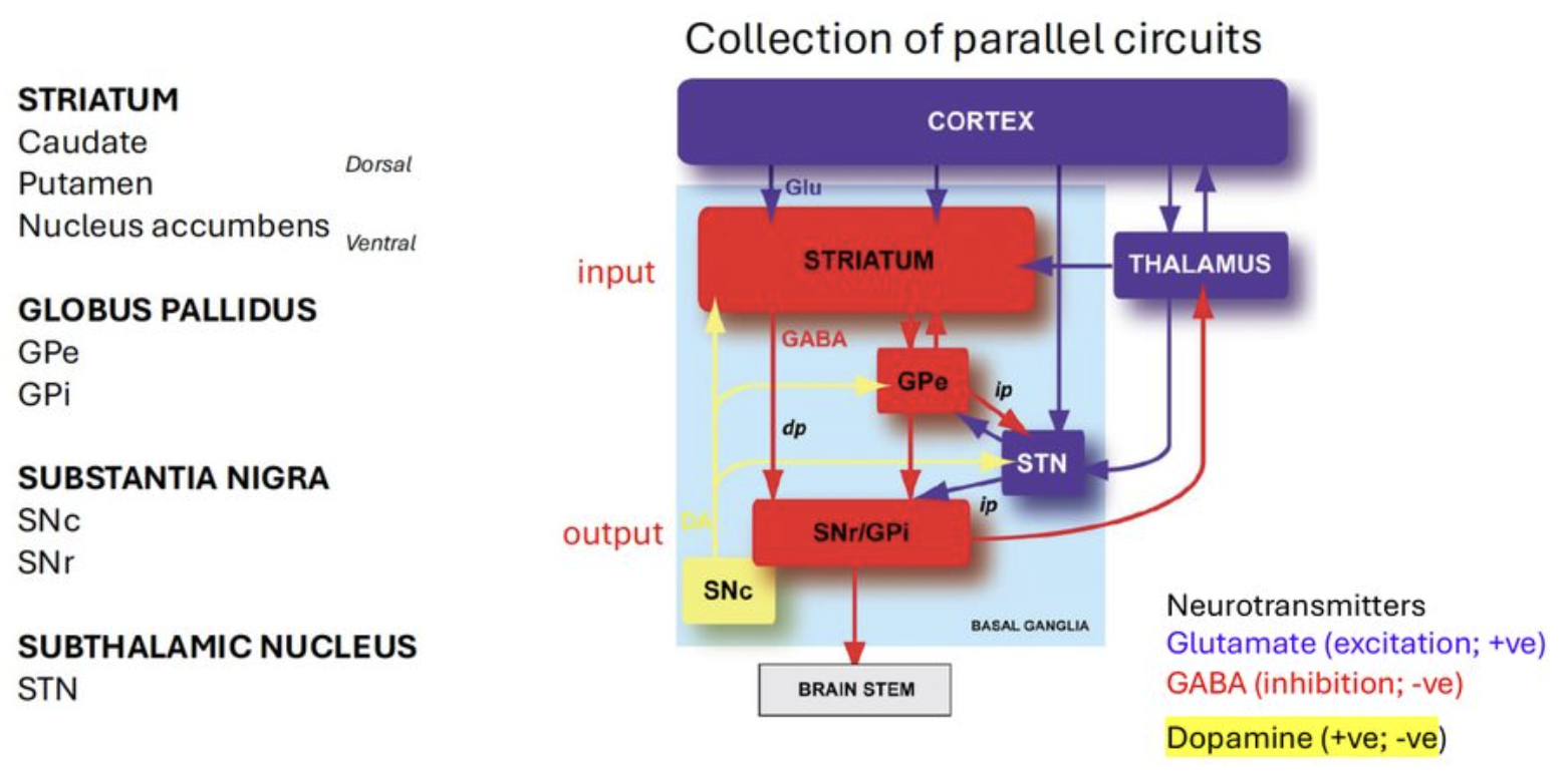
BASAL GANGLIA
The cortex proposes action, the basal ganglia decides which one wins.
The striatum is involved in:
Motor
start walking, reach, stop movement
Cognitive
switch task, make decision, change strategy
Motivation
seek reward, avoid punishment, repeat rewarding behaviour
BASAL GANGLIA CIRCUIT
STRIATUM
caudate
putamen
nucleus accumbens
GLOBUS PALLIDUS
external and internal
SUBSTANTIA NIGRA
Pars compacta
Pars reticulata
NEUROTRANSMITTERS
Glutamate (excit)
GABA (inhib)
Dopamine (excit + inhib)
SUBTHALAMIC NUCLEUS
BASAL GANGLIA - MOTOR LOOP
Motor cortex → putamen → globus pallidus → thalamus
movement execution and selection
BASAL GANGLIA - PREFRONTAL LOOP
prefrontal cortex → caudate → globus pallidus + substantia nigra → thalamus
decision making, switching strategy
BASAL GANGLIA - LIMBIC LOOP
Limbic system → ventral striatum → ventral pallidum → thalamus
emotion, value, reward, motivation
UPPER VS LOWER MOTOR NEURON LESIONS
losing lower MN removes pathway completely
losing upper MN leaves lower MN intact
LOWER LESIONS
weakness or paralysis
severe atrophy
hypoactive superficial and deep reflexes
UPPER LESIONS
weakness
mild or no atrophy
hyperactive deep reflexes after intital period of spinal shock
WHY IS MUSCLE MASS PRESERVED IN UPPER MN LESIONS
Lower MN is intact meaning spinal circuitry can drive flexion/extension even without descending cortical input
This maintains muscle mass, venous return and body heat generation even without voluntary control
SPASTICITY IN UPPER MN LESIONS
losing descending input causes hyperactive reflexes and increased muscle tone
spasticity = caused by loss of inhibitory modulation
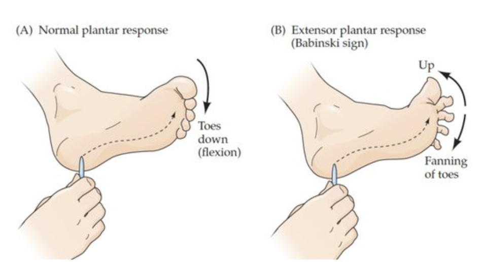
THE BABINSKI SIGN
Damage to corticospinal descending pathways causes extensor plantar response.
indicates incomplete upper MN control of spinal motor circuitry and a clinical sign of corticospinal lesion.
Voltage gated Calcium channels at the neuromuscular synapse are found:
At sites active zones.
MYASTHENIA GRAVIS IS CAUSED BY
A loss of acetylcholine receptors.
MUSCLE SPINDLES ARE INNERVATED BY
gamma motor neurons and type Ia and type II sensory neurons.
If the spinocerebellum division of the cerebellum is damaged, which of the following is likely to be MOST compromised?
Postural control of muscles
THE MAIN INHIBITORY NEUROTRANSMITTER WITHIN THE SPINAL CORD IS
Glycine (spinal cord)
GABA (is the brain)