Final Exam
1/54
Earn XP
Name | Mastery | Learn | Test | Matching | Spaced | Call with Kai |
|---|
No analytics yet
Send a link to your students to track their progress
55 Terms
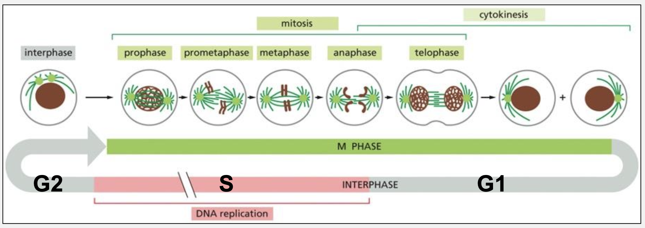
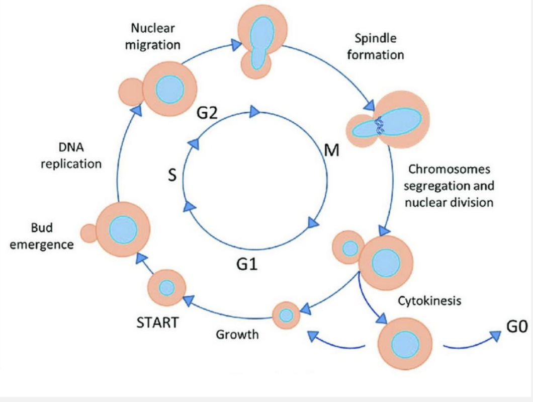
What are the four phases of the cell cycle and the major events in each?
G1: Cell growth, organelle duplication, preparation for DNA replication
S: DNA replication
G2: Preparation for mitosis, quality control
M: Chromosome condensation, spindle assembly, chromosome segregation, cytokinesis
What are the two fundamental tasks of the cell cycle?
Accurate replication of the genome and accurate segregation of chromosomes to daughter cells
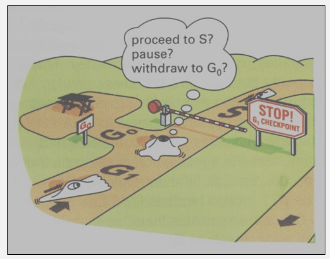
What is the difference between G0 and G1?
G1: Cell is committed to potentially entering S phase
G0: Cell has withdrawn from the cycle; may be reversible (quiescent stem cells) or permanent (neurons)
What are the three major regulatory transitions in the cell cycle?
G1 → S (Start/Restriction Point)
G2 → M
Metaphase → Anaphase
What kinds of cues regulate the three major transitions?
G1/S: Growth factors, nutrients, DNA damage
G2/M: DNA replication completion, DNA damage
Metaphase/Anaphase: Proper spindle attachment (spindle checkpoint)
How were cell cycle control genes identified genetically using yeast?
By isolating temperature‑sensitive (ts) mutants that arrest at specific cell cycle stages at non‑permissive temperatures
Why are temperature‑sensitive mutants essential?
They allow essential genes to function at permissive temperatures but fail conditionally, enabling study of lethal genes.
How are cell cycle mutants distinguished from other essential mutants?
Cell cycle mutants accumulate at a single stage, whereas non‑cycle essential mutants arrest randomly across the cycle
What biochemical approach identified cell cycle regulators?
Purification of M‑phase Promoting Factor (MPF) using activity assays in frog oocytes
What assay was essential for this approach?
An assay that measures induction of M phase (germinal vesicle breakdown)
What are the two subunits of a cyclin–Cdk complex?
Cyclin: Regulatory; levels oscillate
Cdk: Catalytic kinase; levels remain constant
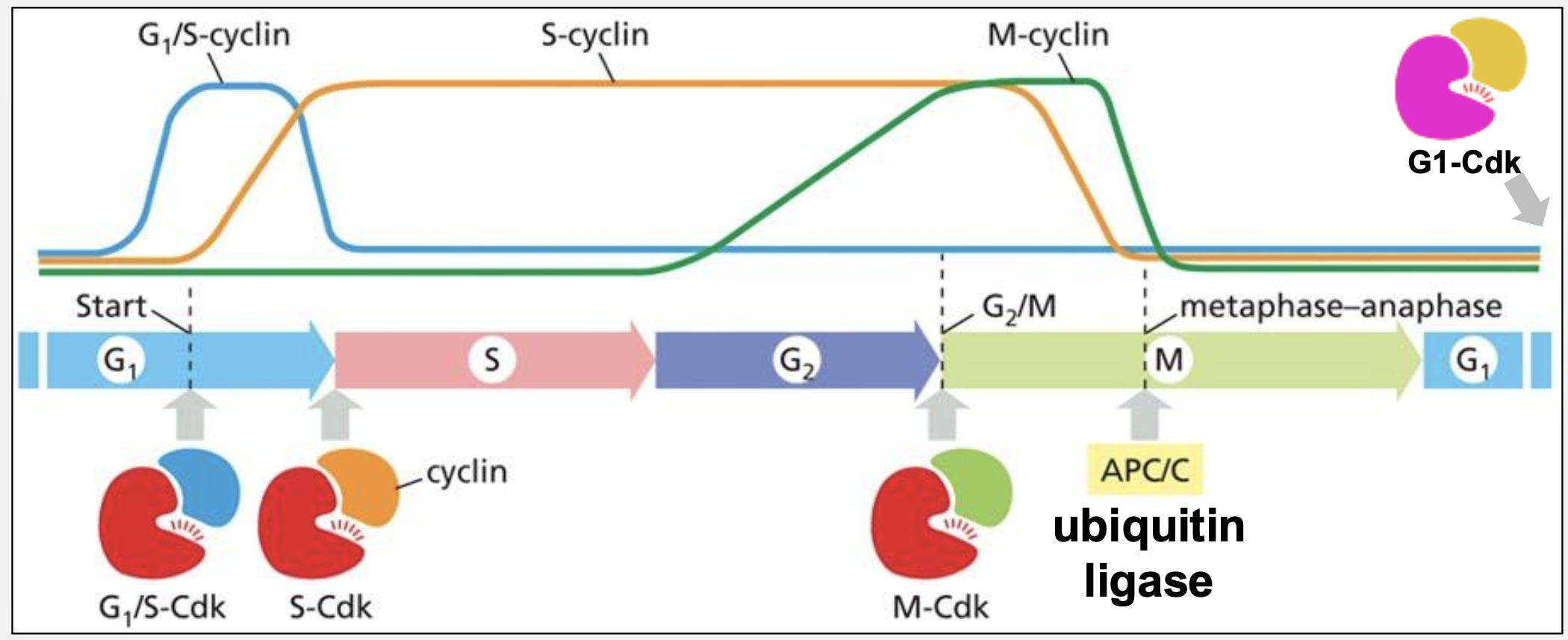
Identify four major cyclin–Cdk complexes and their roles.
G1‑Cdk: Responds to growth cues
G1/S‑Cdk: Commitment to DNA replication
S‑Cdk: DNA replication + early mitosis
M‑Cdk: Entry into mitosis
How does Cdc20‑APC/C drive exit from M phase?
APC/C ubiquitinates M‑cyclin, leading to proteasomal degradation and loss of M‑Cdk activity
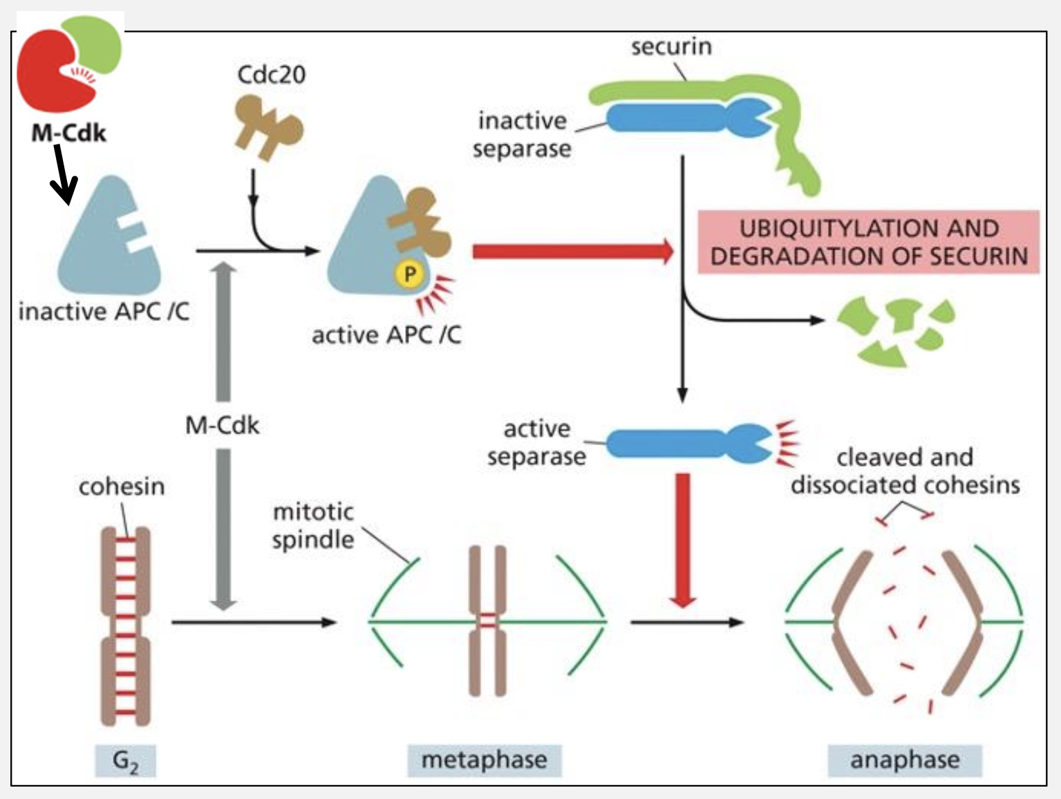
Name two downstream targets of Cdc20‑APC/C.
Securin (its destruction releases separase)
M‑cyclin
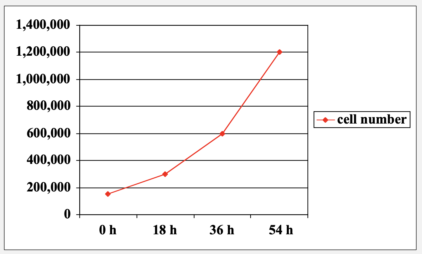
How can total cell cycle length be measured experimentally?
Growth curves using cell doubling time equations
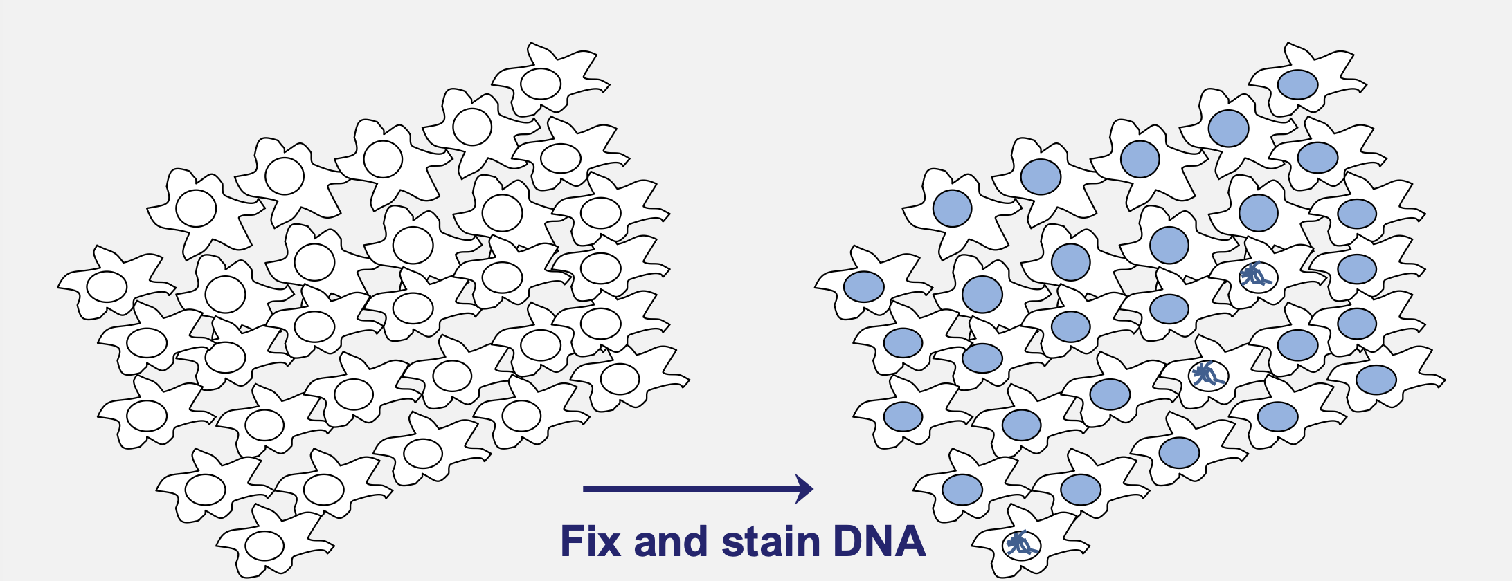
How is M‑phase length estimated?
Fraction of cells with condensed chromosomes × total cycle time
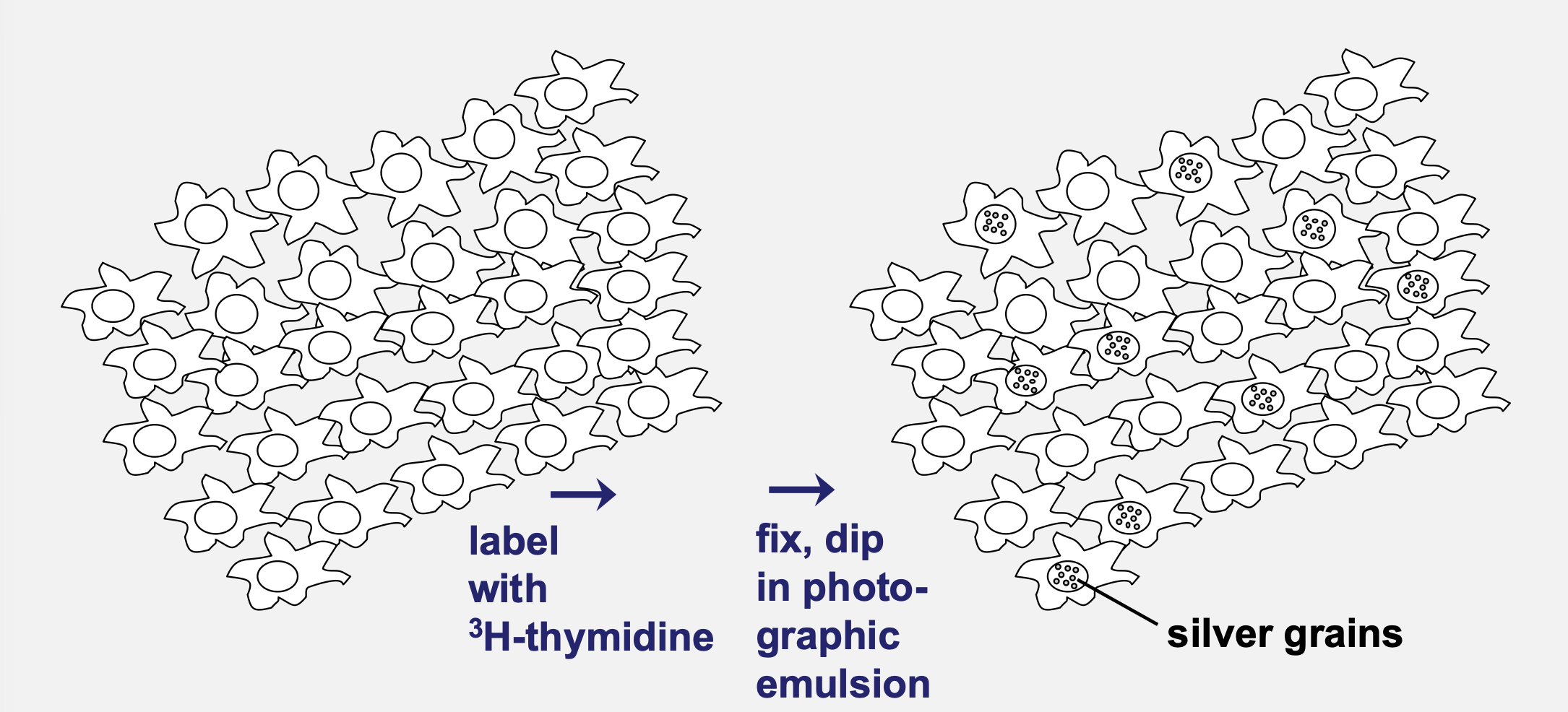
How is S‑phase length measured?
Pulse labeling with ³H‑thymidine
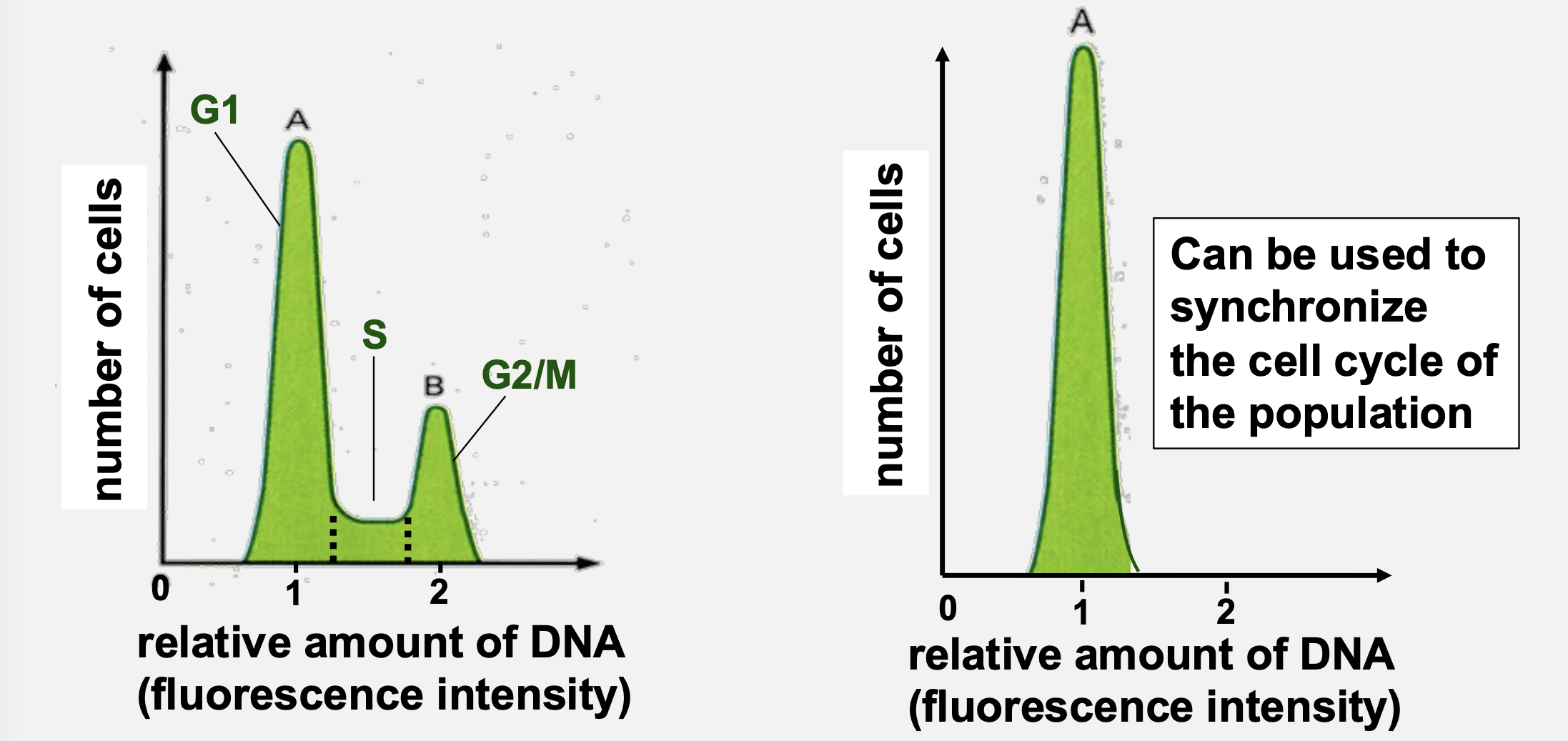
How does flow cytometry distinguish cell cycle phases?
By DNA content (G1 = 1×, S = intermediate, G2/M = 2×)
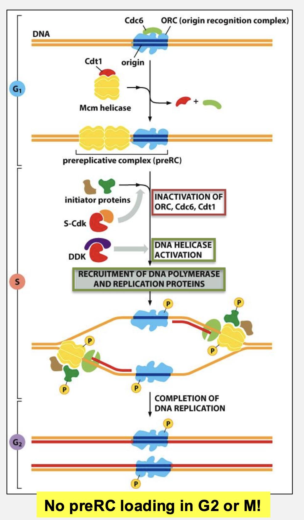
How is DNA replication limited to once per cycle?
Pre‑RCs load only in G1
S‑Cdk activates origins and prevents re‑loading until next G1
Which enzymes regulate the G2/M transition?
Cdc25 (phosphatase) activates M‑Cdk
Wee1 (kinase) inhibits M‑Cdk
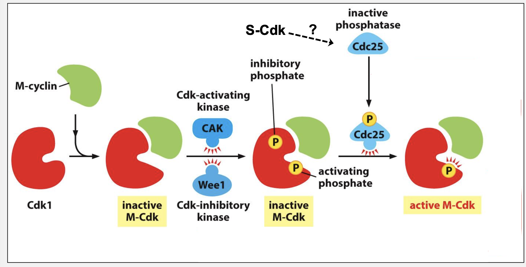
How does positive feedback create a switch‑like entry into mitosis?
Active M‑Cdk activates Cdc25 and inhibits Wee1, rapidly amplifying its own activation
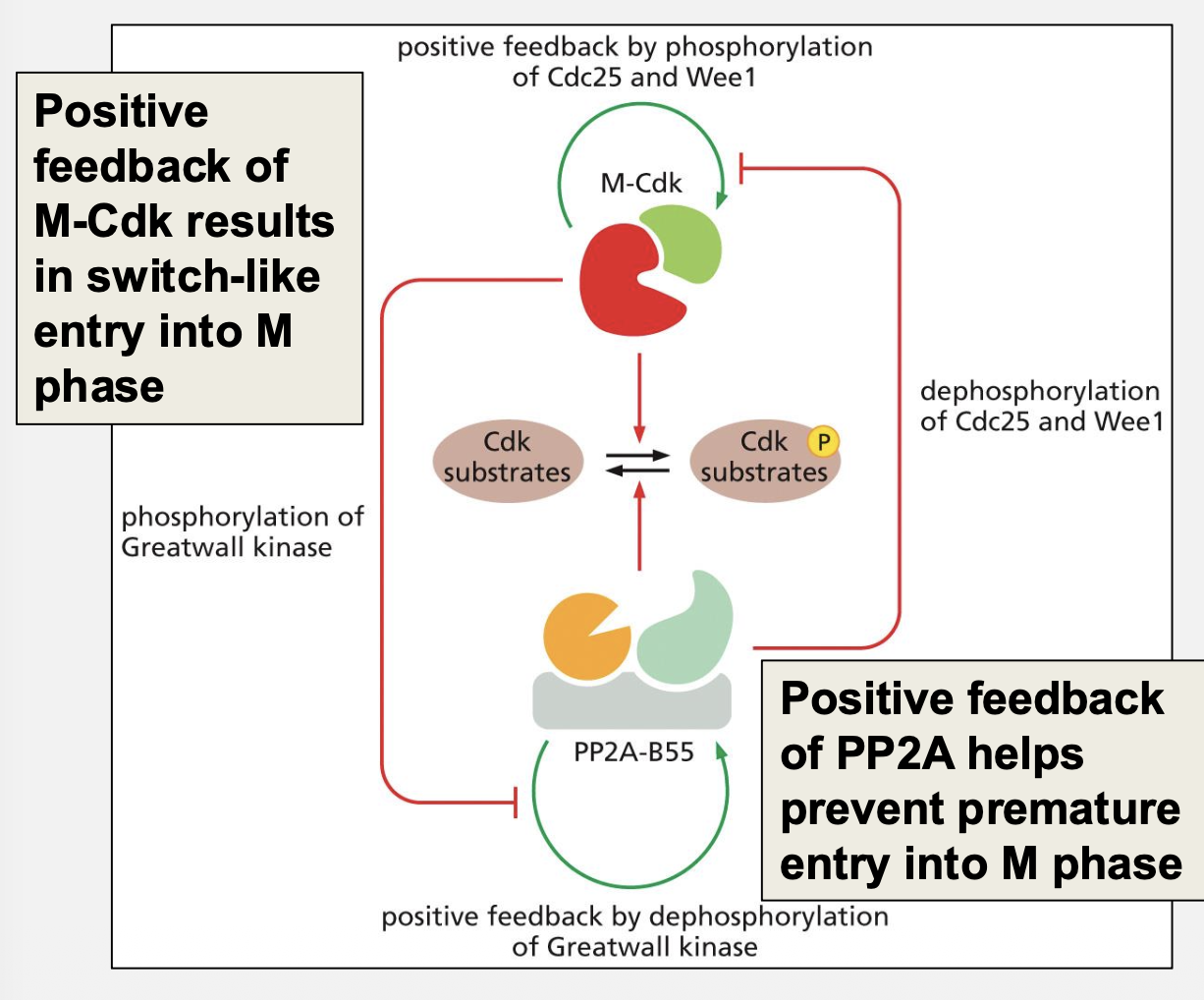
What role does PP2A play?
PP2A counteracts M‑Cdk before G2/M and is inhibited after transition to maintain mitosis
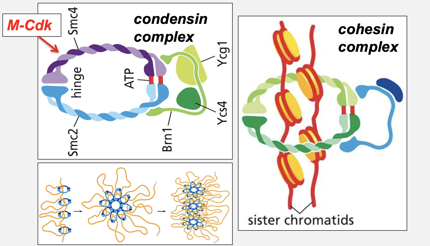
What two related complexes organize chromosomes in M phase?
Condensin: Compacts chromosomes (intra‑molecular)
Cohesin: Holds sister chromatids together (inter‑molecular)
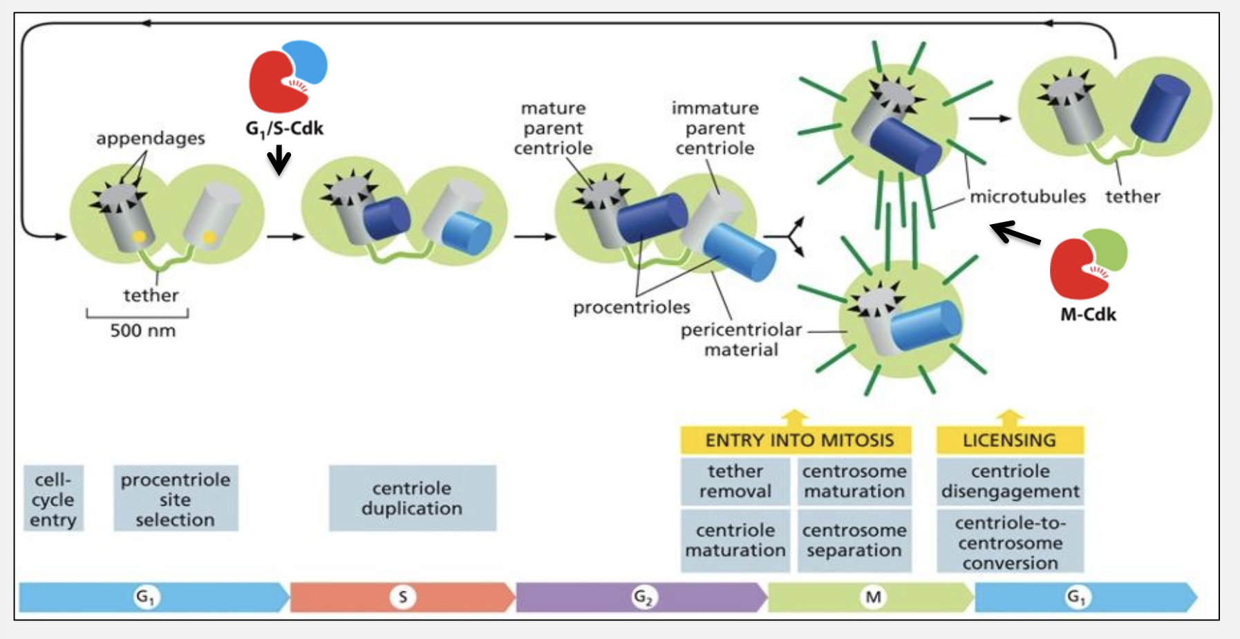
What roles do G1/S‑Cdk and M‑Cdk play in spindle formation?
G1/S‑Cdk: Centrosome duplication
M‑Cdk: Increased microtubule dynamics
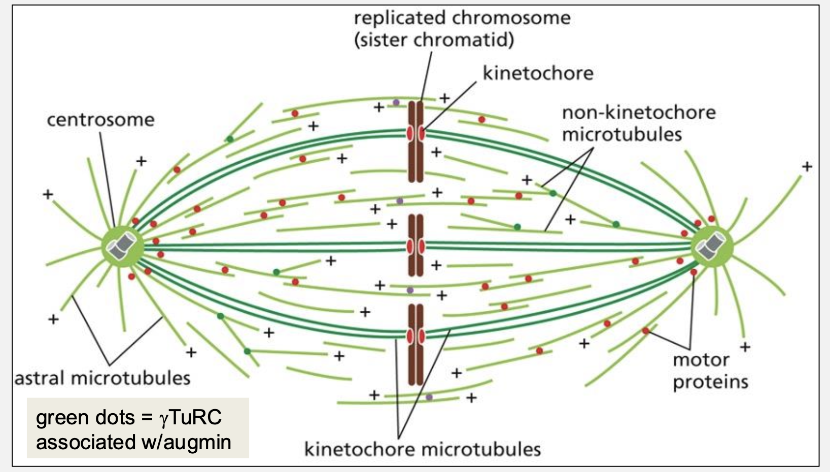
Name and describe the three classes of spindle microtubules.
Kinetochore MTs: Attach chromosomes
Astral MTs: Position spindle via cortex
Interpolar MTs: Push poles apart
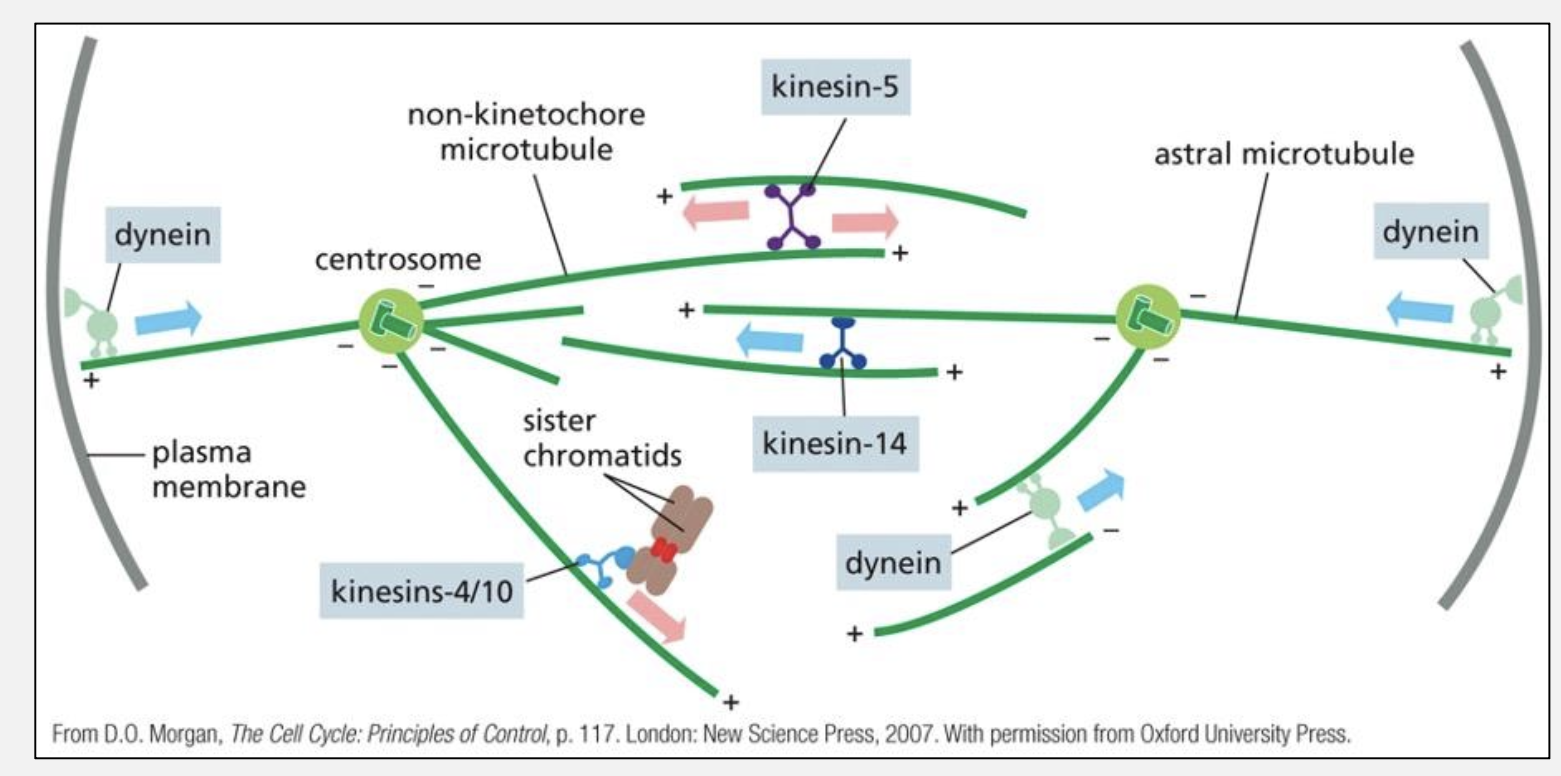
What are the roles of key motor proteins in mitosis?
Dynein: Pulls poles outward from cortex
Kinesin‑5: Pushes antiparallel MTs apart
Kinesin‑4/10: Move chromosomes toward spindle center
Where are kinetochore microtubule + ends located?
At the kinetochore, not the centrosome
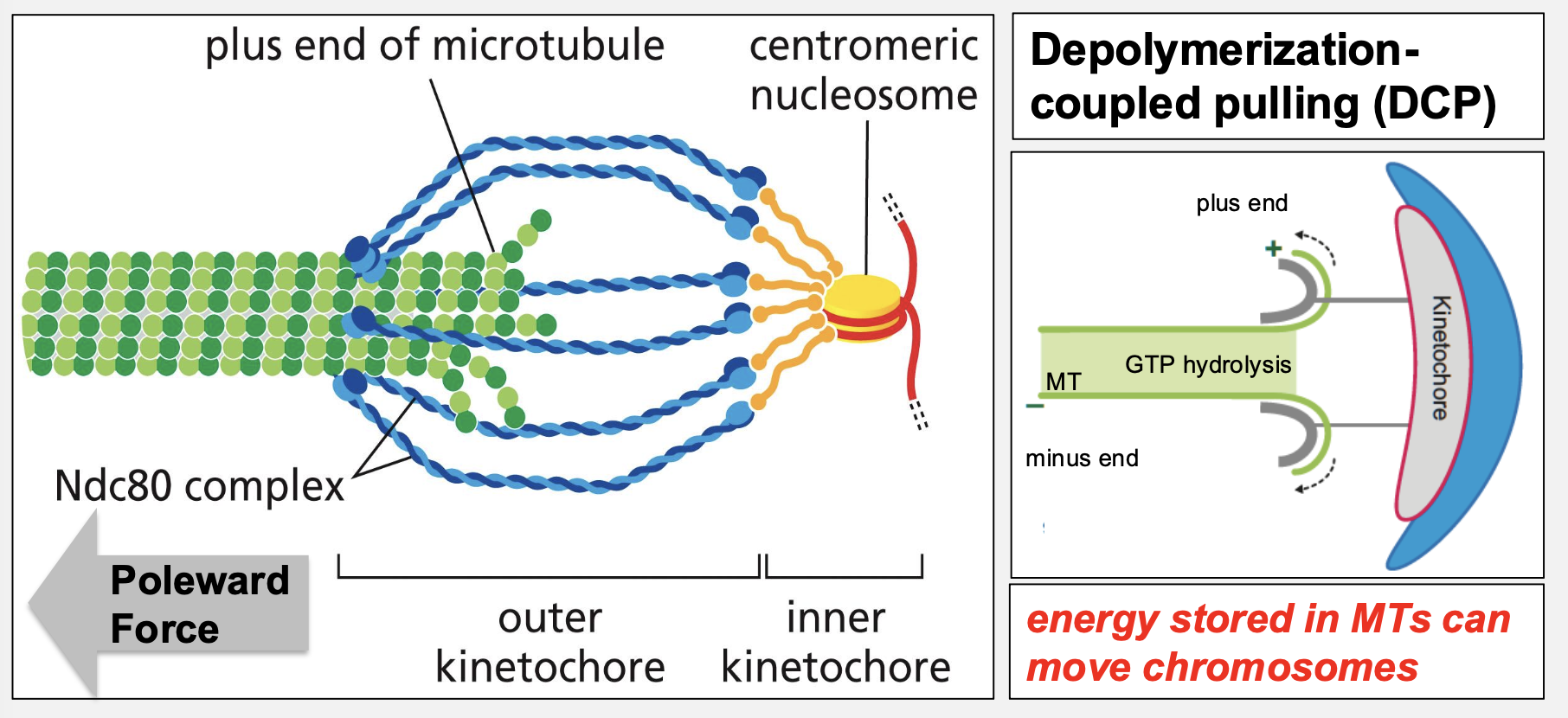
What is depolymerization‑coupled pulling (DCP)?
Energy stored in microtubule depolymerization generates force to pull chromosomes poleward
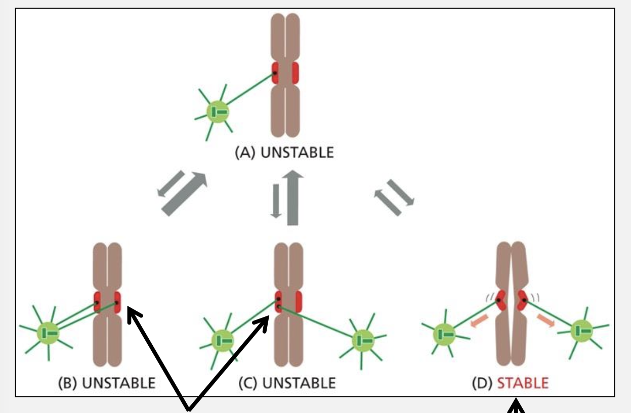
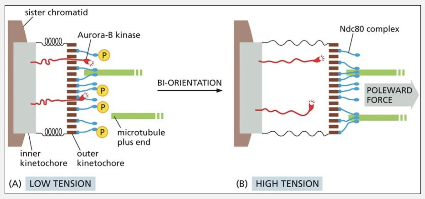
How does the “trial‑and‑error” mechanism ensure bi‑orientation?
Incorrect attachments lack tension and are destabilized by Aurora B, while correct attachments generate tension that stabilizes Ndc80 binding
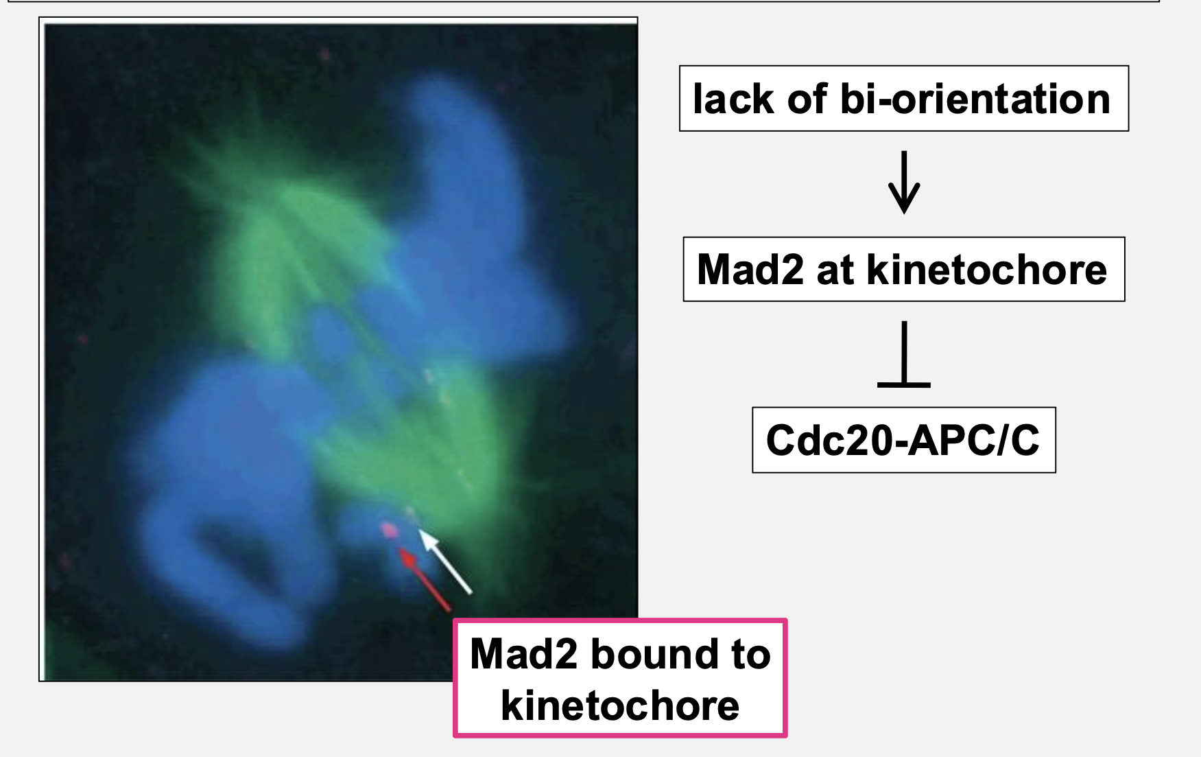
What is the spindle checkpoint?
A quality‑control mechanism that blocks anaphase until all chromosomes are properly attached
What role does Mad2 play?
Mad2 inhibits Cdc20‑APC/C when kinetochores are unattached
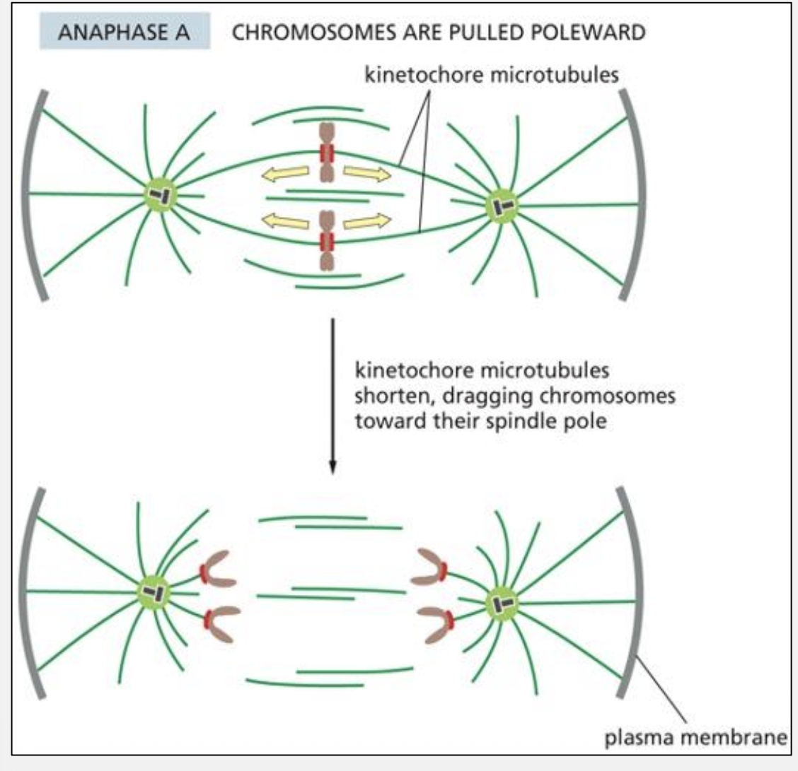
What forces drive Anaphase A?
Kinetochore MT depolymerization + DCP + MT flux
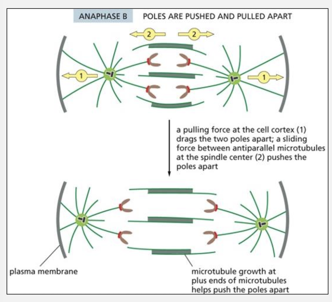
What forces drive Anaphase B?
Dynein pulling on astral MTs + kinesin‑5 sliding antiparallel MTs
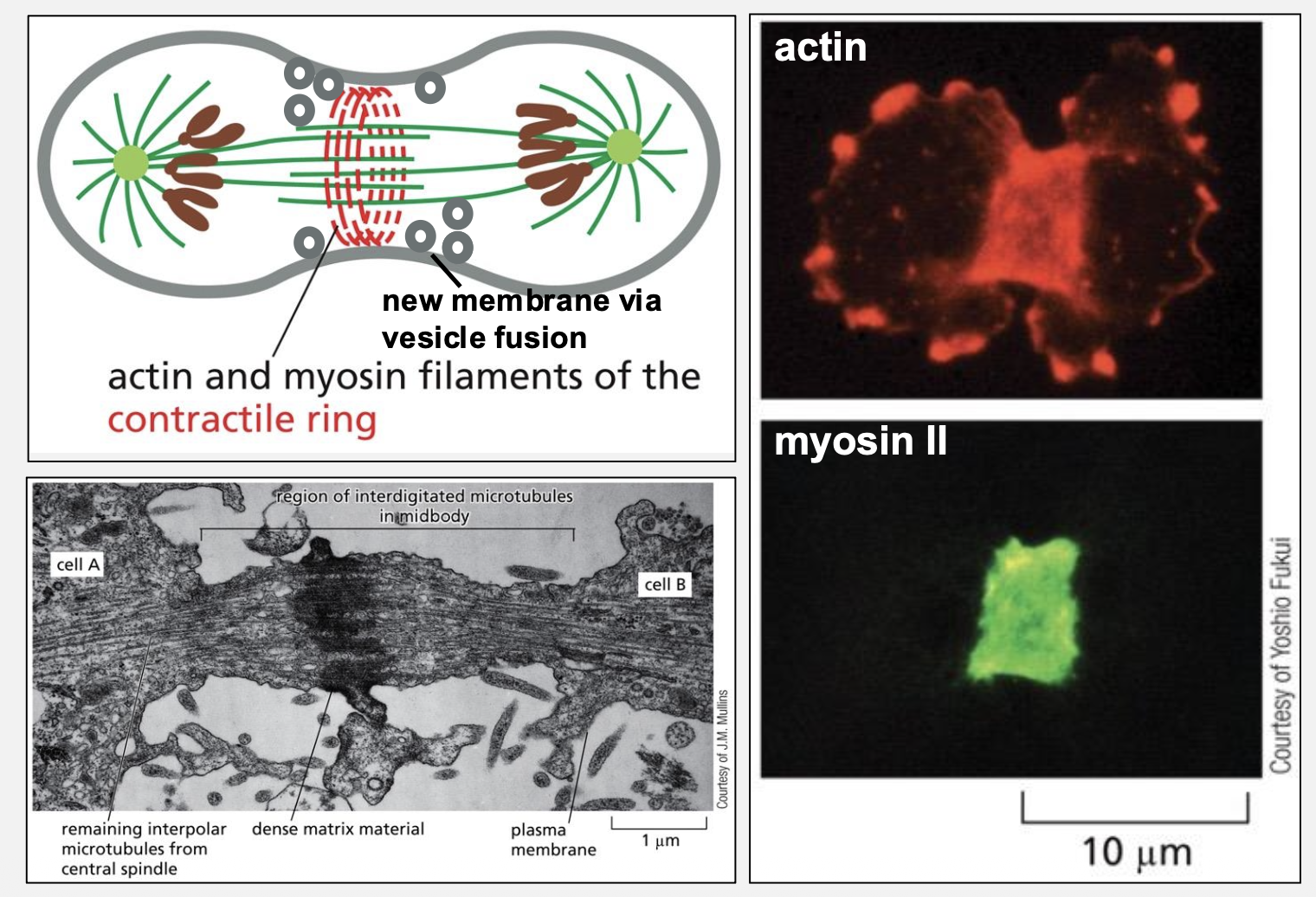
What structure mediates cytokinesis?
Actin‑myosin contractile ring
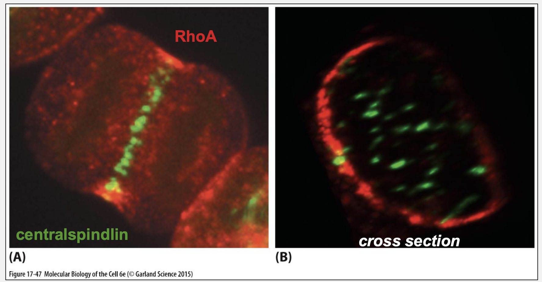
How is the division plane positioned?
Central spindle recruits centralspindlin → RhoGEF → RhoA activation
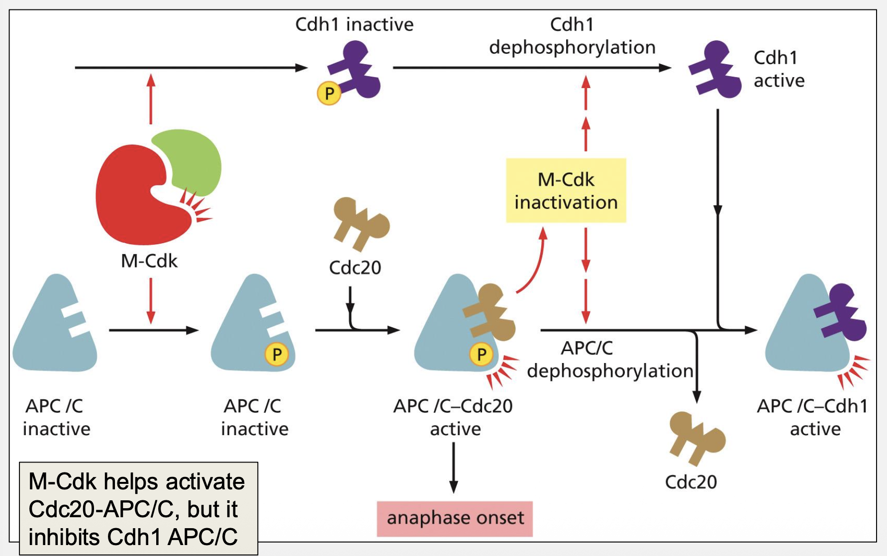
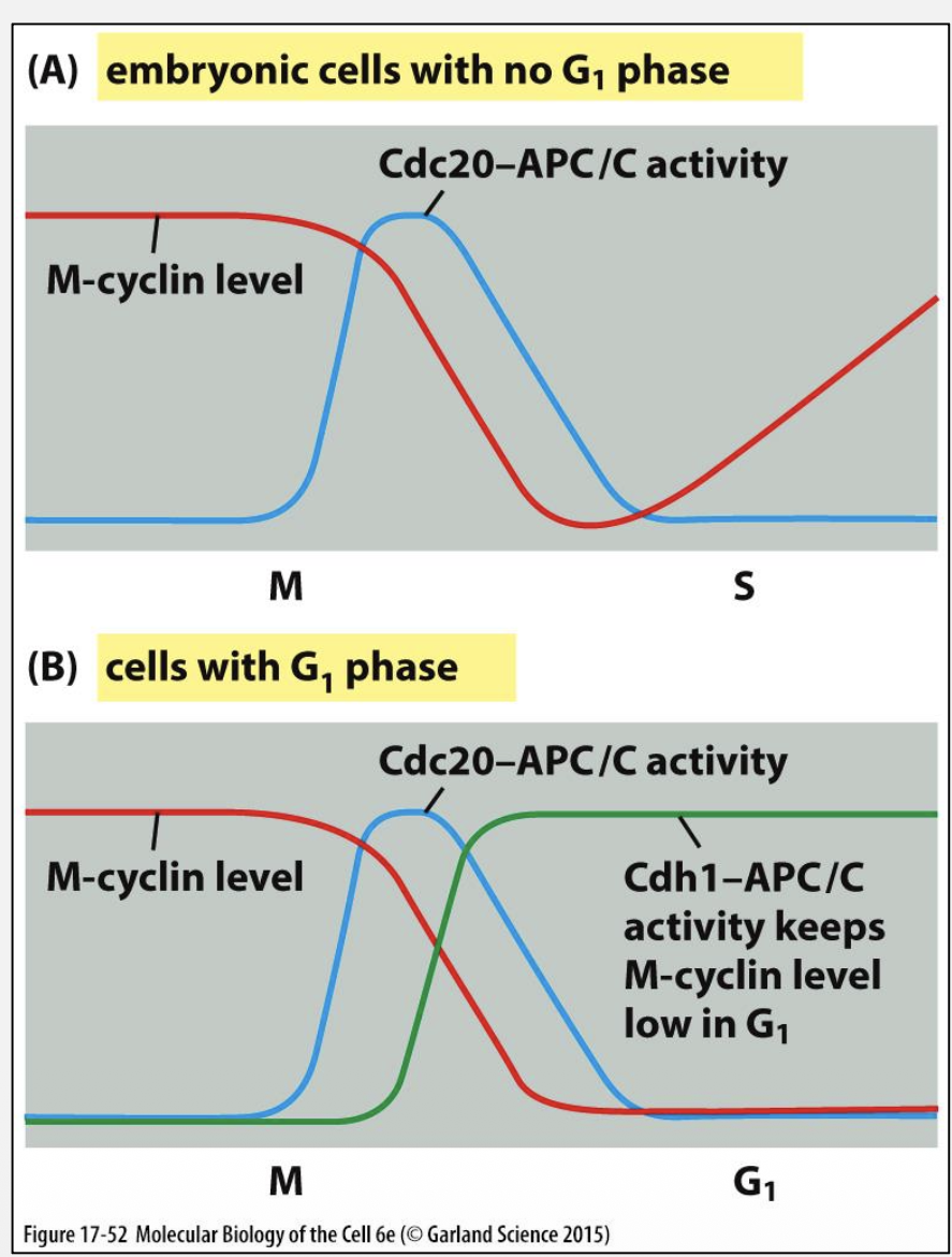
Compare Cdc20‑APC/C and Cdh1‑APC/C.
Cdc20‑APC/C: Activated by M‑Cdk (negative feedback)
Cdh1‑APC/C: Maintains low cyclin levels in G1 (positive feedback)
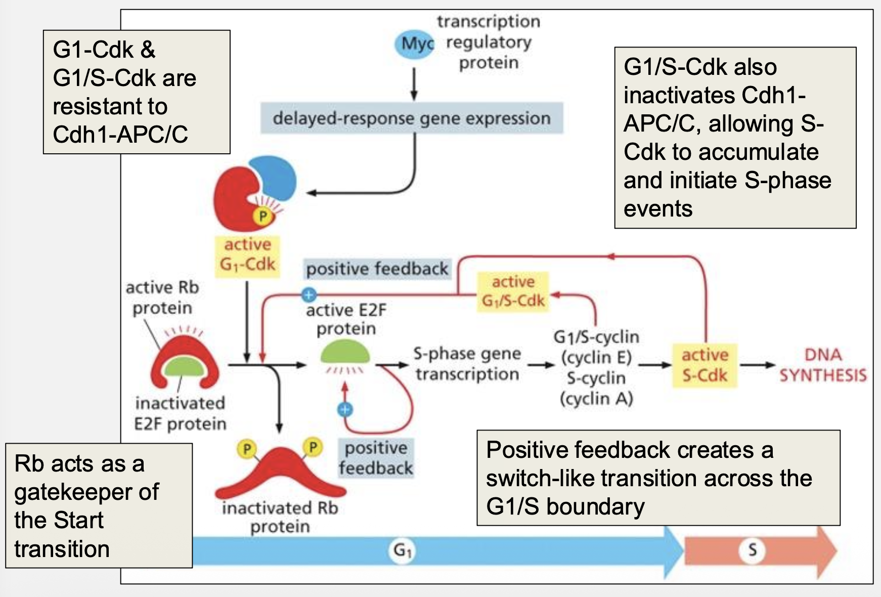
How does RTK signaling promote G1 progression?
Through Ras‑MAPK → Myc → cyclin expression
What is Rb’s gatekeeper role?
Rb inhibits E2F until phosphorylated by G1/S‑Cdk
How does positive feedback drive S‑phase entry?
G1/S‑Cdk inhibits Cdh1‑APC/C, allowing S‑Cdk accumulation
How does DNA damage activate p53?
Chk1/Chk2 phosphorylate p53, preventing Mdm2‑mediated degradation
How does p53 halt the cell cycle?
By inducing p21, a Cdk inhibitor
Difference between proto‑oncogene and tumor suppressor?
Proto‑oncogene: Gain‑of‑function → cancer
Tumor suppressor: Loss‑of‑function → cancer
Why does cancer arise later in life?
Requires accumulation of multiple mutations
What is EMT and why is it important in metastasis?
Transition from epithelial to mesenchymal state increases motility and invasion
Compare epithelial layers vs connective tissue.
Epithelia: Dense cells, strong junctions
Connective tissue: Sparse cells, ECM‑rich
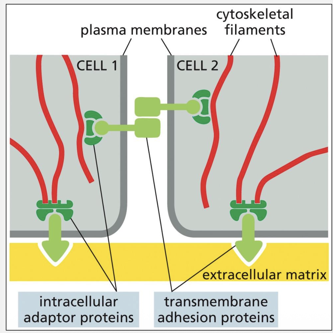
Components shared by all junctions?
Adhesion receptors, cytoplasmic adaptors, cytoskeleton

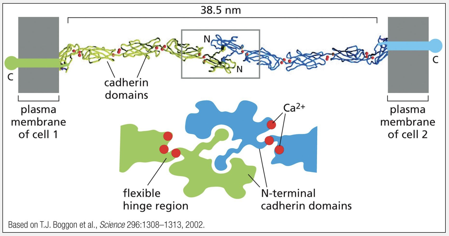
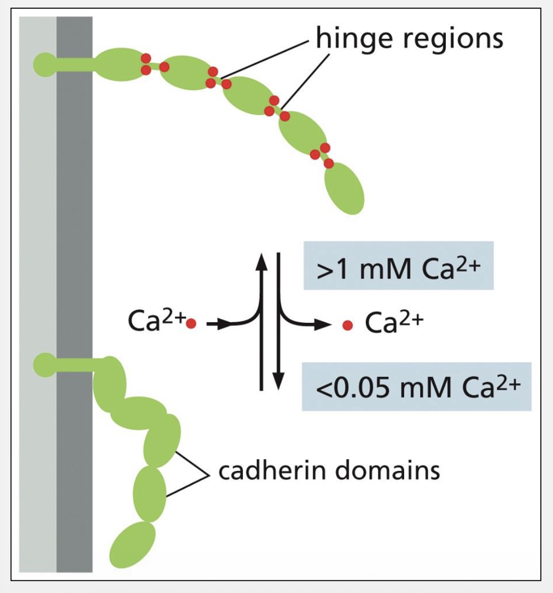
Cadherin adherens junctions link what cytoskeleton?
Actin via catenins
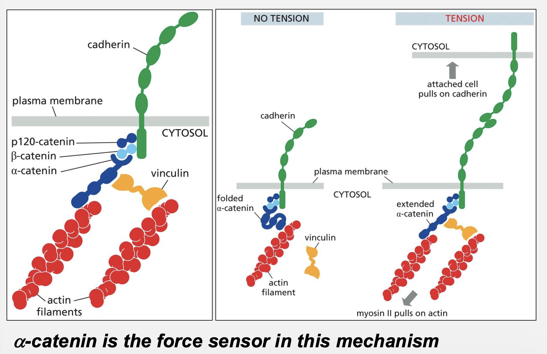
What is the force sensor in cadherin junctions?
α‑catenin

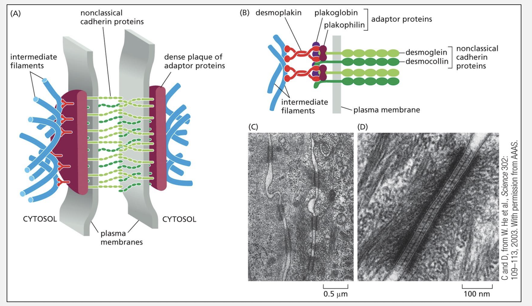
Desmosomes link which cytoskeleton?
Intermediate filaments
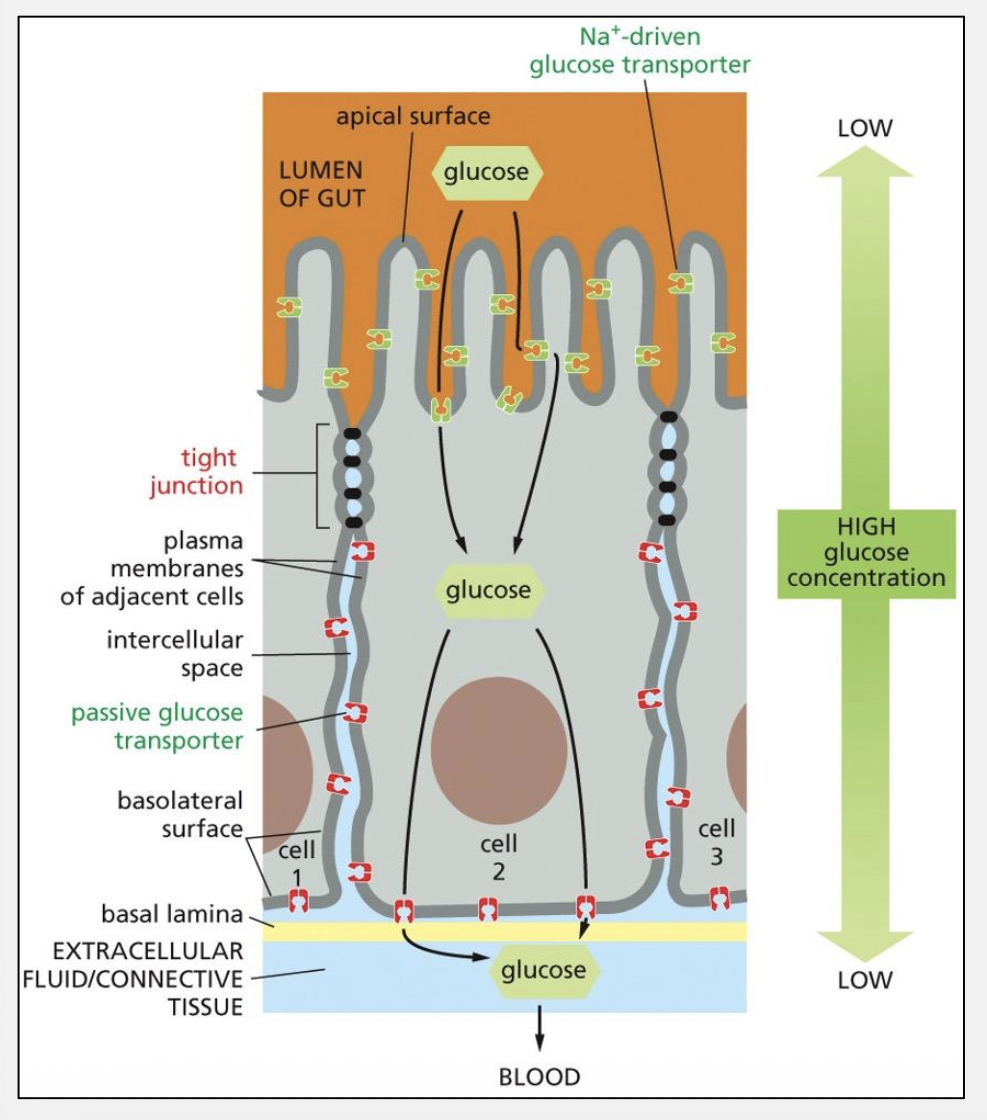
Tight junction barrier vs fence function?
Barrier: Blocks paracellular diffusion
Fence: Maintains membrane polarity
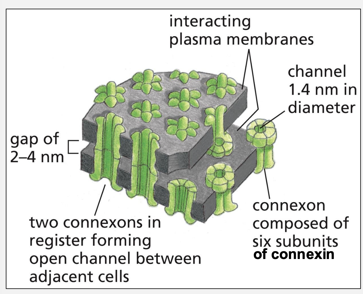
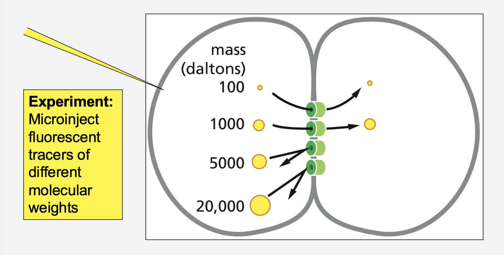
What is the functional unit of a gap junction?
Connexon (6 connexins)
ECM components providing tensile strength, compression resistance, elasticity?
Collagen: Tensile strength
GAGs/PGs: Compression resistance
Elastin: Elastic recoil
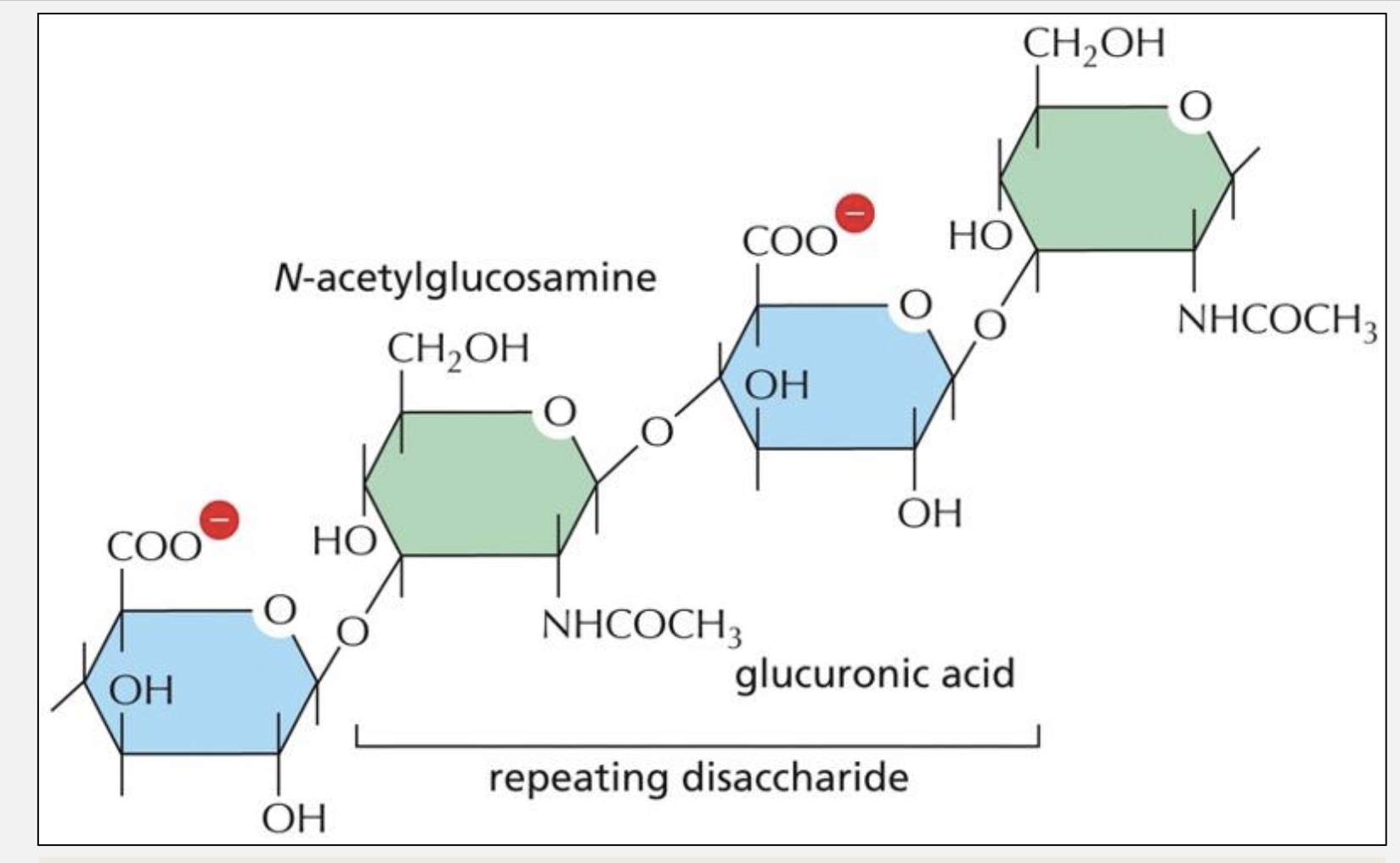
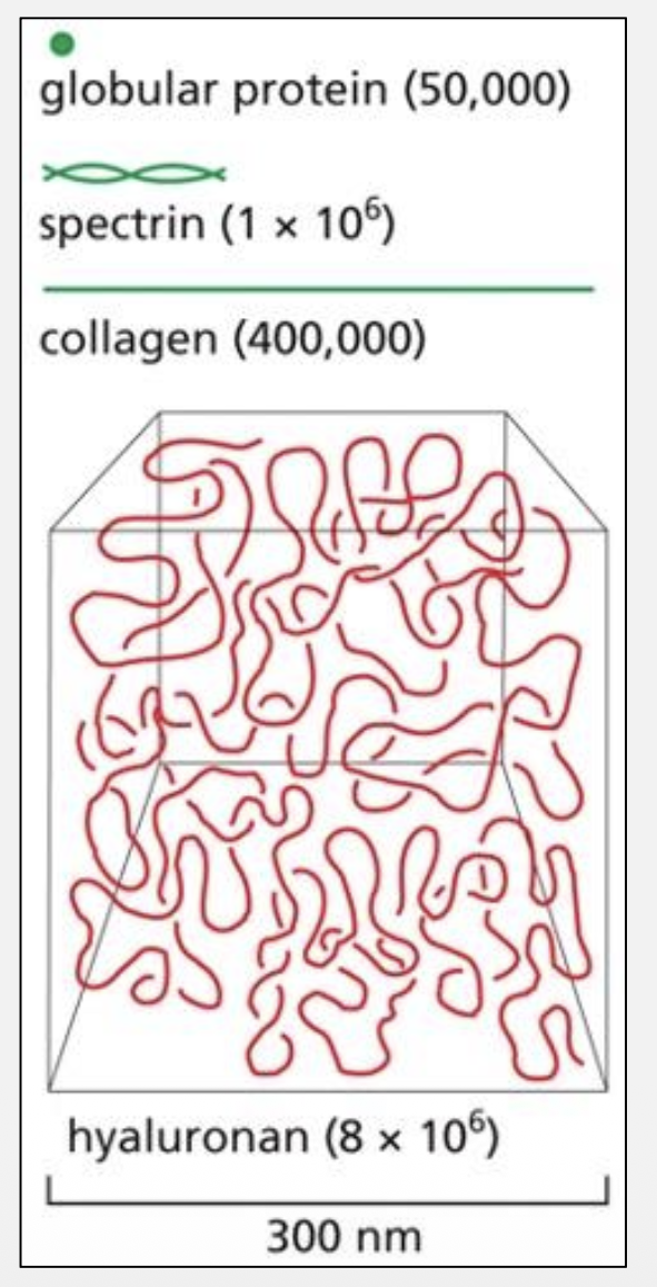
Why does HA fill space so effectively?
Large size, negative charge, hydration shell
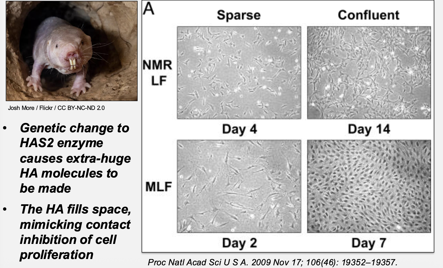
Why are naked mole rats cancer‑resistant?
Extra‑long HA mimics contact inhibition
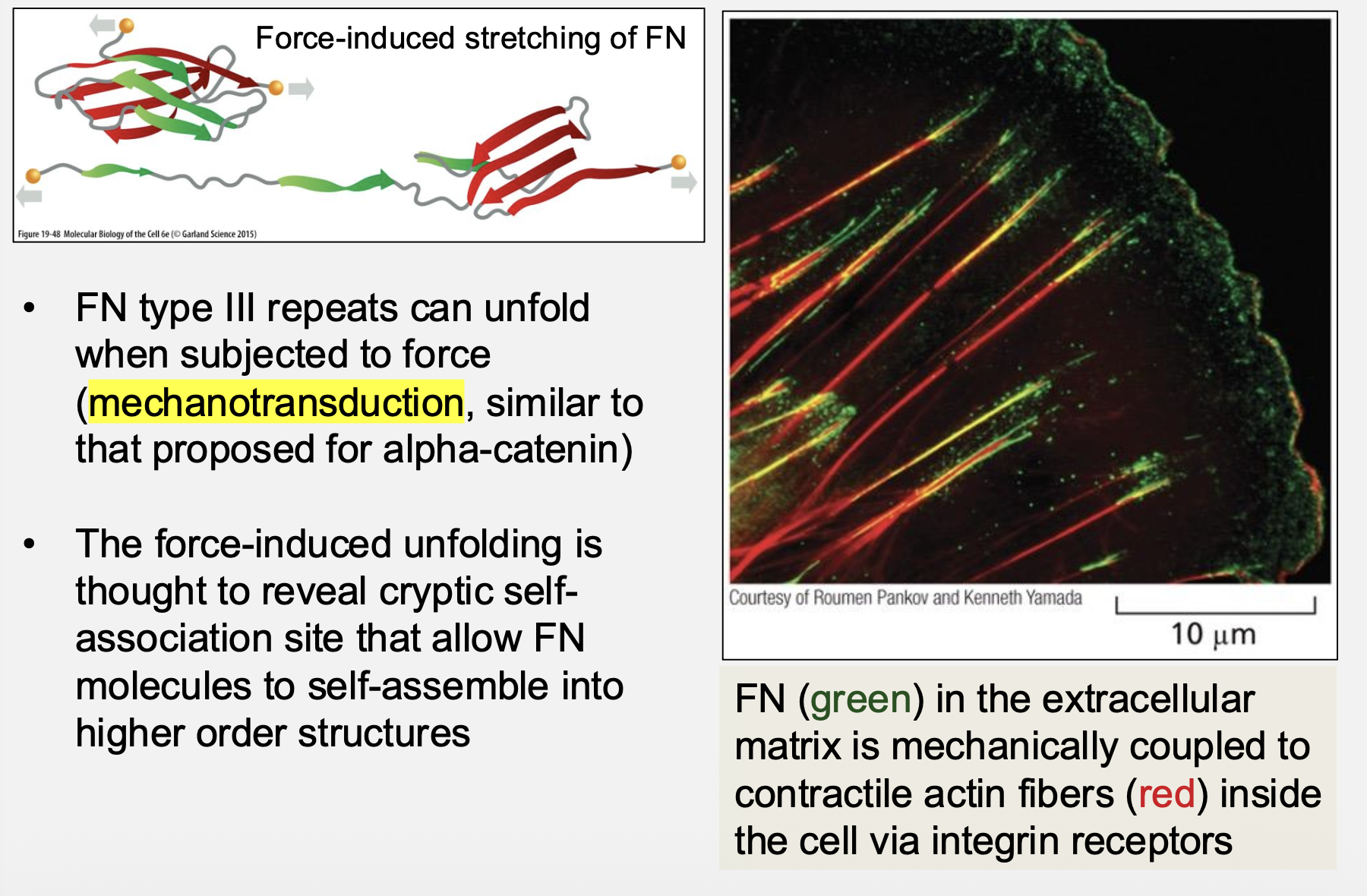
How does fibronectin organize ECM via mechanotransduction?
Force‑induced unfolding reveals cryptic binding sites