Diffuse and focal abnormalities of the spleen
 Call Kai
 Learn
Learn Practice Test
 Spaced Repetition
 Match
Match Flashcards
 Knowt Play
1/65

There's no tags or description
Looks like no tags are added yet.
Name | Mastery | Learn | Test | Matching | Spaced | Call with Kai | Chat |
|---|
No analytics yet
Send a link to your students to track their progress
66 Terms
Most frequent LUQ mass
Splenomegaly
Most common splenic abnormality
Splenomegaly
Splenomegaly is most commonly caused by
Portal HTN
Splenomegaly
Enlarged spleen >13cm in length
Usually caused by portal HTN with cirrhosis
Asher sign - In supine in trans, upper border of spleen extends across the anterior aorta by more than 2cm = splenomegaly
Causes of splenomegaly
Congestive vascular disorder, lymphoma, schistosomiasis, leukemia, infection (AIDS, epstein-Barr) Hemolytic anemia, sickle cell anemia, mets, collagen disease, polycythemia
USA splenomegaly
Length >13cm
Thickness >5cm
Hypoechoic with malignant processes
Hyperechoic with inflammatory processes and in radiation therapy
Enlarges inferiorly and medially
Mild to moderate = infection, portal hypertension and AIDS
Massive = >20cm
Focal lesions may cause splenomegaly
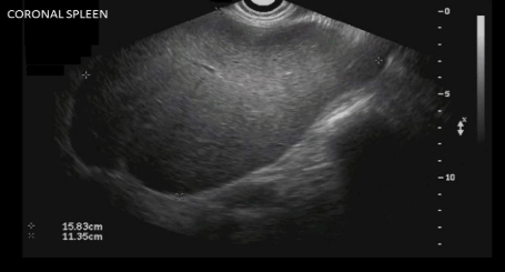
Splenomegaly
Sarcoidosis
Inflammatory disease that causes all clumping in certain organs
The immune system is overactive nodules called granulomas
Multiple hypoechoic nodules throughout the spleen
When granulomas heal, they become calcified
Leads to multiple echogenic foci throughout the spleen, some may cause shadowing
Infections in spleen
Spleen function includes WBC production and filtration
Infection of the spleen may be localized or imported from other areas
Mononucleosis
Mononucleosis
Causes significant increase in spleen sixe
Associated with Epstien Barr infection → FF seen and splenomegaly
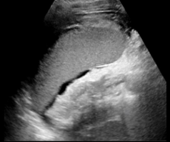
Epstein-Barr infection
AIDS
Moderate splenomegaly in 50-70% of pys
Splenomegaly is noted more in pts with sexually transmitted HIV than in those acquiring the disease through IV use
AIDS is associated with
Hepatomegaly, hepatitis, splenomegaly, candidiasis, pneumocystis carinii (MOST COMMON INFECTION OF AIDS), fatty infiltration of liver, kaposi sarcoma, cholangitis, acalculous cholecystitis, non-hodgkin lymphoma, anemia
Granulomatous infections
Systemic fungal respiratory disease caused by bird, bat and other animal droppings
Associated with splenomegaly, sarcoidosis and brucellosis infections
Granulomatous infections are most commonly caused by
Histoplasmosis and tuberculosis
Granulomatous infections USA
Multiple hypoechoic nodules <1cm in size
When nodules heal, multiple echogenic foci form in the spleen and liver
Portal HTN Calcifications
Increased blood volume in the spleen
Gamma-Gandy bodies are caused by microhemorrhaging and infarction
Organized foci of hemorrhage in the spleen most commonly seen with portal HTN
Contains hemosiderin, fibrous tissue and calcium
Vascular calcifications
Linear calcs that follow course of vessel
USA splenic calcifications
Diffuse echogenic foci within splenic tissue
With or without shadowing
May see twinkle with color
What causes splenic calcs
Granulomatous infections
Portal HTN
Vascular calcs
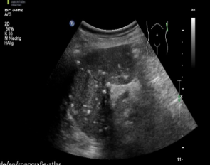
Granulomas
Splenic abscess
Rare
May see them post-splenectomy
Decreased hematocrit, fever, increased WBCs and possible septicemia
Splenic abscess is most commonly associated with
Endocarditis, septicemia, and trauma
Splenic abscess USA
Complex fluid collection with internal echoes
Irregular borders
May see septations and pleural effusion
Dirty shadowing from gas
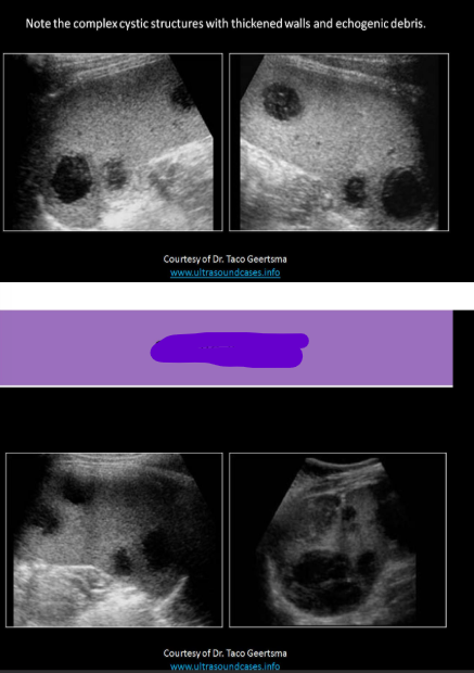
Splenic abscess
Benign neoplasms of spleen are very _____
Rare
Most common benign tumor of spleen
Cystic cavernous hemangioma
Cystic cavernous hemangioma
Composed of high concentration of vascular tissues
Associated with Kasabach - Merritt syndrome and Klippel-Trenaunay-Weber syndrome
Hamartoma
Most commonly INCIDENTAL
Solitary mass but can cause formation of multiple nodules in pts with tuberous sclerosis
Compresses and displaces normal splenic tissue
Hamartoma USA
Round, well-circumscribed, unencapsulated bulging nodules
Hypoechoic or homogeneous
Hypervascular on color
Gaucher disease
Massive splenomegaly due to hypertension
Can see multiple well defined, hypoechoic nodules
Requires splenectomy
Osteoma
Comes from bone
Fibroma
Fibrous tissues
Chondroma
Cartilaginous tumor
Cystic hemangioma
Congenital defect in lymphatic vessels in spleen
Granulomas
Calcifications
Most common splenic malignancy
Lymphoma
Lymphoma
Tumor of lymphatic system
Commonly involves spleen
Marked splenomegaly
Hodgkin
Non-Hodgkin
Hodgkin lymphoma
Most commonly occurs in pts age 15-24 yrs and over 60 yrs
Marked by mature, malignant B cells called Reed-Sternberg cells
Leukocytosis, anemia
More commonly affects lymphnodes in arms, upper chest and neck
Considered one of the most treatable cancers
Non-Hodgkins lymphoma
MOST COMMON TYPE
Average pt age 60 yrs
Elevated lactate dehydrogenase, anemia, leukopenia
No pattern for lymph node involvement; occurs in any lymph nodes throughout body
Much less responsive to treatment than Hodgkin’s
USA lymphoma (4 patterns)
Diffuse, marked splenomegaly, patchy inhomogeneity
Multiple small hypoechoic lesions, miliary nodules
Multiple large lesions 1-10cm
Single bulky mass
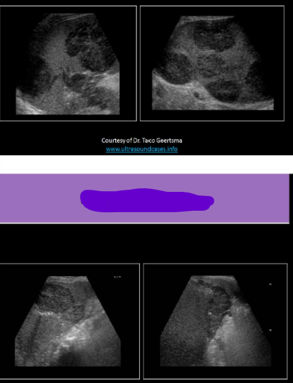
Lymphoma (Hodgkins)
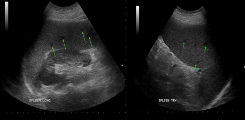
Lymphoma
Leukemia
Cancer of WBCs
Night sweats, fatigue, weakness, unintentional weight loss, painless, swollen lymphnodes, enlargement of liver or spleen, fever, chills
Lymphocytic
Myelogenous
Myelogenous leukemia
Cancer of myeloid cells
Acute or chronic
Acute form is most common leukemia
Lymphocytic leukemia
Cancer of the lymphocytes
Acute or chronic
leukemia Labs
Complete blood count used to eval leukemia
Low platelet count
Low RBC
High WBC
Low sedimentation rate
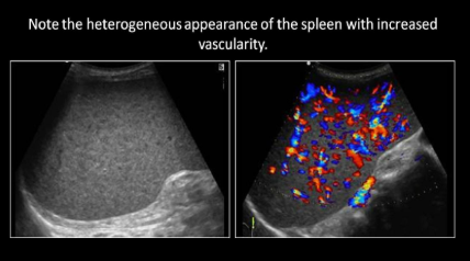
Leukemia
Angiosarcoma
Primary malignant neoplasm of the spleen
Very rare aggressive malignancy
High rate of mets and poor prognosis
Arises from the vascular endothelium
Anemia, LUQ pain, malaise, fever weight loss
High incidence of splenic rupture
Angiosarcoma USA
Splenomegaly
Heterogeneous echotexture
Complex mass with cystic and solid components
Increased vascularity
Most likely primary cancer to spread to spleen
Melanoma
Most common primary cancers seen with splenic mets
Mets from lung or breast
USA mets to spleen
Hypoechoic most common, Bulls eye appearance
Cystic or hyperechoic
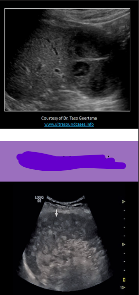
Splenic mets
Perisplenic abnormalities
Lesions of pancreas, left kidney, left adrenal and left lobe of the liver may impinge upon spleen and mimic primary splenic disease
Pancreas
Lesions of body or tail mat mimic primary splenic process
Renal
Upper pole of left kidney impinges on splenic hilum
Renal cyst may indent the splenic parenchyma
Adrenal
Left adrenal gland may be source of tumor or cyst that intrinsically compresses the spleen
A hypoechoic enlarged spleen is typically caused by ______________, while a hyperechoic enlarged spleen is typically caused by_______
Malignancy, infection
Massive splenomegaly (>20cm) can be caused by
Thalassemia major
Lymphoma
Gaucher disease
The most common splenic infection in an AIDS pt is ___________ and the most common malignancy of the spleen in an AIDS pt is ___________
Pneumocystis carinii; Kaposi sarcoma
Gamna Gandy bodies are most commonly seen with
Portal HTN
What splenic abnormality is associated with tuberous sclerosis or Wiskott-Aldrich syndrome
Hamartoma
Hodgkin lymphoma typically causes lymphadenopathy in the ________, while non H lymphoma typically causes lymphadenopathy in the _______
Upper body; Whole body
Pts with angiosarcoma have an increased risk of:
Splenic rupture