Biology of tooth movement and anchorage

0.0(0)
Studied by 0 people
call kaiCall Kai
Locked
learnLearn
examPractice Test
spaced repetitionSpaced Repetition
heart puzzleMatch
flashcardsFlashcards
GameKnowt Play
Card Sorting

1/49

encourage image

There's no tags or description

Looks like no tags are added yet.

Last updated 11:14 AM on 9/7/26
Name
Mastery
Learn
Test
Matching
Spaced
Call with Kai
Chat

No analytics yet

Send a link to your students to track their progress

50 Terms

1
New cards

Composition of bone? (3 main components)

  • Matrix - 35% organic phase mainly collagen type 1 and other proteins such as osteonectin, osteocalcin, GAG’s, proteoglycan, GF’s

  • 65% Mineral phase - HAP, calcium, phosphate

  • Cellular - osteoblasts, osteoclasts, osteocytes

  • osteoblasts - mesenchymal cells

  • osteoclasts - macrophage lineage

  • Mature osteoblasts in calcified bone matrix lying in lacunae and communicate with each other via canaliculi


2
New cards

Bone strucutre?

  • Cortical/compact bone - hard dense bone - 80-90% calcified - lamellar organisation of collagen in mature bone matrix, parallel or concentric organisation - surrounding channel centred on blood vessel known as haversion system

  • Cancellous/spongy bone - 15-25% - containing bone marrow/connective tissue and blood vessels in the spaces


<ul><li><p>Cortical/compact bone - hard dense bone - 80-90% calcified - lamellar organisation of collagen in mature bone matrix, parallel or concentric organisation - surrounding channel centred on blood vessel known as haversion system</p></li><li><p>Cancellous/spongy bone - 15-25% - containing  bone marrow/connective tissue and blood vessels in the spaces</p></li></ul><p></p>
3
New cards
<p>Which is compact and which is cancellous bone?</p>

Which is compact and which is cancellous bone?

knowt flashcard image
4
New cards

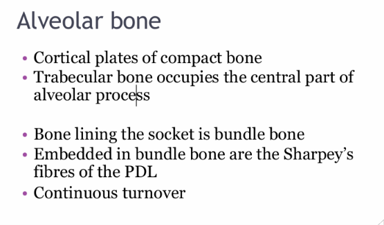
What is the structure of alveolar bone?

comment on the turnover of bone

  • Cortical plates of compact bone

  • Trabecular bone occupies the central part of alveolar process

  • Bone lining the socket is bundle bone

  • Embedded in the bundle bone are the Sharpey’s fibres of the pDL

  • there is continuous turnover


<ul><li><p>Cortical plates of compact bone</p></li><li><p>Trabecular bone occupies the central part of alveolar process</p></li><li><p>Bone lining the socket is bundle bone</p></li><li><p>Embedded in the bundle bone are the Sharpey’s fibres of the pDL </p></li><li><p>there is continuous turnover</p></li></ul><p></p>
5
New cards

Again what are the 3 bone cells?

  • Osteoblasts -

  • osteoclasts - bone lining cells

  • osteocytes - encased in bone matrix


6
New cards
<p>Where in this photo are the osteoblasts situated?</p>

Where in this photo are the osteoblasts situated?

knowt flashcard image
7
New cards

Osteoblasts synthesis: (what type of organic protein, other proteins, an important indicator of bone metabolism and receptors for (4))

  • Type 1 collagen

  • Bone matrix proteins - ostecalcin/pontin, bone sialoprotein

  • alkaline phosphatase

  • PTH, vitamin D3, growth factors, cAMP response to PTH


<ul><li><p>Type 1 collagen</p></li><li><p>Bone matrix proteins - ostecalcin/pontin, bone sialoprotein</p></li><li><p>alkaline phosphatase </p></li><li><p>PTH, vitamin D3, growth factors,  cAMP response to PTH</p></li></ul><p></p>
8
New cards
<p>Where are the osteocytes?</p>

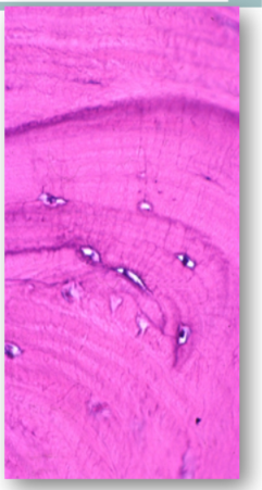
Where are the osteocytes?

knowt flashcard image
9
New cards

Osteocytes

abundance?

arrangement and role?

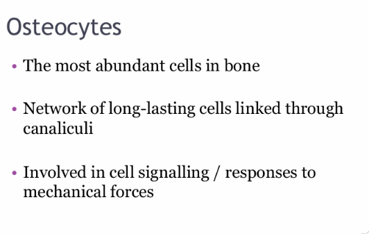
  • The most abundant cells in the bone

  • Network of long-lasting cells linked through canaliculi

  • Involved in cell signalling and response to mechanical forces

<ul><li><p>The most abundant cells in the bone</p></li><li><p>Network of long-lasting cells linked through canaliculi </p></li><li><p>Involved in cell signalling and response to mechanical forces</p></li></ul><p></p>
10
New cards
<p>label</p>

label

Osteocytes are arranged around the vascular canals forming the harversion systems

<p>Osteocytes are arranged around the vascular canals forming the  harversion systems</p>
11
New cards
<p>What are these? where are the osteoclasts in the top image?</p>

What are these? where are the osteoclasts in the top image?

Osteoclasts

<p>Osteoclasts</p>
12
New cards

Morphology of osteoclasts and where they are usually located?

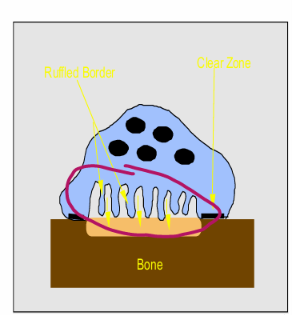
  • Multinucleated with ruffled border

  • Adjacent to resorbed bone

  • Bone lining cells, in contact with calcified bone surface and in lacunae

  • 4-20 nuclei, giant cells


<ul><li><p><strong>Multinucleated with ruffled border</strong></p></li><li><p><strong>Adjacent to resorbed bone</strong></p></li><li><p>Bone lining cells, in contact with calcified bone surface and in lacunae</p></li><li><p>4-20 nuclei, giant cells</p></li></ul><p></p>
13
New cards

Osteoclasts produce what important enzyme? have what receptors that reduced their activity? osteoclasts are responsive to what hormones for what response?

  • TRAP - differentiation, activation and proliferation of OC’s

  • Calcitonin receptors - reduces their activity

  • Responsive to calcitropic hormones - bone growth and remodelling


<ul><li><p><strong>TRAP -</strong> differentiation, activation and proliferation of OC’s</p></li><li><p><strong>Calcitonin receptors -</strong> reduces their activity</p></li><li><p>Responsive to<strong> calcitropic</strong> hormones - bone growth and remodelling </p></li></ul><p></p>
14
New cards

Mechanism of bone resorption:

Osteoclasts are recruited through?

They attach to what?

What does the osteoclast secrete?

What can the osteoclasts resorb and not resorb and how is this managed?

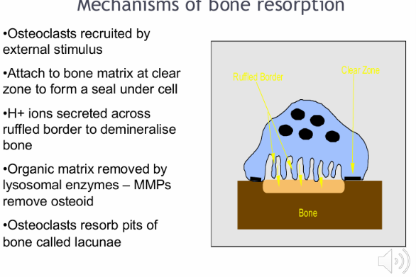
  • External stimulus

  • Bone matrix at clear zone to form a seal under cell

  • Secrete hydrogen ions across ruffled border to demineralise bone

  • Cannot break down unmineralized organic matrix aka osteoid so MMPs and other collagenase enzymes must remove the osteoid first to allow it to resorb the mineralised bone

  • Osteoclasts resorb pits of bone called lacunae

<ul><li><p>External stimulus</p></li><li><p>Bone matrix at clear zone to form a seal under cell</p></li><li><p>Secrete hydrogen ions across ruffled border to demineralise bone</p></li><li><p>Cannot break down unmineralized organic matrix aka osteoid so MMPs and other collagenase enzymes must remove the osteoid first to allow it to resorb the mineralised bone </p></li><li><p>Osteoclasts resorb pits of bone called lacunae</p></li></ul><p></p>
15
New cards
<p>Explain how osteoclastic activation required Osteoblasts?</p>

Explain how osteoclastic activation required Osteoblasts?

  • Formation of osteoclast progenitors or precursor cells in hematopoietic tissues from a macrophage lineage

  • and the development of resting pre-osteoclasts and osteoclasts in the bone itself

  • 2 Preparation of the bone surface, osteoblasts facilitate the removal of unmineralized osteoid layer by producing enzymes such as MMPs and other collagenases

  • This allows osteoclasts access to the mineralised bone and ensuing resorption

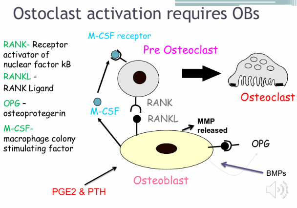
  • Osteoblasts have receptors for certain bone resorption agents such as prostaglandins, vit D3 and PTH

  • Osteoblasts then transmit resorptive signals to the osteoclast to activate them:

  • RankL on osteoblasts connects with RANK receptor on Pre osteoclasts and promotes osteoclast differentiation

  • As a control mechanism, OPG is a decoy receptor and binds RANKL and inibits OC activations

  • Also, M-CSF - stimulates the differentiation and proliferation of pre-osteoclasts

  • Osteoblasts stimulated by bone resorbing hormones causes release of MMP and allow for osteoclastic activity to ensue by removing osteoid layer

  • 3 - Osteoclasts resorb bone, ruffled border with finger like projections to create increased SA to facilitate exchanges of materials

  • 4 - OC activity decreases and osteoblast precursor cells appear in area of resorption and differentiate locally into osteoblasts - mediated by BMPs and growth factors the cells become completely embedded to become osteocytes until which they produce collagen/osteoid

  • TNF released from local cells inhibit continued production of bone

<ul><li><p><strong>Formation of osteoclast progenitors or precursor cells in hematopoietic tissues from a macrophage lineage</strong></p></li><li><p>and the development of resting pre-osteoclasts and osteoclasts in the bone itself</p></li><li><p>2 Preparation of the bone surface, osteoblasts facilitate the removal of unmineralized osteoid layer by producing enzymes such as MMPs and other collagenases</p></li><li><p>This allows osteoclasts access to the mineralised bone and ensuing resorption </p></li><li><p><strong>Osteoblasts have receptors for certain bone resorption agents such as prostaglandins, vit D3 and PTH</strong></p></li><li><p>Osteoblasts then transmit resorptive signals to the osteoclast to activate them:</p></li><li><p><strong>RankL on osteoblasts connects with RANK receptor on Pre osteoclasts and promotes osteoclast differentiation </strong></p></li><li><p><strong>As a control mechanism, OPG is a decoy receptor and binds RANKL and inibits OC activations </strong></p></li><li><p><strong>Also, M-CSF - stimulates the differentiation and proliferation of pre-osteoclasts </strong></p></li><li><p><strong>Osteoblasts stimulated by bone resorbing hormones causes release of MMP and allow for osteoclastic activity to ensue by removing osteoid layer</strong></p></li><li><p>3 - Osteoclasts resorb bone, ruffled border with finger like projections to create increased SA to facilitate exchanges of materials </p></li><li><p>4 - OC activity decreases and osteoblast precursor cells appear in area of resorption  and differentiate locally into osteoblasts - mediated by BMPs and growth factors the cells become completely embedded to become osteocytes until which they produce collagen/osteoid</p></li><li><p>TNF released from local cells inhibit continued production of bone</p></li></ul><p></p>
16
New cards

Again, how do OBs regulate OCs?

what acts against the OCs but acting on the OBs?

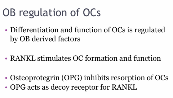
  • Differentiation and function is regulated through OB derived factors (RANKL M-CSF)

  • RANKL stimulated formation and function of OCs

  • OPG by acting as a decoy receptor for the RANKligan

<ul><li><p>Differentiation and function is regulated through OB derived factors (RANKL M-CSF)</p></li><li><p>RANKL stimulated formation and function of OCs</p></li><li><p>OPG by acting as a decoy receptor for the RANKligan</p></li></ul><p></p>
17
New cards

What is the with of the PDL?

What are some functions of the PDL? (5)

what is it primarily composed of?

What 2 undifferentiated cells does it contain and these are responsible for what 2 processes?

What property does the pdl have? (one word)

  • 0.5 mm

  • Tooth support, cementum ligament and bone repair, sensory: pressure pain proprioception, orthodontic tooth movement, dissipation of forces, shock absorption

  • Collagen type 1

  • Undifferentiated fibroblasts and osteoblasts

  • Ligament cementum and bony socket remodelling

  • Fluid visco-elastic properties


<ul><li><p>0.5 mm</p></li><li><p>Tooth support, cementum ligament and bone repair, sensory: pressure pain proprioception, orthodontic tooth movement, dissipation of forces, shock absorption</p></li><li><p>Collagen type 1</p></li><li><p>Undifferentiated fibroblasts and osteoblasts</p></li><li><p>Ligament cementum and bony socket remodelling </p></li><li><p>Fluid visco-elastic properties </p></li></ul><p></p>
18
New cards

What is the zone of equilibrium?

Teeth appear to lie in a position of balance between the tongue on one side and lips/cheeks on the other

19
New cards

How does the PDL influence this position of balance?

  • The PDL has an intrinsic force which needs to be overcome before teeth move

  • Loss of PDL e.g in periodontal disease, means less force and therefore uncontrolled movement of teeth

  • Lack of PDL means there is a lack of resistance to constant soft issue forces on teeth with influence on tooth position as a result


<ul><li><p>The PDL has an intrinsic force which needs to be overcome before teeth move</p></li><li><p>Loss of PDL e.g in periodontal disease, means less force and therefore uncontrolled movement of teeth</p></li><li><p>Lack of PDL means there is a lack of resistance to constant soft issue forces on teeth with influence on tooth position as a result</p></li></ul><p></p>
20
New cards
<p>What is this picture showing?</p>

What is this picture showing?

Zone of equilibrium (depiction of forces applied to teeth externally)

21
New cards

What happens when light forces are applied to teeth?

1-2 sec

5 sec

minutes

hours

2-3 days

5-14 days

14-30 days

  • pdl fluid expressed

  • pdl fibres and cells compressed

  • altred blood flow - pg an ck released

  • metabolic changes - ob and oc activity

  • frontal resorption and slight tm

  • lamina dura resorption - no otm until this is removed

  • otm occurs again - resorption an deposition


  • Blood flow altered due to compression and tension forces and distortion of pdl fibres and cells

  • oxygen levels altered signalling proteins such as ck and pg are released

  • metabiotic changes occur over next few hours, enzyme levels change and chemical changes

  • cellular diff occurs within pdl and days later oc and ob are required and tooth movement occurs

  • teeth move by frontal resorption on the compression side

  • 4 week period is required for removal of hyalinised bone and for pdl to recover

  • so pts are reviews on avg every 4-6 weeks to allow for tm to occur in the appropriate time required for bone and pdl turnover


<ul><li><p>pdl fluid expressed</p></li><li><p>pdl fibres and cells compressed</p></li><li><p>altred blood flow - pg an ck released</p></li><li><p>metabolic changes - ob and oc activity</p></li><li><p>frontal resorption and slight tm</p></li><li><p>lamina dura resorption - no otm until this is removed</p></li><li><p>otm occurs again - resorption an deposition </p></li><li><p></p></li><li><p></p></li><li><p>Blood flow altered due to compression and tension forces and distortion of pdl fibres and cells</p></li><li><p>oxygen levels altered signalling proteins such as ck and pg are released</p></li><li><p>metabiotic changes occur over next few hours, enzyme levels change and chemical changes</p></li><li><p>cellular diff occurs within pdl and days later oc and ob are required and tooth movement occurs</p></li><li><p>teeth move by frontal resorption on the compression side</p></li><li><p>4 week period is required for removal of hyalinised bone and for pdl to recover</p></li><li><p>so pts are reviews on avg every 4-6 weeks to allow for tm to occur in the appropriate time required for bone and pdl turnover </p></li></ul><p></p>
22
New cards

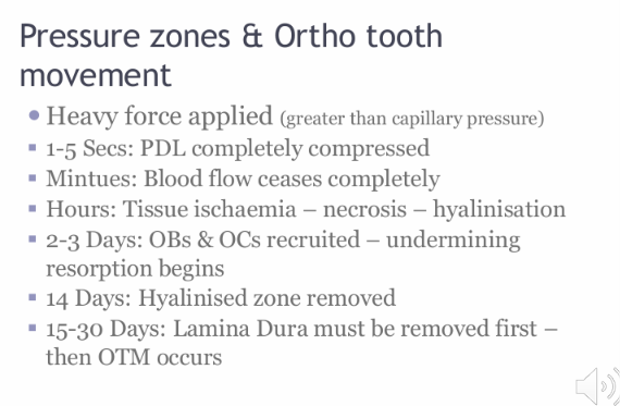
What happens when heavy forces (greater than capillary pressure) areapplied to tooth?

1-5 second

minutes

hours

2-3 days

14 days

15-30 days

  • pdl completely compressed

  • blood flow completely ceases

  • tissue ischaemia and necrosis - hyalinisation

  • ob/oc requited mediating undermining resorption (in adj area)

  • hyalinsation zone is removed

  • OTM occurs

  • in first few seconds blood flow completely occluded to the local area

  • hours later ischaemia results in cell death and tissue necrosis or areas of hyalinisation

  • OC and OB activity cannot occur in this area

  • days later these cells are recruited to adjacent bone areas to initiate undermining resorption

  • weeks later lamina dura adjacent to pdl is removed and tooth movement finally occurs

<ul><li><p>pdl completely compressed</p></li><li><p>blood flow completely ceases</p></li><li><p>tissue ischaemia and necrosis - hyalinisation</p></li><li><p>ob/oc requited mediating undermining resorption (in adj area)</p></li><li><p>hyalinsation zone is removed</p></li><li><p>OTM occurs</p></li><li><p>in first few seconds blood flow completely occluded to the local area</p></li><li><p>hours later ischaemia results in cell death and tissue necrosis or areas of hyalinisation </p></li><li><p>OC and OB activity cannot occur in this area</p></li><li><p>days later these cells are recruited to adjacent bone areas to initiate undermining resorption  </p></li><li><p>weeks later lamina dura adjacent to pdl is removed and tooth movement finally occurs</p></li></ul><p></p>
23
New cards

What are the optimal forces required for OTM? as it reduces risk of what?

  • Just greater than resting capillary pressure

  • to reduce risk of tissue and cell necrosis, pulp damage, pt discomfort

  • for optimal OTM


<ul><li><p>Just greater than resting capillary pressure</p></li><li><p>to reduce risk of tissue and cell necrosis, pulp damage, pt discomfort</p></li><li><p>for optimal OTM</p></li></ul><p></p>
24
New cards

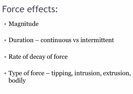
force effects depend on what? (4)

  • Magnitude

  • Rate of decay of the force

  • Duration - Continuous vs intermittent

  • Type of force - tipping, intrusion, extrusion, bodily, torque

<ul><li><p>Magnitude</p></li><li><p>Rate of decay of the force</p></li><li><p>Duration - Continuous vs intermittent</p></li><li><p>Type of force - tipping, intrusion, extrusion, bodily, torque</p></li></ul><p></p>
25
New cards
<p>What does centre of resistance mean?</p>

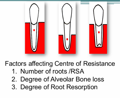
What does centre of resistance mean?

  • It is the point on the tooth when a single force is passed through it, would bring about its translation along the line of action of the force


<ul><li><p>It is the point on the tooth when a single force is passed through it, would bring about its translation along the line of action of the force </p></li></ul><p></p>
26
New cards

What are factors that affect the centre of resistance? (3)

  • Number of roots/root surface area

  • Degree of alveolar bone loss

  • Degree of root resorption


<ul><li><p>Number of roots/root surface area</p></li><li><p>Degree of alveolar bone loss</p></li><li><p>Degree of root resorption </p></li></ul><p></p>
27
New cards
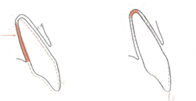
<p>What type of force is being applied in this image?</p><p>how many areas of compression?</p>

What type of force is being applied in this image?

how many areas of compression?

  • Tipping

  • The force is not applied through the centre of resistance

  • Therefore the rotation of the tooth is around the point at which the force is applied

  • 2 areas of compression


28
New cards
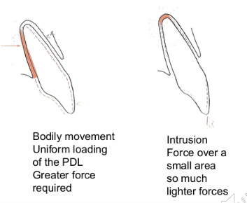
<p>What types of forces are these?</p><p>how much force does each need and what is the general rule?</p>

What types of forces are these?

how much force does each need and what is the general rule?

  • Bodily and intrusion forces

  • the more pdl surface involved the more force required to generate the desired tooth movement

<ul><li><p>Bodily and intrusion forces</p></li><li><p>the more pdl surface involved the more force required to generate the desired tooth movement</p></li></ul><p></p>
29
New cards

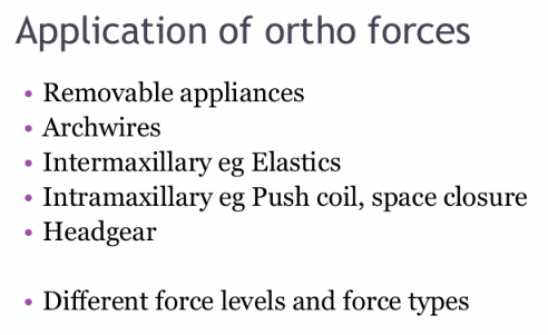
What can be used to apply ortho forces? (appliances) (5)

  • Removable appliance

  • Archwires (fixed)

  • Headgears

  • Intermaxillary - elastics

  • Intramaxillary - push coild


<ul><li><p>Removable appliance</p></li><li><p>Archwires (fixed)</p></li><li><p>Headgears</p></li><li><p>Intermaxillary - elastics</p></li><li><p>Intramaxillary - push coild</p></li></ul><p></p>
30
New cards

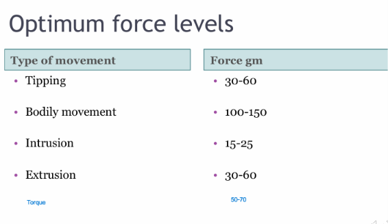
Optimum force levels for tipping, bodily, intrusion, extrusion and torque?

  • bodily movement

  • torque

  • tipping , extrusion

  • intrusion


<ul><li><p>bodily movement</p></li><li><p>torque</p></li><li><p>tipping , extrusion</p></li><li><p>intrusion</p></li><li><p></p></li></ul><p></p>
31
New cards

Optimum force levels are determined by what? (4)

  • Type of force

  • PDL/surface area

  • Bone levels

  • RSA and root number


<ul><li><p>Type of force</p></li><li><p>PDL/surface area</p></li><li><p>Bone levels</p></li><li><p>RSA and root number </p></li></ul><p></p>
32
New cards

What are the ideal visit intervals?

  • 4 weeks minimum to allow repair mechanisms for hyalinisation ears


<ul><li><p>4 weeks minimum to allow repair mechanisms for hyalinisation ears </p></li></ul><p></p>
33
New cards

What is anchorage?

  • The resistance to unwanted tooth movement

based on newtons third law that for every action there is an equal and opposite action

34
New cards

Why is anchorage important?

what can be used (4)

  • To present wastage of space gained from xla not being used to align the teeth

  • Transpalatal arch, nance appliance, tad and headgear


<ul><li><p>To present wastage of space gained from xla not being used to align the teeth</p></li><li><p>Transpalatal arch, nance appliance, tad and headgear </p></li></ul><p></p>
35
New cards

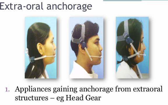
Headgear

what is it attached to?

what 2 things its used for?

  • extra oral anchorage

  • Kloehn bow attaches to headgear tubes on upper 6’s bands to prevent mesial molar movement

  • can also be used to distalise molars by increasing the forces to create space


<ul><li><p>extra oral anchorage</p></li><li><p>Kloehn bow attaches to headgear tubes on upper 6’s bands to prevent mesial molar movement</p></li><li><p>can also be used to distalise molars by increasing the forces to create space </p></li></ul><p></p>
36
New cards
<p>What are these headgears called? </p>

What are these headgears called?

  • cervical pull or Low pull head gear

  • Occipital head gear - high pull

  • Combination pull head gear


<ul><li><p>cervical pull or Low pull head gear</p></li><li><p>Occipital head gear - high pull</p></li><li><p>Combination pull head gear</p></li></ul><p></p>
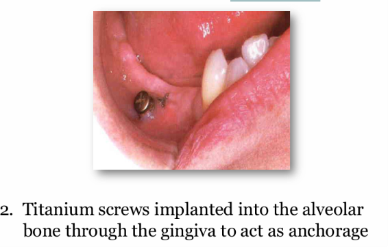
37
New cards
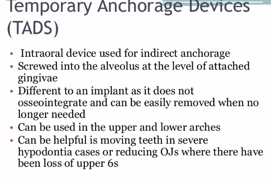
<p><u>Temporary anchorage devices:</u></p><p>What type of anchorage do they provide?</p><p>How is it attached?</p><p>How is it different to an implant?</p><p>what arches can it be used in?</p><p>What is it used in?</p>

Temporary anchorage devices:

What type of anchorage do they provide?

How is it attached?

How is it different to an implant?

what arches can it be used in?

What is it used in?

  • Intraoral device used for indirect anchorage

  • Screwed into the alveolus at the level of the attached gingivae

  • It doesn’t Osseointegrate and can be easily removed when no longer needed

  • upper and lower

  • moving teeth in severe hypodontia, reducing OJs in loss of 6’s

<ul><li><p>Intraoral device used for indirect anchorage</p></li><li><p>Screwed into the alveolus at the level of the attached gingivae</p></li><li><p>It doesn’t Osseointegrate and can be easily removed when no longer needed</p></li><li><p>upper and lower</p></li><li><p>moving teeth in severe hypodontia, reducing OJs in loss of 6’s</p></li></ul><p></p>
38
New cards

Intraoral anchorage:

How is the anchorage value for a tooth calculated?

  • Anchorage value of a tooth is proportional to the surface area of the root

  • The tooth with larger root surface area requires greater force to move an therefore higher anchor value


<ul><li><p>Anchorage value of a tooth is proportional to the surface area of the root</p></li><li><p>The tooth with larger root surface area requires greater force to move an therefore higher anchor value </p></li></ul><p></p>
39
New cards

anchorage value of each tooth

knowt flashcard image
40
New cards

What 2 conditions affecting a tooth can make them good sources of anchorage?

Difference in Crown root angulation - dilaceration

ankylosis - fused to bone

<p>Difference in Crown root angulation - dilaceration</p><p>ankylosis - fused to bone</p>
41
New cards

What is a common risk factor associated with ortho tx?

Root resorption

42
New cards

How much mm of root from the apex is usually lost

1-2 mm

43
New cards
<p>What are some risk factors associated with root resorption? (10)</p>

What are some risk factors associated with root resorption? (10)

  • long time

  • class 2 elastics

  • roots into cortical plate

  • nail biting

  • history of trauma

  • atopic pts

  • blunt, short and pipette roots

  • previous ortho tx

  • previous radio therapy

  • family history


<ul><li><p>long time </p></li><li><p>class 2 elastics</p></li><li><p>roots into cortical plate</p></li><li><p>nail biting</p></li><li><p>history of trauma</p></li><li><p>atopic pts</p></li><li><p>blunt, short and pipette roots</p></li><li><p>previous ortho tx</p></li><li><p>previous radio therapy</p></li><li><p>family history </p></li></ul><p></p>
44
New cards

Where does the root resorption occur

be specific about tooth structures and cells

  • Cementum adjacent to the hyalinised pdl undergoes resorption by cementoclasts

  • this can progress overtime to the extend of the dentine

  • Once ortho forces are removes, repair occurs by the deposition of new cementum in the aea of previous destruction

  • dentine once lost will not be replaced


<ul><li><p>Cementum adjacent to the hyalinised pdl undergoes resorption by cementoclasts</p></li><li><p>this can progress overtime to the extend of the dentine</p></li><li><p>Once ortho forces are removes, repair occurs by the deposition of new cementum in the aea of previous destruction</p></li><li><p>dentine once lost will not be replaced</p></li></ul><p></p>
45
New cards

Resorption categorisation?

Slight blunting - very common

Moderate

Severe

can either be localised or generalised

<p>Slight blunting - very common</p><p>Moderate</p><p>Severe</p><p>can either be localised or generalised </p>
46
New cards

Moderate generalised resorption:

what happens in this?

risk factors?

What teeth are mostly affected?

  • most teeth show some loss of root length - mostly clinically insignificant

  • longer duration of tx

  • maxillary incisors and first molars


<ul><li><p>most teeth show some loss of root length - mostly clinically insignificant</p></li><li><p>longer duration of tx</p></li><li><p>maxillary incisors and first molars</p></li></ul><p></p>
47
New cards

Severe generalised resorption?

risk factors?

  • thyroid deficiency

  • atopic e.g asthamtics


<ul><li><p>thyroid deficiency</p></li><li><p>atopic e.g asthamtics</p></li></ul><p></p>
48
New cards

Severe localised resorption

causes? (3)

greater risk for what teeth?

  • excessive forces and prolonged duration of tx

  • if roots of maxillary incisors are forced against the lingual cortical plate

  • greater for maxillary incisors


<ul><li><p>excessive forces and prolonged duration of tx</p></li><li><p>if roots of maxillary incisors are forced against the lingual cortical plate</p></li><li><p>greater for maxillary incisors</p></li><li><p></p></li></ul><p></p>
49
New cards

Pulp damage?

increased risk in? (1)

knowt flashcard image
50
New cards

What are medical inhibitions of OTM? (4 main)

  • NSAIDS and TAD - inhibit COZ patway meaining OC signalling is reduced

  • But LOX pathway can take over


  • Bisphosphonates - promotes bone deposition and OB activity but inhibits OC and resorption so necrotic bone is not removed


  • Reduced oestrogen levels - influence bone remodelling


  • Bone disorders: OP/osteopetrosis - impact bone remodelling


<ul><li><p>NSAIDS and TAD - inhibit COZ patway meaining OC signalling is reduced</p></li><li><p>But LOX pathway can take over</p></li></ul><p></p><ul><li><p>Bisphosphonates - promotes bone deposition and OB activity but inhibits OC and resorption so necrotic bone is not removed</p></li></ul><p></p><ul><li><p>Reduced oestrogen levels - influence bone remodelling </p></li></ul><p></p><ul><li><p>Bone disorders: OP/osteopetrosis - impact bone remodelling </p></li></ul><p></p>