IMED1004 - Introduction to Pathology (L2)
1/42
There's no tags or description
Looks like no tags are added yet.
Name | Mastery | Learn | Test | Matching | Spaced |
|---|
No study sessions yet.
43 Terms
Pathology
- patho: disease or suffering
- logy: study
- the study of disease or disordered function by scientific methods
- structural, biochemical, and functional changes in cells, tissues and organs in disease
Disease
- disease = lack of ease
- abnormal variation in structure or function of part of the body which often originate from alteration of a survival mechanism or response
WHO definition of Health
state of complete physical, mental and social well-being and not merely the absence of disease or infirmity
What Pathology Underpins
All aspects of medicine:
- Investigating the causes of disease
- Analysis of blood, body fluids and tissues
- Making a diagnosis
- Role in disease prevention
- Treatment decisions and monitoring
- Determining the cause of death
- Medical research
Pathology and Life
Pathology intersects science and medicine
- Pathology and our life cycle: Preconception, Throughout gestation and growth, development, aging and death
- Pathologists are specialist medical practitioners who study the cause of disease and the ways in which diseases affect our bodies. they examine changes in tissues, blood and body fluids

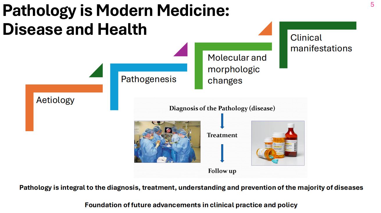
Pathology: Framework to understand disease process
DIAGRAM ON SLIDE 6
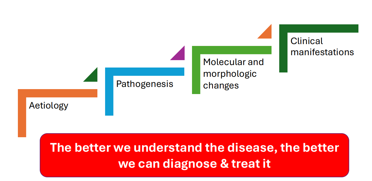
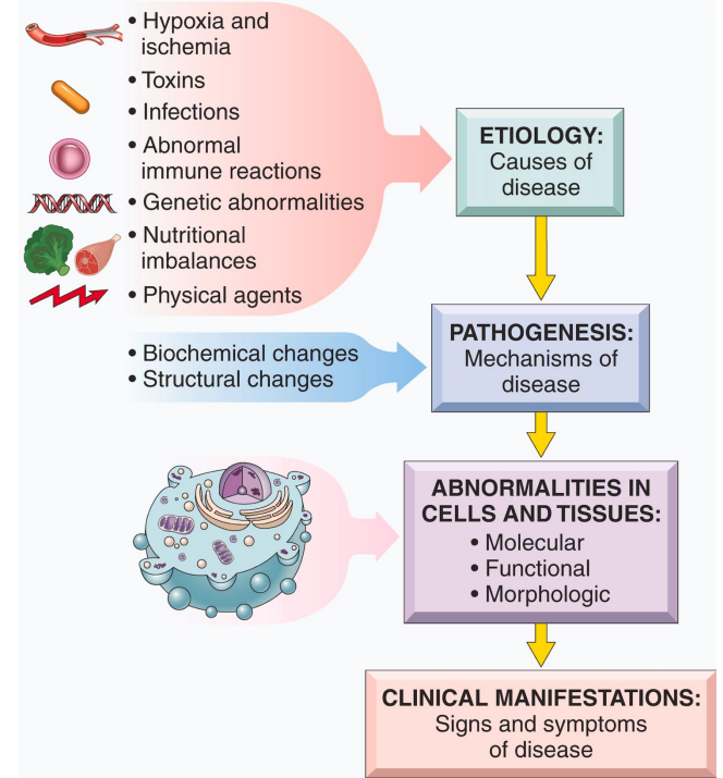
Aetiology
- the cause, set of causes, or manner of causation of a disease or condition
- benefits of knowing the aetiology: management and prevention
- major etiologic factors: genetic and acquired
Pathogenesis
- mechanisms of development/progression of disease. Following factors of etiology
- Pathogenesis leads to morphologic and molecular changes
- Leads clinical manifestations - structural and functional abnormalities
Morphologic Changes
structural alterations in cells or tissues observed following pathogenesis:
- Naked eye: gross morphologic changes (gross lesions) (we can see surface level stuff)
- Microscope: microscopic changes (microscopic lesions)
- morphologic changes are used to diagnose a disease (seen by a pathologist)
- can lead to functional alteration and to the clinical signs and symptoms
Functional Derangements and Clinical Manifestations
- genetic, biochemical and structural changes in cells/tissues
- the morphologic changes in the organ influence the normal function of the organ leading to the disease
- Determine: clinical features - signs and symptoms, course and prognosis of disease
How does Pathology affect the patient
- Symptoms: things the patient feels (a departure from normal function or feeling. Noticed by the patient. e.g temperature, lump, rash, weight loss)
- Signs: Physical Findings (objective evidence of disease, noticed by patient or doctor on clinical examination. e.g heart rate, enlarged organs)
- Investigations: pathology and radiology (structural changes)
Pathology Pathway Diagram
DIAGRAM ON SLIDE 14


Acquired Cases and Genetic Disorders
DIAGRAM ON SLIDE 15
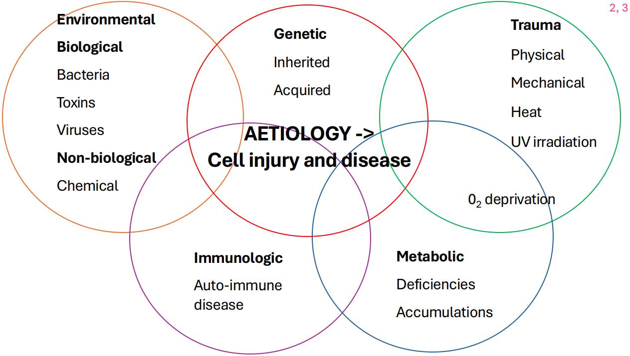
Aetiology Diagram
DIAGRAM ON SLIDE 16

Disease Aetiology and Paths of Pathogenesis
1. Inheritied and congenital malformations
2. Acquired:
- Vascular events
- inflammatory/infective
- trauma
- autoimmune
- metabolic disorders
- nutrition and the environment
- Iatrogenic/idiopathic
- Neoplastic
- Degenerative
Iatrogenic
produced by a physician (the unexpected results from a treatment prescribed by a physician) (e.g chemo gone wrong)
Idiopathic
pertaining to disease of unknown origin
Healthy Cell Pathogenesis Diagram
DIAGRAM ON SLIDE 18

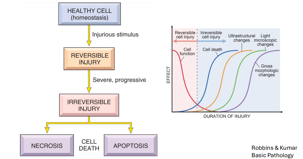
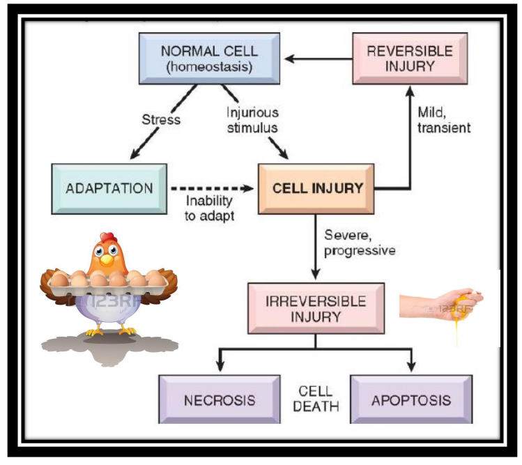
Cell Injury
- if the cell fails to adapt under stress, they undergo certain changes called cell injury
Reversible and Irreversible Cell Injury
- the affected cells may recover from the injury (reversible) or may die (irreversible)
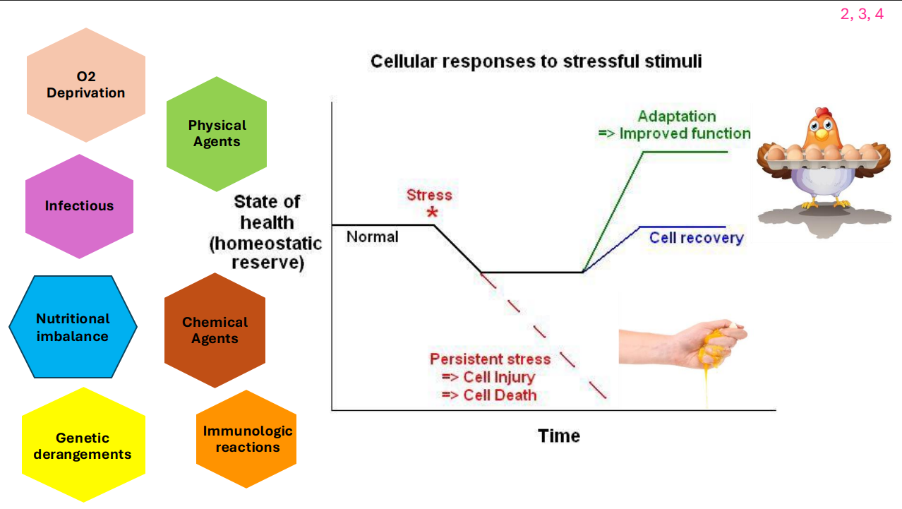
Cellular Responses to Stressful Stimuli
DIAGRAM ON SLIDE 21

Reversible Cell Injury
- swelling
- cytoplasmic inclusions
- steatosis (fatty change)
- Pigments
Towards Irreversible/Irreversible Cell Injury
- Abnormal mitochondria
- irregular cell contours/blebbing
- Eosinophilia (pink appearance)
- Nuclear deformities
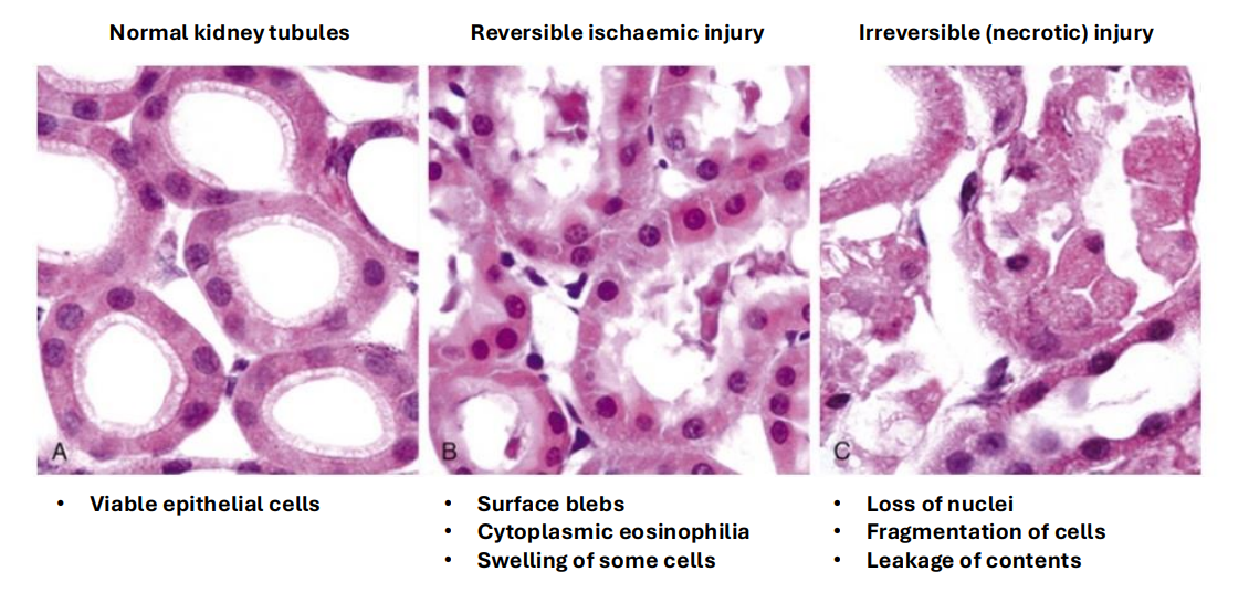
Example of Histological Change (Kidneys)
DIAGRAM ON SLIDE 23

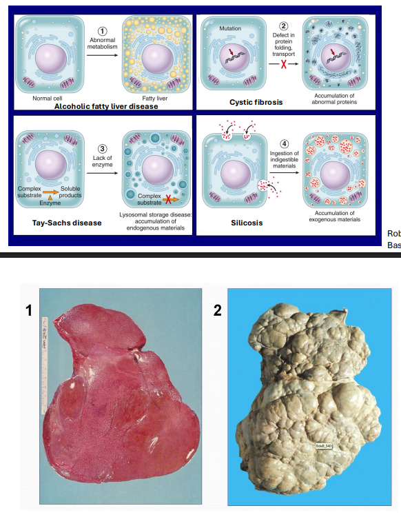
Intracellular Accumulations
DIAGRAM ON SLIDE 24 and 25


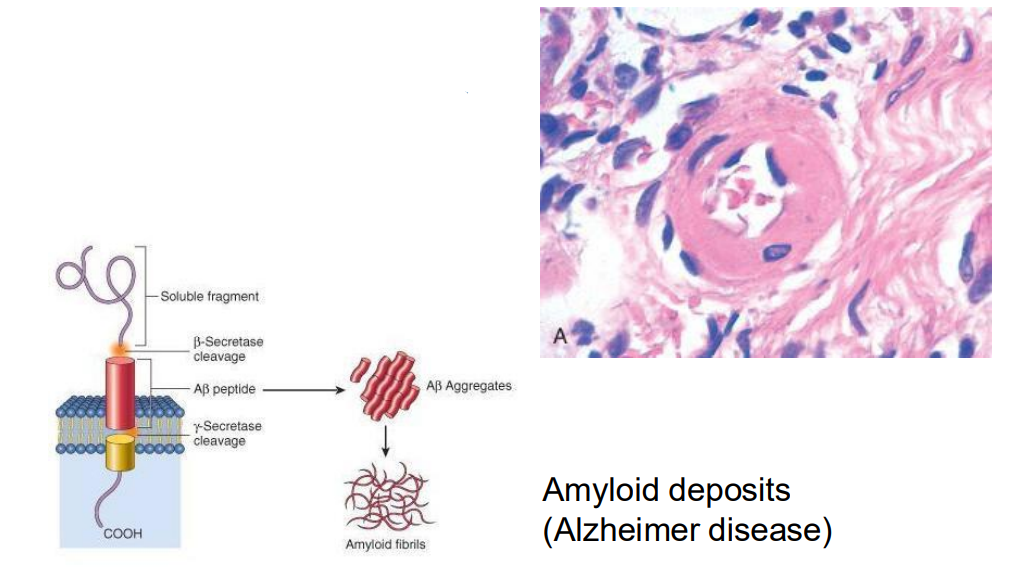
Extracellular Accumulations
- abnormal products build up in the interstitium
- hyaline and/or fibrinoid deposits around blood vessels (acute hypertension) (in the diagram)
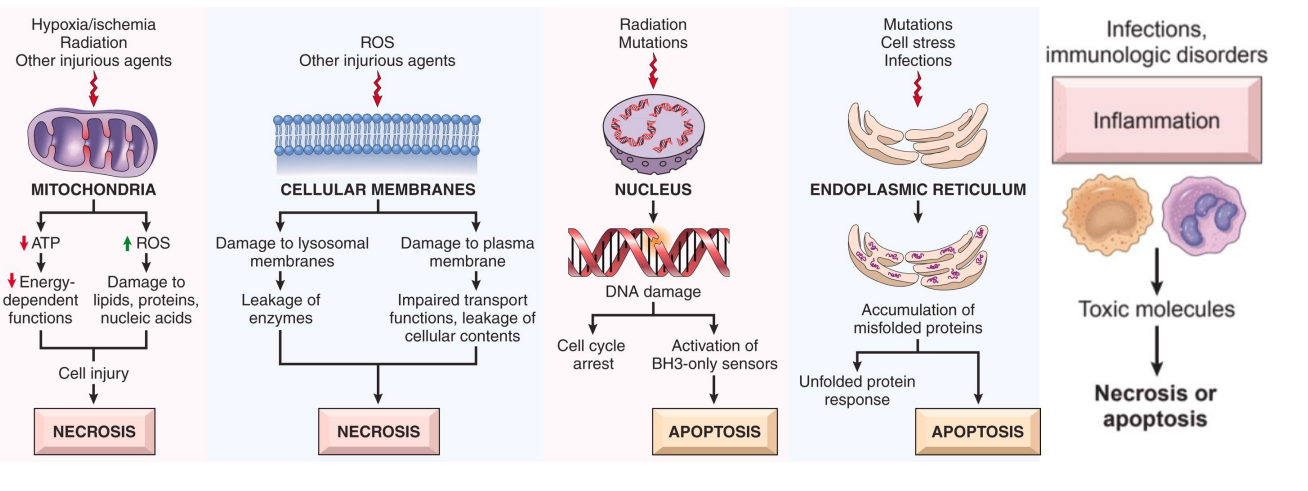
Irreversible Cell Injury
- persistent or excessive injury
- no point of return
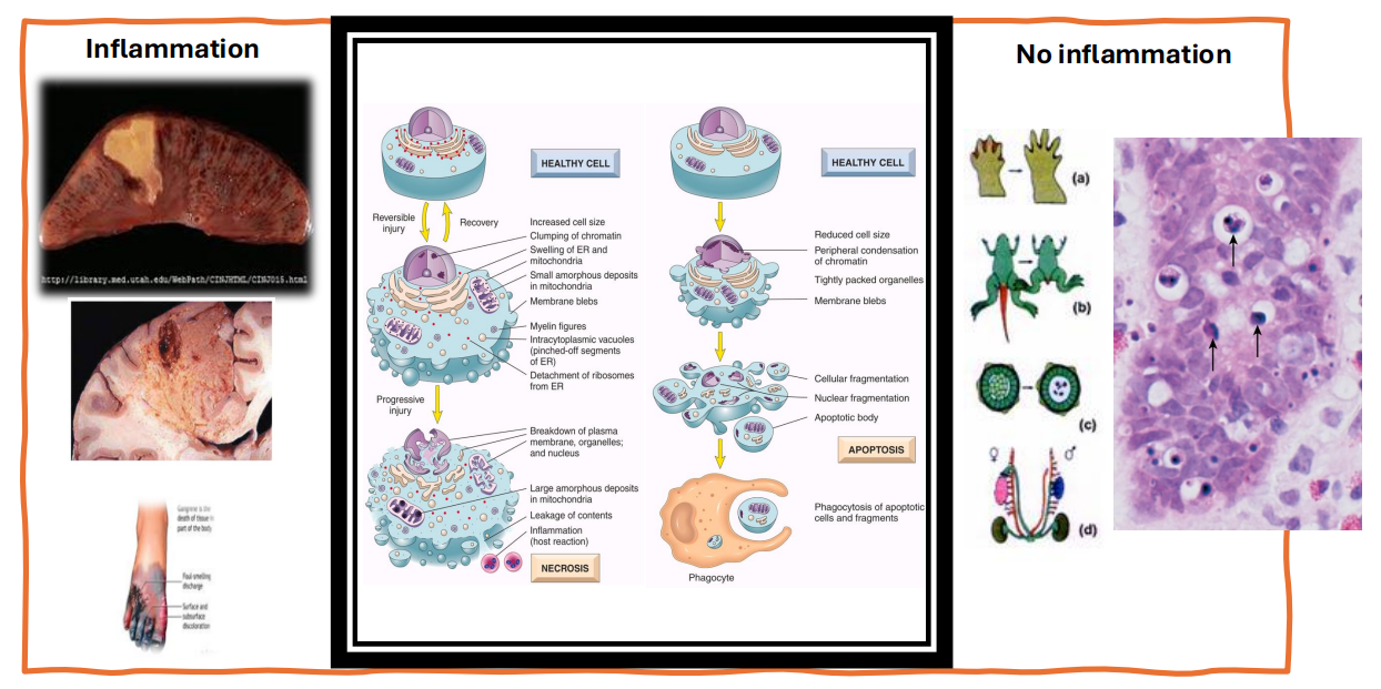
- Can either undergo necrosis (premature death of cells in living tissue) or apoptosis (programmed cell death)
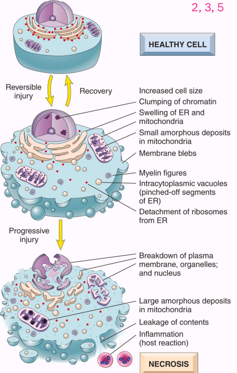
Necrosis
- accidental cell death
- cell membranes fall apart, cell enzymes leak and digest cell
- elicits a local host reaction - inflammation
- cytoplasmic and nuclear changes
- necrotic cells may persist for some time or may be digested by enzymes and disappear
- leakage of intracellular proteins into circulation allows detection of tissue-specific necrosis using blood or serum (cardiac muscle -> unique isoform of the enzyme creatine kinase and of the contractile protein troponin)
- irreversible cell injury and cell death in this tissue elevates the serum levels of these proteins which makes them clinically useful markers of tissue damage


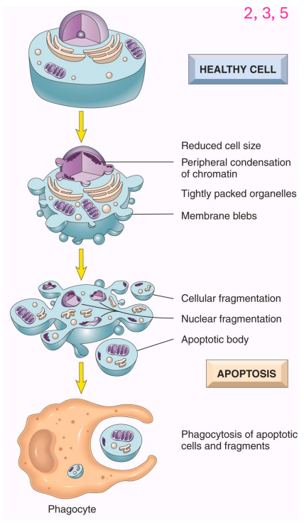
Apoptosis
- occurs in many normal situations to eliminate: potentially harmful cells, cells that have outlived their usefulness or irreperably damaged cells
- activation of cellular enzymes (caspases) leads to degredation of nuclear DNA and cytoplasmic proteins
- fragments (apoptotic bodies) of the cells break off
- plasma membrane remains intact but is altered and apoptotic bodies are consumed by phagocytes
- little leakage of cell contents and thus no inflammation
.
- no inflammation of the cell, no swelling like necrosis

Cell with Inflammation vs without inflammation
DIAGRAM ON SLIDE 30

Pathways of cell injury and death
DIAGRAM ON SLIDE 31
Ischemic heart Disease: Coagulative necrosis in myocardial infraction
Stroke: liquefactive necrosis in brain infarcts
Cancer: apoptosis induced by chemotherapy
Congenital Disease
presnt at birth
Genetic Disease
caused by chromosome or gene defects
Inherited Disease
- passed from parent to offspring
Teratogen
- exposure that irreversibly affects the normal growth, structure or function of a developing embryo or foetus

Flow chart of Cell Damage
DIAGRAM ON SLIDE 33
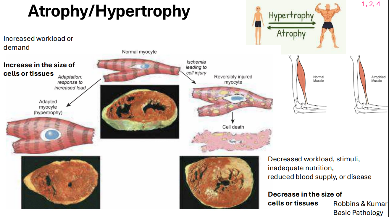
Atrophy/Hypertrophy
- one of the ways that cells adapt to injury
- increased workload or demand results in increase in the size of cells or tissues

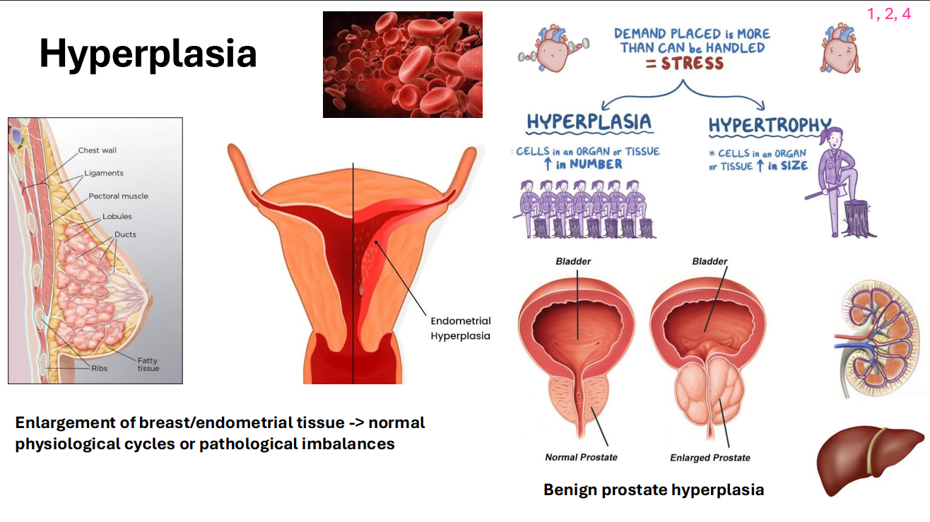
Hyperplasia
- another way that cells adapt to injury
- it is the increase in the number of cells
- enlargement of breast/endometrial tissue -> normal physiological cycles or pathological imbalances


Hyperplasia and Atrophy example
DIAGRAM ON SLIDE 36

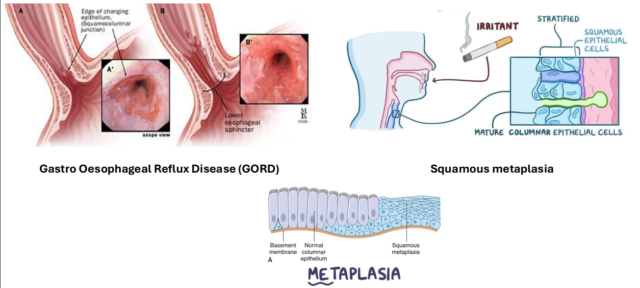
Metaplasia
- one type of mature cell is replaced by another type of mature cell that is not normal for that tissue = adaptive response
- e.g stomach lining in gastric reflux disease, acid damages lining. so the cells change from squamous columnar to ciliated columnar cells, since these cells are more resilient
DIAGRAM ON SLIDE 37


Metaplasia Examples
DIAGRAM ON SLIDE 38

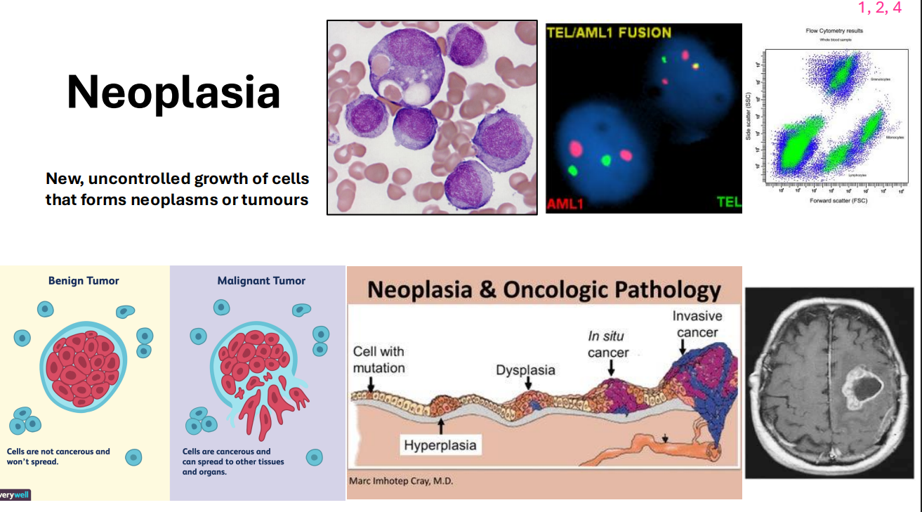
Neoplasia
- new, uncontrolled growth of cells that forms neoplasms or tumours
- e.g skin cancers

Pathology SUMMARY
- Diagnosis of pathology -> Treatment -> Follow up