TRAMUA
 Call Kai
 Learn
Learn Practice Test
 Spaced Repetition
 Match
Match Flashcards
 Knowt Play
1/353

There's no tags or description
Looks like no tags are added yet.
Name | Mastery | Learn | Test | Matching | Spaced | Call with Kai | Chat |
|---|
No analytics yet
Send a link to your students to track their progress
354 Terms
Complete or incomplete disruption in continuity and structure of the bone and/or cartilage. Creates an insult to bone marrow, periosteum and soft tissues
Fracture
Bones are __________ meaning unequal in strength, and therefore can withstand mechanical forces differently
Anisotrophic
____________ bone is stronger on compression and less resilient to distraction, most vulnerable to shearing forces
Cortical
Surgical pine holes or a site of bone resection may weaken the bone, known as:
Stress raiser
3 types of incomplete fractures that are seen in children
Torus
Green Stick
Plastic deformity
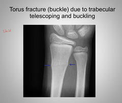
Incomplete fracture seen in children which is cortical buckling due to compression
Torus
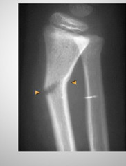
Incomplete fracture seen in children that results in incomplete fracture on tensino
Green stick
Incomplete fracture seen in children which results from bending of the bone without angular break and remodeling
Plastic deformity
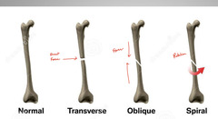
3 types of complete fractures
transverse, oblique, spiral
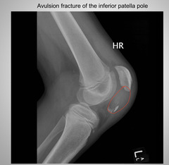
Type of fx where a fragment of bone is being detached by the tension from the muscles or ligaments
Avulsion fracture
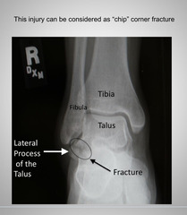
Type of Dx that presents as usually a corner fracture that is chipped rather than avulsed
Chip fracture
Type of fx that results in telescoping of osseous trabeculae. No typical radiolucent line is seen on radiographs and instead a zone of sclerosis or condensation may be present
Impaction (Compression) Fracture
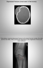
Type of Fx usually in the calvaria and occasionally on the tibial plateau
Depression fracture
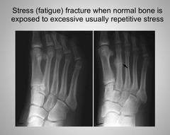
Type of Fx that results from repeated stress applied to normal bone leading to bone marrow hyperemia and bone resorption (ex: march fx of 2nd or 3rd metatarsal bones, tibial stress fx in runners, etc)
Stress (fatigue) fracture
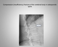
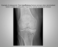
Type of Fx that results from normal stresses like normal weight bearing or walking but applied to osteoporotic bone. Ex: Vertebral osteoporosis, sacral insufficiency Fx
Insufficiency Fracture
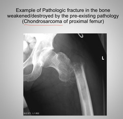
Type of Fx that results from weakened bone by pathology such as neoplasms, infection, congenital defect of collagen, etc
Pathologic Fracture
Type of fracture that has more than 2 segments, has subtypes: Segmenetal and Butterfly
Comminuted Fracture
Subtype of comminuted Fx in which 2 separate fracture lines produce an isolated segment of bone
Segmental Fracture
Subtype of comminuted fracture in which there is a wedge shaped fragment created at the apex of the maximal force
Butterfly Fractuer
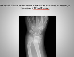
When skin is intact and no communication with the outside air is present, a fracture is considered:
Closed
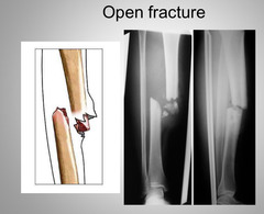
When skin is broken and communication between bone and outside air is present, a fracture is considered:
Open
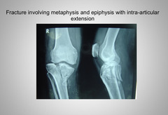
It must be stated when a fracture extends into the joint creating an _______-_________ fracture. Greater complications can occur like delayed healing, Chondroblasts, secondary OA
Intra-articular
Fractures that cause a complete loss of apposition due to forced traction
Distraction
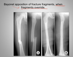
When a fracture causes one fragment of bone to overlap another this is called:
Bayonet apposition
Alignment of a fracture is considered ________ if the distal portion of bone deviates away from midline (Laterally)
Valgus
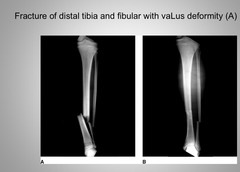
Alignment of a fracture is considered _______ if the distal portion of bone deviates towards the midline (medially
Varus
_________ means that fractures maintain alignment, can be lost in laterality or with anterior-posterior displacmenet
apposition
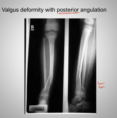
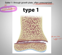
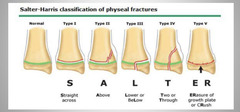
Type of salter Harris fracture that goes through the growth plate itself but is often unrecognized because of minimal displacement. Prognosis is fine if diagnosed early
Salter Harris type 1
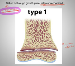
What is one clinical example of salter Harris type 1 fractures?
SCFE
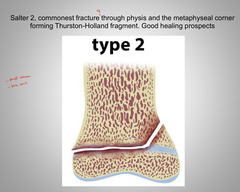
Type of salter Harris fracture that is the most common (>75%), fracture through the physis and a part of the metaphysis forming a thurston-holland fragment. Fx is proximal to the epiphysis. Good healing prospects
Salter Harris type 2
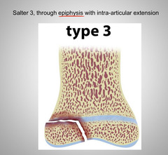
Type of salter Harris fracture that goes through the plate and into the epiphysis. Distal to epiphyseal line (physis)
Salter Harris type 3
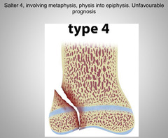
Type of salter Harris fracture that transversus the metaphysis, physis and into the epiphysis
Salter Harris type 4
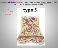
Type of salter a Harris fracture that is a crush injury to the growth plate. Often unrecognized or confused with type 1 but essentially damages physeal blood supply. Very bad because can cause premature fusion of growth plate resulting in stunted growth
Salter Harris type 5
What types of salter Harris fractures are rarest but show the highest complications leading to premature plate closure, limb deformities, shortening and other sequela
Salter Harris type 4 & 5
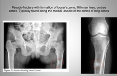
Sub type of insufficiency fracture that develops in bones with insufficient osteoid. Most often seen in rickets and osteomalacia, hyperparathyroidism, renal osteodystrophy, pages disease and other conditions that disturb normal bone pathology
Pseudo fracture
What bones most often have stress fractures?
Lower extremity
Occasionally on UE associated with repetitive activities
These fractures show very characteristic appearance on xrays as widened transverse radiolucent lines oriented at the right angle typically to the medial cortex
Pseudofractures
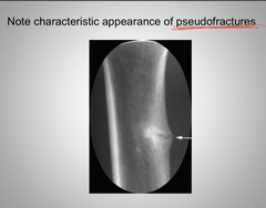
Where are pseudofractures most often found?
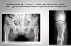
Medial aspect of the cortex of long bones
During bone bruise injuries, cell damage creates what?
Intra-osseous edema
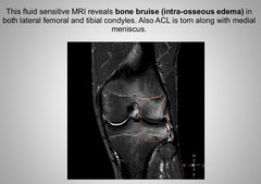
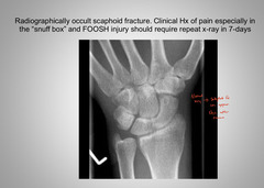
Fractures that occur when intra-osseous edema and injury have occured. Osteoclasts will become activated by inflammatory mediators released from injured cells. As a result, more significant bone resorption will occur and what was initially undetected on xrays as bone bruise may progress to fracture several days later. Best example is scaphoid fractures
Occult fractures
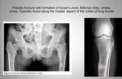
Often referred to as:
Looser zones
Milkman lines
Umbau zones
Pseudofractures
Complete loss of articular contact/alignment with resultant injury to periarticular restraints
Dislocation
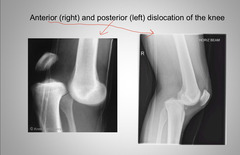
Partial loss of articular alignmnet
Subluxation
Separation of fibrous joints or fibrocartilagenous joints often seen as suture diastasis in the scull and symphysis pubis
Diastasis
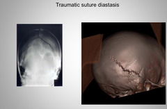
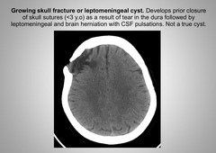
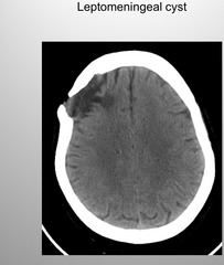
Aka growing skull fracture, develops prior to closure of the skull sutures (
Leptomeningeal cyst
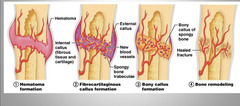
3 main phases of fracture healing
Inflammatory: 48hrs
Repair: 7-14 days
Remodeling: 9-24 months
Phase of healing in which hematoma and inflammatory mediators within the firs t48 hrs initiate chemotaxis with phagocytes and repair cells being drawn to fracture site (shortest phase)
Inflammatory phase
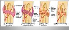
Phase of healing in which cells involved during initial inflammation will gradually begin to form granulation tissue and remove unwanted material and damaged cell. During htis phase within 7-14 days hematoma becomes vascularized and may appear more translucent on xrays
Repair phase
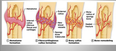
Fracture callus created during the repair phase of healing is still very vulnerable to _________ forces but may be better against ___________ forces
Shearing, axial
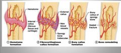
Phase of healing in which population of cells will sufficiently evolve into fibroblasts, chondrocytes and osteoblasts and bone mineralization will continue. Takes 9-24 months
Remodeling phase
Due to vascularity of the _________, pediatric fractures heal quicker
Periosteum
Type of disturbed fracture healing that takes twice as long as physiologically expected
Delayed union
Type of disturbed fracture healing where there is no healing for >9 months
Non-union
Type of disturbed fracture healing where there is a week callus with insufficient vacularization and new bone formation
Hypotrophic
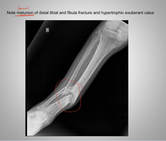
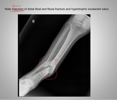
Type of disturbed fracture healing where there is abnormal exuberant callus
Hypertrophic
Type of disturbed fracture healing in which there is absent callus often with synovial fluid or infected exudate intervening between fracture ends
Atrophic
_________ complications of fracture include neurological and vascular injury, acute compartment syndrome and renal failure, pulmonary fat embolism and gas gangrene
Immediate
_________ complications of fracture include osteomyelitis, sepsis, complex regional pain syndrome, nonunion/malunion
Intermediate
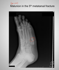
Healing occured in abnormally positioned fracture ends
Malunion
_________ complications of fracture include ischemic necrosis (AVN), secondary OA
Late
Associated fractures from this may sometimes cause lead toxicity. If bullet is lodged in synovial or serous cavity, hyaluronic acid will chemically remove lead and cause its reabsorption into general circulation potentially causing lead toxicity.
Gun shot wound (GSW)
Result of head injury that must be evaluated with highest level of clinical suspicion
Skull Fx
May present significant life threatening complication with head injury
Intra-cranial hemorrhage
Advanced imaging like what must be used to evaluate head injuries?
CT or MRI
Conventional radiography of evaluation of head injuries has become relatively:
Obsolete, non-existant
Occasionally __________ Sculls may be evaluate by plain film radiographs due to high radiation dose of CT
Pediatric
Type of imaging used most often to evaluate head injury?
CT without contrast
MC type of skull Dx that accounts for 80% of all skull Fx
Linear
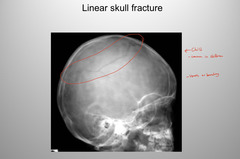
Where are linear skull fractures most often found?
Parietal and temporal bones
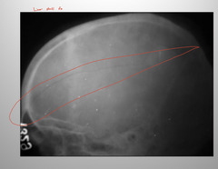
How do you identify linear skull fractures?
Radiolucent lines that CROSS SUTURES
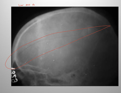
Type of skull Fx that is considered an open Fx, 15% of all skull fxs
Depressed skull Fx
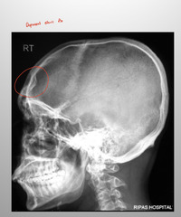
Depressed skull fractures occur almost always where?
Cranial vault
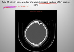
Best modality for imaging depressed skull fxs?
CT scanning
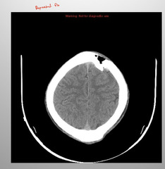
In depressed skull fx, if depressed fragments extend > _________cm into the cranium neurosurgical referral is crucial
0.5-1
Occasionally named pond skull Fx, variation in depressed skull fx that may be seen in newborn infants as an inward buckling of the calvaria without osseous fragments
Ping-Pong Fx
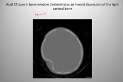
Type of skull fxs that happen during labor due to forceps possibly, or direct blunt trauma like dropped infant
Ping-Pong Fx
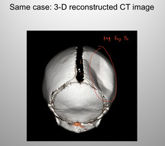
Type of skull fracture that may be traumatic or non-traumatic due to pathology or rarely physiological changes related to rapid growth spurts
Sutural diastasis
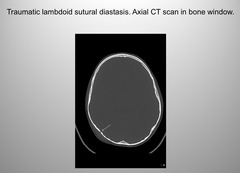
At birth, diastatic fxs are considered if suture is wider than _______cm
1
At age 2 diatstatic fxs are considered if suture si wider than _______ mm
3
At age 3 diastatic fxs are considered if suture is wider than ____mm
2
Traumatic sutural separation may be seen in
5
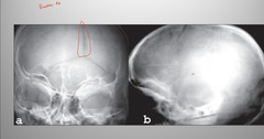
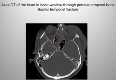
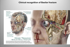
Type of skull fx that is better diagnosed clinically than radiographically. Sphenoid and temporal bones may be involved in >75% of cases with complete or partial obstruction of the sphenoid sinus. Best detected on lateral skull projections
Basilar skull FX
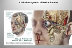
_________ may be noted on skull xrays with basilar fractures
Pneumocephalus
Tearing of the _________ is common in basilar skull fxs
Meninges
Who are at highest risk for subdural/subarachnoid hematoma?
Elderly, children
CSF rhinorrhea and otorrhea
Battle sign
Raccoon eyes are all signs of what?
Basilar skull fx
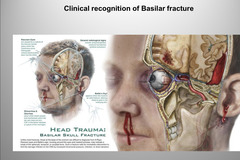
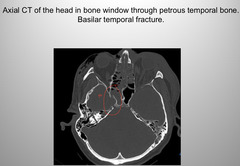
Best modality to evaluate basilar skull fxs?
CT
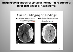
__________ hematoma may develop rapidly with acute rise in intracranial pressure, cerebral edema and transtentorial and or subfalcine brain herniation
Epidural
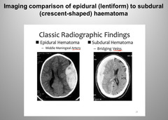
Middle meningeal artery damage can commonly cause what?
Epidural hematoma
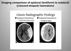
Type of hematoma that is caused by affecting the bridging veins
Subdural (subarachnoid) hematoma
Basilar Skull fx have a _________ probability of intracranial hemorrhage and meningitis, making this a medical emergency
High
Subarachnoid hemorrhage are identified in ______% of fatal head injuries due to trauma
30
Mostly found in an pediatric skull
Leptomeningeal Cyst- Growing fracture
Most common of all facial bone fractures accounting for 45% of total
Nasal fractures
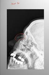
Most nasal fractures are induced with the ______ force component and show laterally displaced component
Lateral
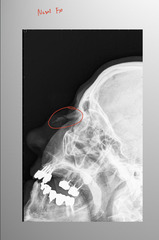
Conventional radiographs are _____% sensitive to identifying nasal fractures (on lateral nasal view)
80
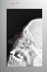
Best imaging modality for nasal fractures, 100% sensitive
CT
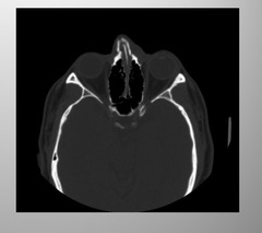
__________ nasal fractures may not require treatment
Undisplaced
_________ nasal fractures may lead to nasal difficulty breathing, occasionally septal necrosis may develop
Displaced
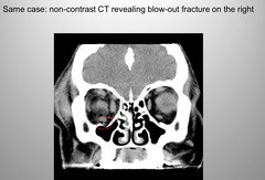
Most cases of this fracture will force orbital fat and sometimes inferior rectus muscle into the maxillary sinus
Orbital Blow out fracture