5: Axilla and brachial plexus
 Call Kai
 Learn
Learn Practice Test
 Spaced Repetition
 Match
Match Flashcards
 Knowt Play
1/68

There's no tags or description
Looks like no tags are added yet.
Name | Mastery | Learn | Test | Matching | Spaced | Call with Kai | Chat |
|---|
No analytics yet
Send a link to your students to track their progress
69 Terms
aching pain, numbness and tingling in his right arm. On taking his history, patient had spent the weekend replacing a ceiling in his flat. On examination, the doctor noticed cyanosis and venous distension in the distal part of his patient's right arm, a weak radial pulse (when compared to the left), some weakness of flexion of his elbow and his grip was impaired in the right hand. What is cyanosis? Explain the cyanosis and venous distension of veins Cyanosis
development of bluish culour resulting from build up of deoxygenated haemoglobin.
compressed of axillary vessels = ischaemia and distension (swelling) of superficial veins
aching pain, numbness and tingling in his right arm. On taking his history, patient had spent the weekend replacing a ceiling in his flat. On examination, the doctor noticed cyanosis and venous distension in the distal part of his patient's right arm, a weak radial pulse (when compared to the left), some weakness of flexion of his elbow and his grip was impaired in the right hand. which major artery was affected and explain weakness of radial artery pulse and how will right ulnar pulse be effected
compression of subclavian artery compromised blood supply to UL. blood supply to both radial and ulnar arteries come from subclavian, these pulses will be weak.
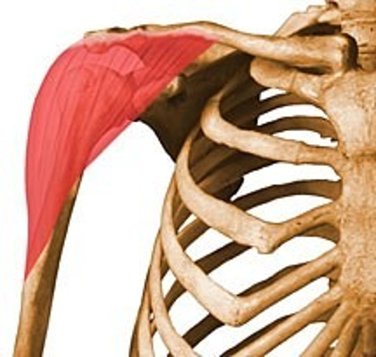
deltoid muscle function + innervation
Abducts upper arm. axillary nerve c5 and 6.
consider bone movement in clavicular fractures and explain why pt having fractured a clavicle will support upper limb with left hand
both medial and lateral segments move. medial moves superiorly due to pull of sternocleidomastoid muscle and lateral moves inferiorly and medially due to the weight of the arm being too much for the trapezius
- to neutralize gravity and prevent the weight of the arm from pulling the shoulder downward
what parts of clavicle (immediately posterior to usual frature point) can be at risk
trunks and divisions of brachial plexus.
- immediately posterior to usual fracture site = brachial plexus since it’s behind the clavicle
how would you test damage to brachial plexus after clavicle fracture
sensory testing only. test radial, ulnar, median, musculocutanous (lateral antebrachial cutaneous nerve)
how would you determine if a vessle has been damaged from clavicle fracture
subclavian passes under mid 1/3 to become axillary artery: closed fracture with rapidly expanding swelling from arterial blood. show pulsatile release of blood from open wound. pt unconscious or dying.
subclavian vein: non-pulsatile, pt conscious, haematoma
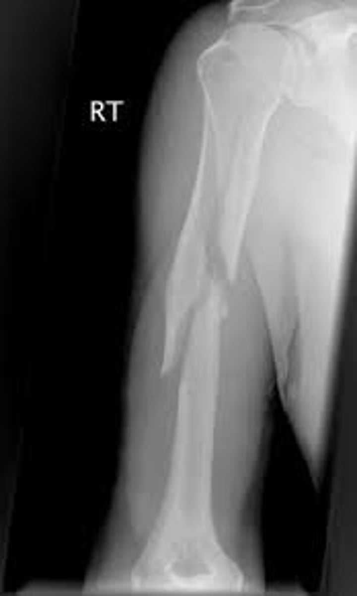
Mid shaft humerus fracture structures
radial nerve and profunda brachii run immediately against it in spiral groove
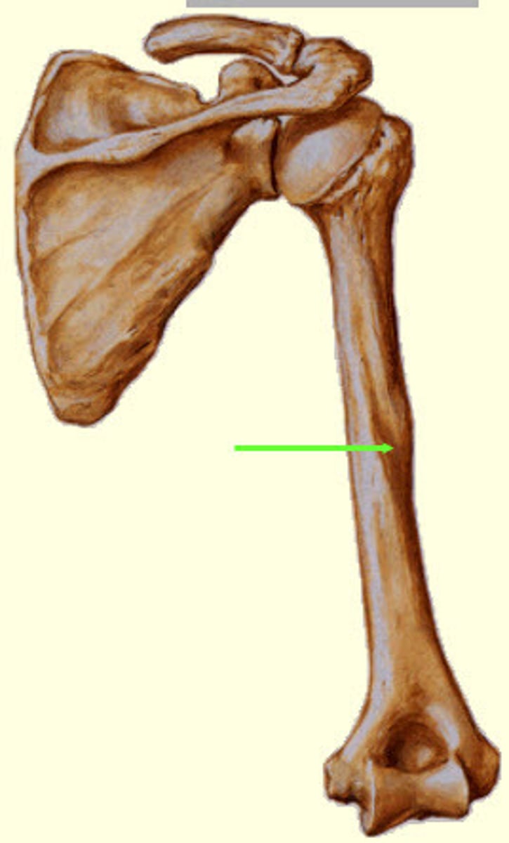
Where is the spiral groove of humerus, and what lies in it?
inferolaterally around posterior humerus.
centred on deltoid tuberosity .
radial nerve, profunda brachii. separating the origin of the lateral head of the triceps above and the origin of the medial head below.
spiral groove fracture muscle groups damaged
radial nerve injury leads to partial loss of triceps innervation, total loss of innervation to posterior forearm compartment. weakened elbow extention, loss of wrist extention, weak thumb abduction, lack of digit extention.
supination of forearm despite mid shaft fracture
means that supinator (radial nerve) + biceps brachii (musculocutanous) still working
mid shaft humerus fracture but can still extend elbow
triceps innervated by radial nerve at different levels. partial loss of triceps innervation but triceps recieve innervation prior to spiral groove
How to test Radial Nerve
1st interosseous muscle on dorsal surface of hand
- sensory + motor tests checking the 1st interosseous musc. on dorsal surface of hand
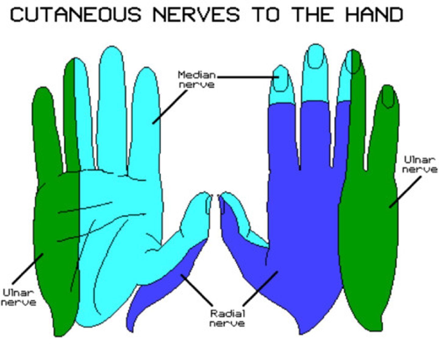
Horner's syndrome
Sympathetic trunk/cervical ganglion damage (affects eyes), results in ptosis (drooped upper eyelid), miosis (pupil constriction), vasodilation, and anhidrosis (absence of sweating)
pancoast tumour caused horners syndrome how
pancoast tumour eroded the BP + sympathetic chain which causes parasympathetic constriction of pupils.
sympathetic supply to tarsal muscle of eye and levator palpebrae causing eye not to be opened widely.
sympathetic responsible for sweating so loss of sweating
Pancoast tumours can cause Horner's syndrome . what loss will there be
c8-t1. ulnar aspect of forearm and hand. wasting and weakness of intrinsic muscles of hand, loss of abduction and adduction of hands, adduction of thumb and powergrip. median and radial nerve share c8,t1 nerve roots so may be affected.
fibrous axillary sheath
axillary artery, vein and chords and branches of BP
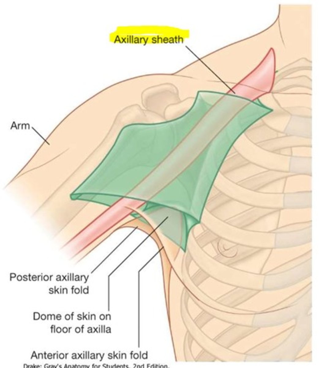
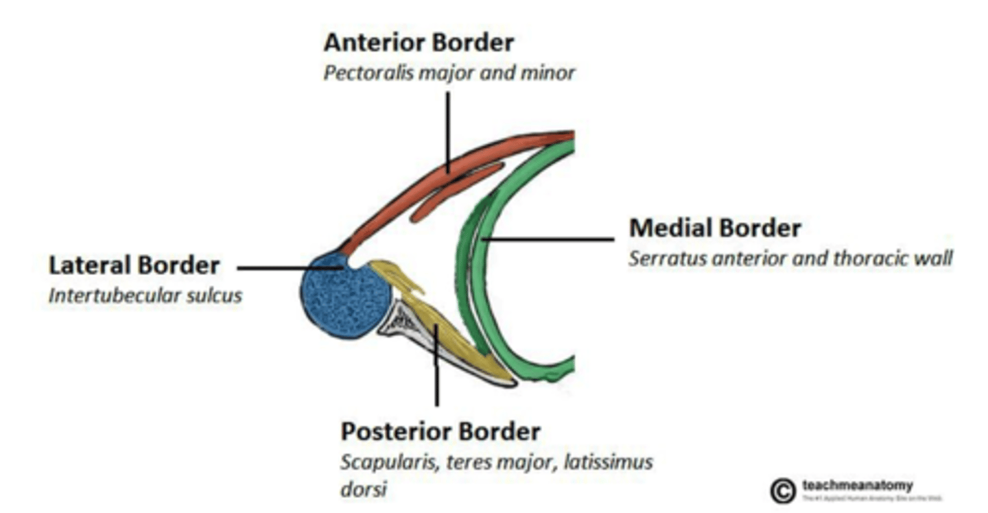
what muscles form anterior and posterior folds of axilla
anterior: pectoralis major and minor more superiorly
posterior wall: teres major, latissimus dorsi (subscapularis and scapula more superiorly)
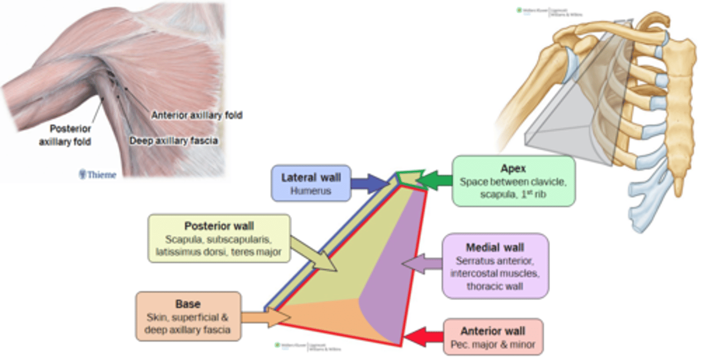
medial wall of axilla formed by what muscles
intercoastal muscles and serratus anterior
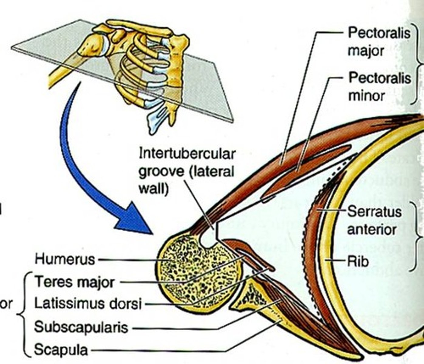
biceps brachii insertion
radial tuberosity and bicipital aponeurosis
biceps brachii attachment (proximal)
long head: supragenoid tubercle of scapula
short head: coracoid process of scapula
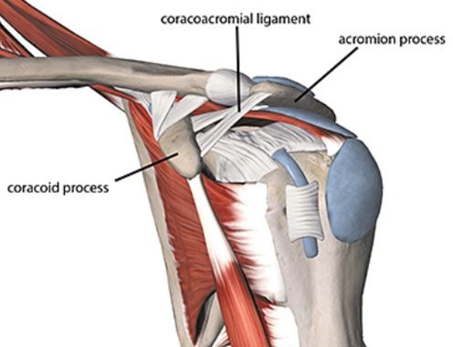
coracoacromial arch - what is found in it + function
acromion process, coracoacromial ligament, and coracoid process
Prevents superior translation of the humerus
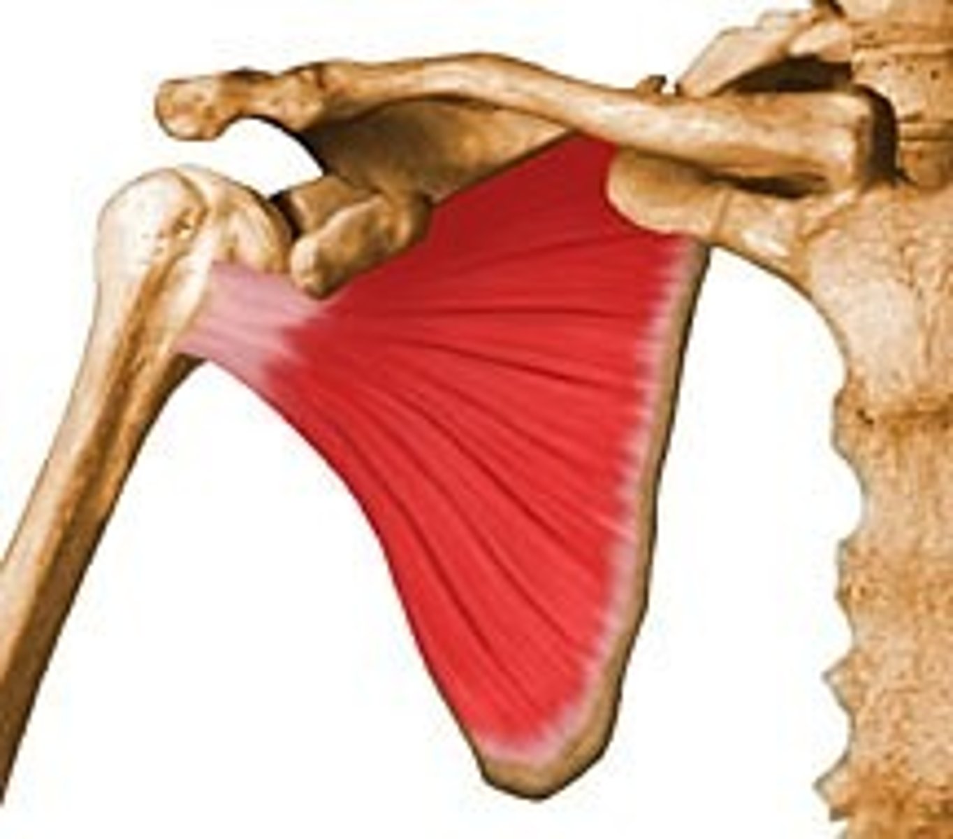
Subscapularis muscle - functions
Internal/medial rotation and shoulder adduction
stabilizer to keep humeral head centered in glenoid socket
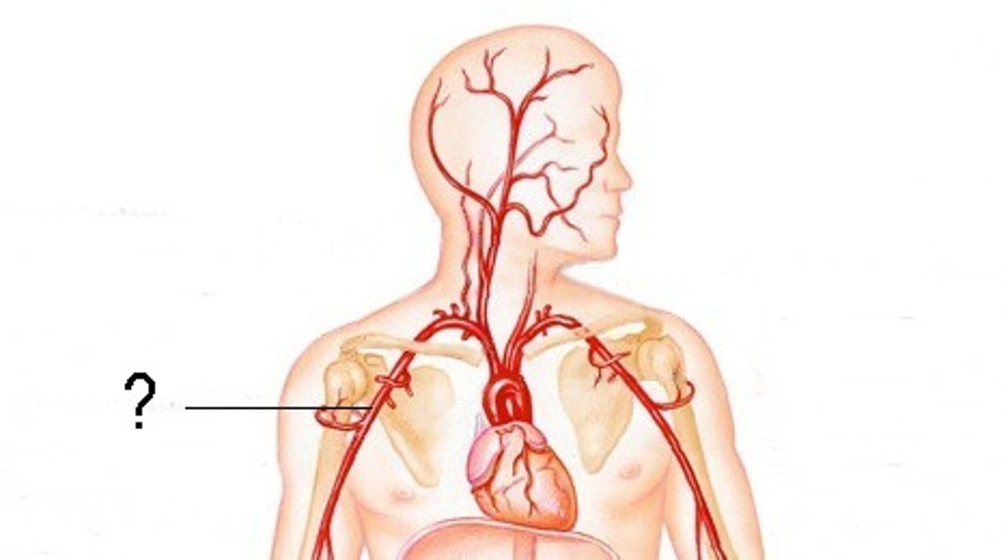
lateral, medial and posterior cords of the brachial plexus are named with respect to an important blood vessel
axillary artery
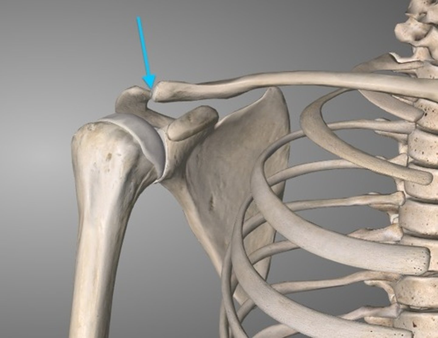
acromioclavicular joint
the joint where the acromion and the clavicle meet
vein immdediately posterior to sternoclavicular joint
brachiocephalic
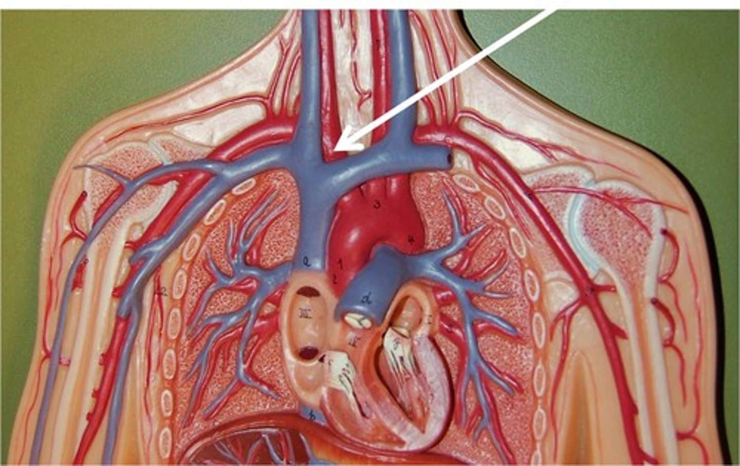
anatomical function of clavicle
suspends scapula + upper limb to keep away from thorax for maximum freedom of motion
transmits forces from upper limb to axial skeleton
1st rib
under clavical
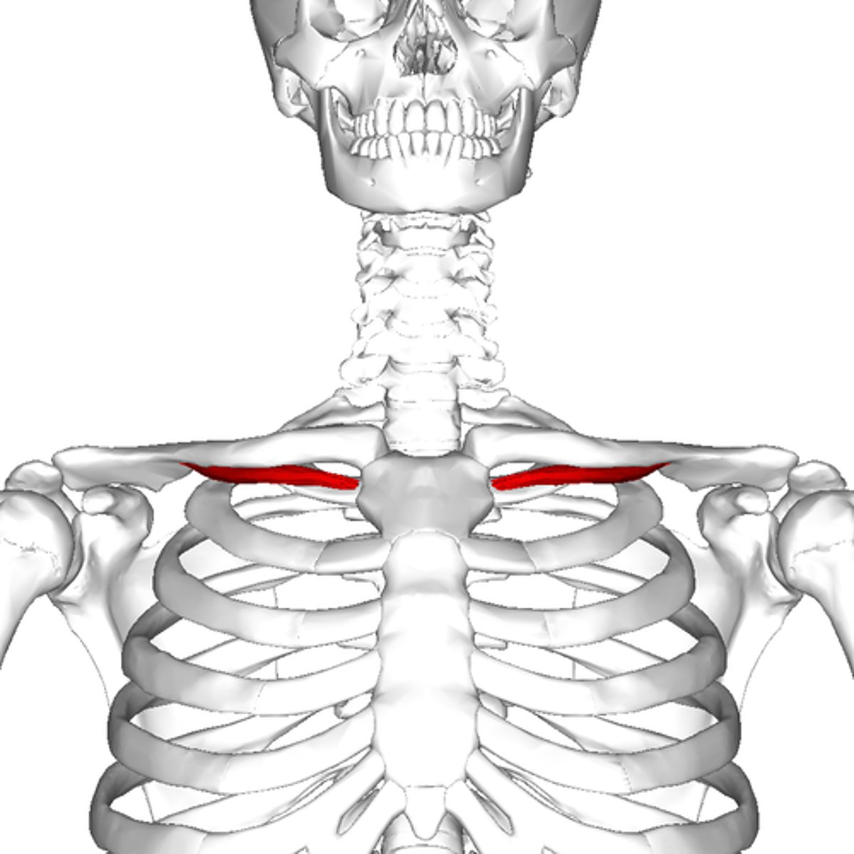
position of arm in clavicle fracture
sternocleidomastoid muscle elevates medial segment because trapezius muscle unable to hold lateral segment up and because of weight of upper limb, shoulder drops and arm is pulled medially by adductor muscles of the arm
Waiter's tip hand
upper limb adducted, internally rotated and flexed at the wrist.
indicates damage to upper trunk of brachial plexus.
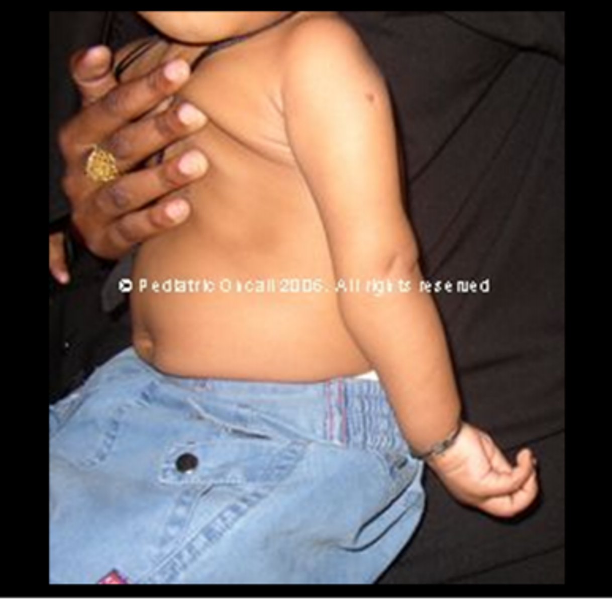
describe the artery direction of upper limb
subclavian passes behind middle of clavicle and crosses first rib. supply structures in neck and thoracic wall and entire UL. axillary artery continuation of subclavian beginning at lateral border of first rib. brachial artery is a continuation of axillary artery beginning at lateral, lower border of teres major. in cubital fossa, brachial artery bifurcates into the ulnar and radial arteries
osteoblasts - how do they form bone?
secrete type I collagen to produce a matrix to be calcified
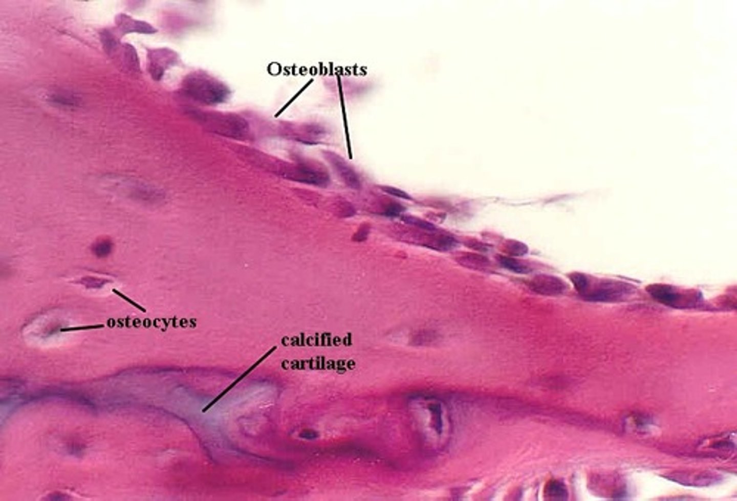
what are osteocytes and what do they do?
former osteoblasts that have become trapped in the matrix forming tight junctions across the matrix and control bone remodelling
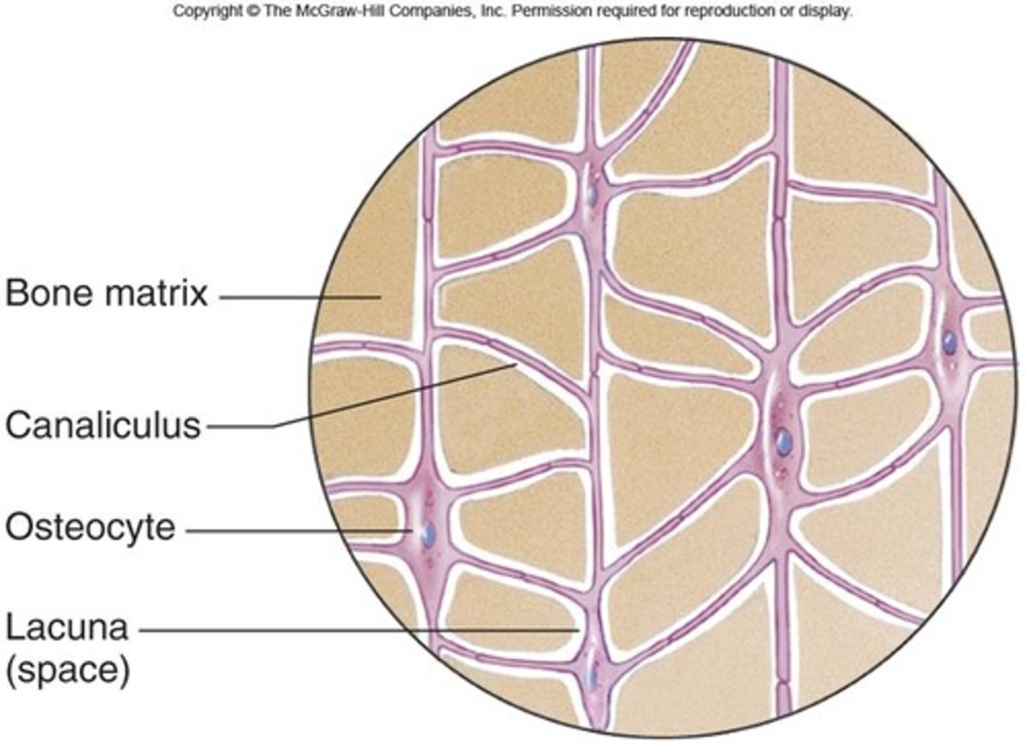
osteoclasts
bone breaking cells
hypocalcaemia dangerous
small decreases of calcium in plasma and ECF can cause nervous system to be more excitable because of increased neural membrane permeability with resultant muscle spasm.
hypercalcaemia dangers
body fluid depresses nervous system and causes muscles to become sluggish and weak → effects calcium on muscles plasma membranes
bones and homeostasis
osteoclasts and blasts maintain the levels of calcium and phosphates to regulate the amount and consistency of extracellular fluid by reducing or adding to the calcium
nerve that supplies anterior flexor compartment of arm
musculocutaneous nerve. terminal branch of posterior cord.
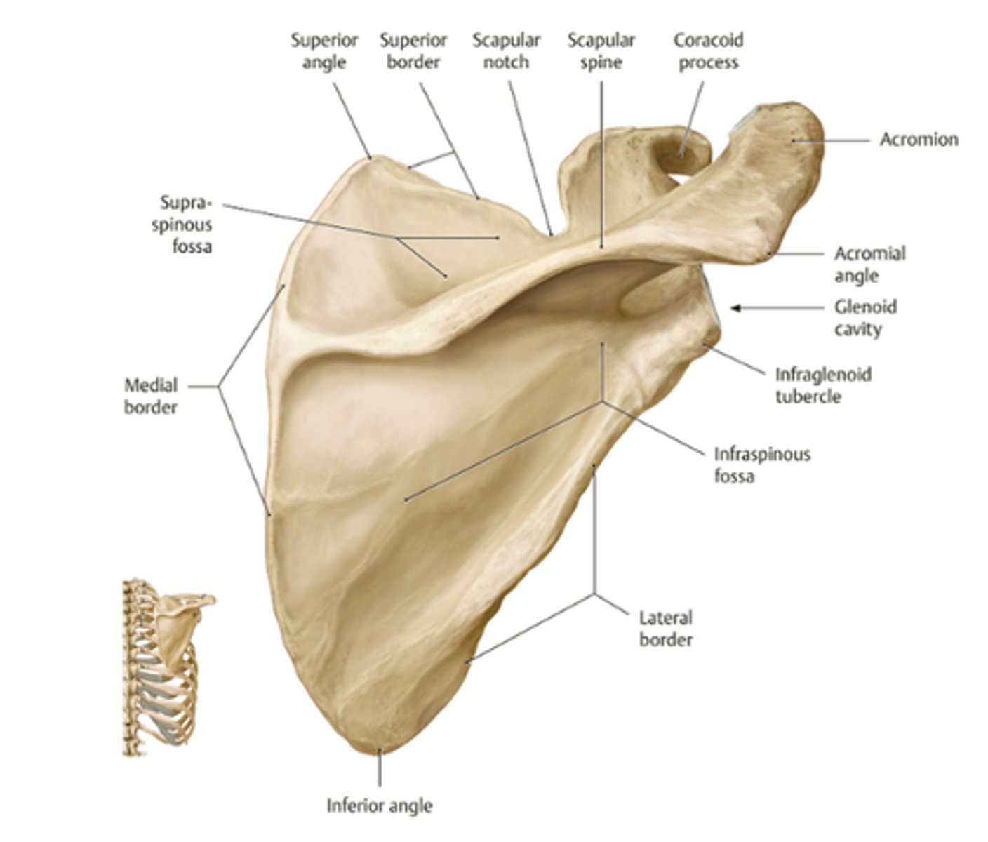
labelled scapula
labelled humerus
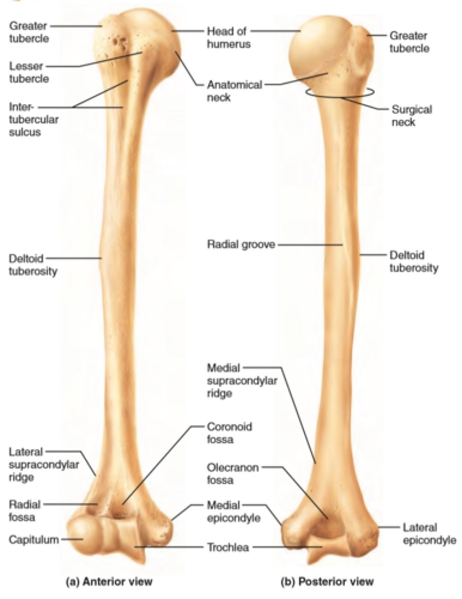
surgical neck fracture
affects axillary nerve. loss of sensation around regimental badge area. deltoid and teres minor weak abduction upper limb and external rotation. runs with circumflex artery around humeral neck.
teres minor muscle functions + innervation
adduction and lateral rotation of arm. axillary nerve
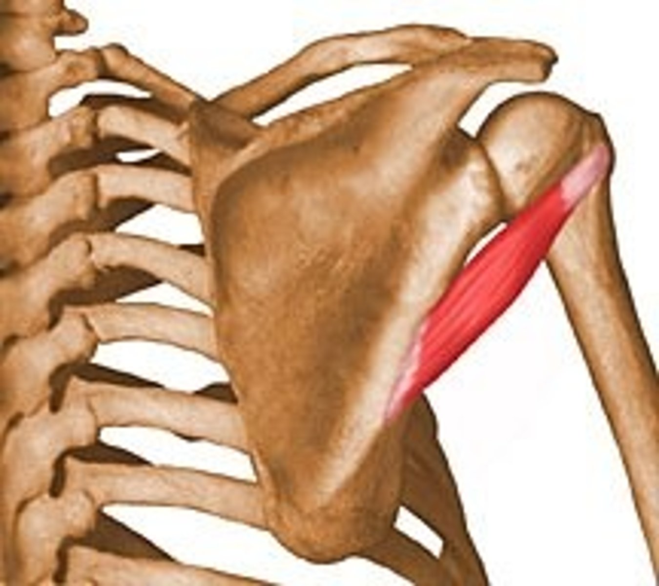
muscle flexes both shoulder and forearm and why
biceps brachii. origin and insertion crosses both shoulder and elbow so acts on both joints
posterior compartment of arm blood supply
profunda brachii/deep brachial artery
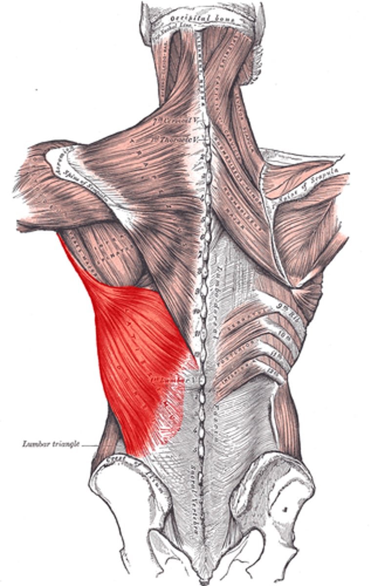
latissimus dorsi
posterior wall of axilla.
Plexuses are formed by
ventral rami
muscles that have their origin at coracoid process
short head of biceps, pec minor, coracobrachialis
A 30-year old farmer suffers severe traumatic injury to his upper right arm. There is profuse bleeding with fracture of the surgical neck of humerus.Which one of the following nerves is at the greatest risk of injury in this case?
axillary + bits of radial
On examination, a patient experienced pain when abducting their shoulder between 60 and 120 degrees. The tendon for which one of the following muscles is most likely to be inflamed in this case?
supraspinatus impingement syndrome. inflamed tendon presing against acromion
Following mastectomy and axillary clearance, a 45-year old female is noted to be developing a winged scapula. Which one of the following nerves is most likely to be injured in this case?
long thoracic nerve
A man suffers a penetrating wound through the posterior axillary fold, with resulting damage and weakness in shoulder adduction and medial rotation. Which one of the following muscles is most likely to have been damaged:
latissimus dorsi
nerve supplying triceps arises from which cord of brachial plexus
posterior
innervation of triceps brachii
radial nerve
What does accessory nerve CNXI innervate?
trapezius and sternocleidomastoid muscle
which two of rotator cuff muscle have opposing actions at shoulder joint
infraspinatus and subscapularis
The anterior and posterior folds of the axilla form the anterior and posterior wall of the axilla. Which muscles make up the anterior and posterior wall of the axilla?
anterior: pec major and minor
posterior: latissimus dorsi, teres major and subscapularis
Which muscle is a major abductor of the arm, and can also be a flexor of the arm, or an extensor of the arm? B) What nerve innervates this muscle?
deltoid. axillary nerve
testing biceps nerve roots
c5 and 6.
An injury to the long thoracic nerve can cause loss of function to the serratus anterior muscle. Which clinical condition might the patient present with and WHY?
winged scapula. serratus anterior attaches to ventral medial border of scapula preventing it from being pushed off posterior thoracic cage when upper limb pushed posteriorly.
deltoid can bring about abduction, internal rotation and external rotation. what movement can't it do
adduction
describe axillary nerve location
posterior cord of brachial plexus + pass around surgical neck of humerus below the acromion
part of clavicle that articulates with manubrium
sternal/medial end
contents of axilla
axillary artery
axillary vein
axillary lymph nodes
part between spine and superior border of scapula
supraspinous fossa
rotator cuff muscles
supraspinatus, infraspinatus, teres minor, subscapularis
posterior wall of axilla
latissimus dorsi, teres major, subscapularis
describe coracoid process
short, hook-like bony process of scapula. palpable below lateral third of clavicular shaft
glenohumeral ligament - how many are there and what are their names
anterior side - superior, middle, inferior
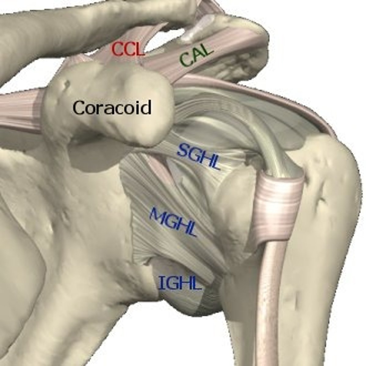
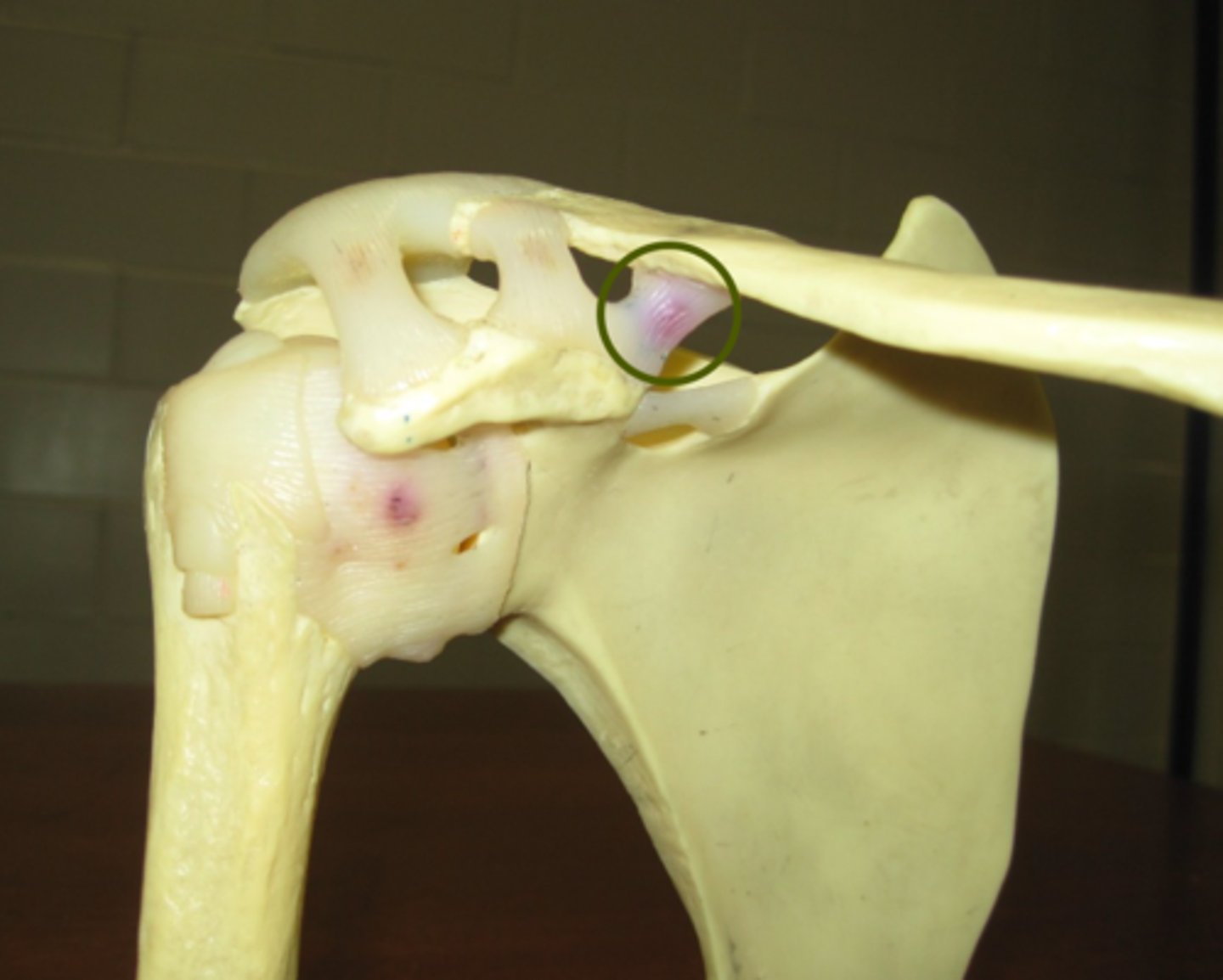
conoid part of coracoclavicular ligament
most posteromedial