BIOPSYCHOLOGY
1/78
There's no tags or description
Looks like no tags are added yet.
Name | Mastery | Learn | Test | Matching | Spaced | Call with Kai |
|---|
No analytics yet
Send a link to your students to track their progress
79 Terms
the nervous system
A complex network of nerve cells that carry messages to/from the brain to/from the rest of the body.
The central nervous system
Made up of brain + spinal chord. Controls behaviour + controls regulation of physiological processes in the body. Brain - keeps us alibe e.g breathing, swallowing. Spinal chord - Relays info from brain to rest of body, allowing brain to regulate bodily processes, also contains nerve cells responsible for reflexes.
Peripheral nervous system
Made up of all nerves outside of the CNS. Relays nerve impulses from CNS to the rest of the body. Divided into 2 main parts:
Somatic nervous system
Made up of cranial nerves (emerge from underside of brain), spinal nerves (emerge from spinal chord) which both have sensory(to CNS) and motor neurons(from CNS). It is in charge of voluntary actions.
Autonomic nervous system
Governs actions performed without conscious awareness. Necessary so vital bodily functions can work effectively. Further divided into 2 parts:
Sympathetic nervous system (fight or flight)
Helps deal with emergencies by activating fight or flight e.g by dilating pupils, increasing heart rate and bp.
Parasympathetic nervous system (rest and digest)
Relaxes an individual when an emergency passes e.g contracting pupils, decreasing heart rate and bp.
Neurons
Brain is made up of them - specialised cells that move chemical and electrical impulses to and from the CNS.
Motor neuron
Directly and/or indirectly control muscles, and carry messages away from the brain through axons located outside the CNS. Also form synapses with muscles to control contractions.

Relay neuron
Connect motor neurons to other ones, allowing them to communicate. Only in the brain and the spinal chord.
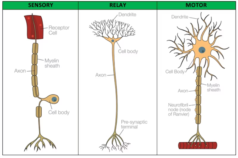
Sensory neuron
Carry nerve impulses from sensory receptors e.g vision to spinal chord and brain. Found in eyes, tongue, skin. Convert info from these receptors into neural impulses which are converted to sensations when they reach the brain.

Cell body
A control panel which contains the nucleus which contains DNA (chromosomes)
Dendrites
Branch-like structures that extend from the cell body, recieving signals/impulses from other neurons and send towards the cell body.
Axon
Impulses are carried along it, and it extends the neuron carrying impulses away
Myelin Sheath
A fatty subtsance that covers the axon to protect it and increase the speed that impulses travel along it.
Nodes of ranvier
Breaks in the myelin sheath of 0.2-2.2mm, action potentials jump from node to node, speeding up their travel.
Axon terminal
Communicates with other neurons
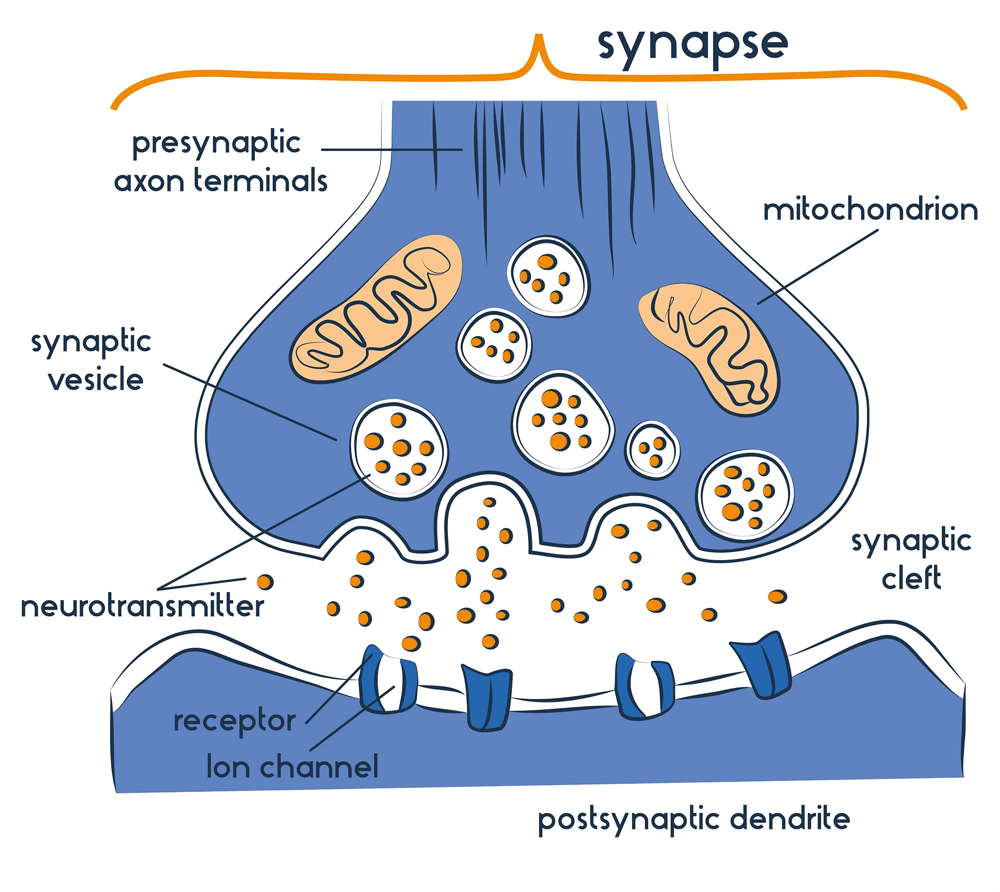
Action potential (synaptic transmission)
An electrical signal that travels through the dendrites of other neurons aswell as a single neuron, then down the cell body and to an axon.
The transportation of nerve signals
1) Action potential reaches terminal button of pre-synaptic neuron. 2)As this happens, vesicles containing neurotransmitters release their contents. 3)These cross synaptic cleft(gap) to the dendrites of the post synpatic neuron and bind to receptors. 4)Then summation occurs, inhibitory being a negative charge meaning the signal wont fire (e.g seretonin). Excitatory being a positive charge meaning the signal will fire(e.g dopamine).
The endocrine system
Controls vital bodily functions with a network of glands that secrete hormones(chemical messengers) which release into the bloodstream. Here, the hormones affect target cells and trigger a psychological reaction with enough stimulation.
Pituitary gland
Located in the brain, controlling the release of hormones from all other glands. ‘master gland’
Adrenal gland
Sits on top of kidneys and secretes adrenaline for fight or flight.
Thyroid
Located in the neck, producing thyroxine which controls growth and metabolism.
Ovaries
Located in female reproductive system, releasing oestrogen and progesterone responsible for fertility.
Testes
Located as part of the male reproductive system, producing testosterone which develops male characteristics e.g facial hair. Also plays a role in sex drive, sperm production, muscle strength.
Pineal gland
Located in the brain, producing melatonin responsible for quality and duration of sleep.
Example of how the endocrine system works:
A signal is sent from hypothalamus to pituitary gland to release a hormone. This stimulates a gland to release a hormone. As these stimulate enough target cells in the bloodstream, it creates a physiological change.
The fight or flight response
When threatened by immediate dangers, our SNS works antagonistically to our PNS to allow us to fight or escape.
The amygdala
Associates sensory signals with the emotions of fight or flight, sending a distress sgnal to the hypothalamus, which communicates with the rest of the body. Stress can come in two forms: acute - sudden e.g attack. chronic - over time e.g stressful job
The SAM pathway (response to acute stressor)
1)Amygdala senses danger, alerts the hypothalamus which triggers the SNS. 2)SNS sends signal to adrenal medulla, releasing adrenaline. 3)This causes physiological changes e.g increasing stuff. 4)When threat passes, PNS calms us down.
Fight or flight (evaluation)
-Can be neg responses to it. stressors can trigger response repeatedly, which leads to physical dmg of humans e.g heart disease. Shows its dangerous. So not useful.
-Partial expla. Gray states first phase of reaction is ‘freeze’.This is so we can stop, process through looking/listening. So isnt wholely internally valid.
-Individual differences(genetics). Lee + Harley found SRY gene promotes aggression in males, resulting in response, but lack of gene in women may prevent respone. Suggests is not credible. So may devalue the detail of response.
-Individual differences(gender). Women ‘tend and befriend’, protecting themselves and their young through nuturing and forming alliances with other women. Suggests women may not respond in a fight or flght way. So lacks external valid as cant be applied to all people.
Localisation
The idea that specific aread of the brain are associated with specific functions.
Cerebrum
Outer layer of brain divided into 4 lobes, is 1.5-5mm thick and consists of neurons and cell bodies.
Frontal lobe
Both hems, at front, more vunerable to injury. Respsonible for consciousness as controls functions that make us uniquely human. e.g language, personality, morals.
Broca’s area
Left frontal lobe, named after Paul Broca, he had patient ‘Tan’ who could only say this word, but could understand language. Studied 8 other ppl with this, and all had post-mortems which found they all had lesions to this part of frontal lobe. Area that produces speech. Dmg = brocas aphasia (slow speech lacking fluency)
Motor cortex
Both hems, back of frontal lobe. Plans and executes voluntary movements, arranged logically e.g region that controls leg is next to region that controls foot. Damage = loss in control of fine movement.
Sensory cortex
Both hems, behind motor cortex as parietal lobe. Detects info related to touch from regions of body, uses info to provide sensations e.g hot/cold. Damage = decrease in sensory thresholds (cant determine hot/cold.)
Occipital lobe
Both hems, back of brain. Processes visual stimuli and allows us to assign meaning +remember visual perception through nerve impulses that travel via optic nerve from retina to thalamus, relaying info to visual cortex. Damage = full/partial blindness
Temporal lobe
Both hems, under other lobes. Processes auditory info through cochlea in ear, sound waves convert to nerve impulses which travel to thalamus via auditory nerve, relaying to auditory cortex. Damage = full/partial hearing loss
Wernicke’s area
back left of temporal lobe. Understands language, named after Carl Wernicke, patients with lesions here could speak fluently, but it was meaningless. (Wernicke’s aphasia)
Localisation:evaluation
+Case study evidence. phineus cage, exlosion at railroad causes iron rod through left frontol lobe, lead to changhe in personality. suggests frontal lobe responsible for personality. so credible. However, case study, lacks external.
-ignored individual diff (gender). Harasky et al. found women have larger Brocas + Wernickes areas, because they use language more. suggests theory has beta bias, gender diff ignored. theory lacks external valid.
-Contradicting evidence. Lashley, equipotentiality theory, basic motor/sensory functions localised but higher mental functions not. removed 10-50 perc of cortex in rats who learned maze and no cortex was more important in ability to learn maze.suggests localisation distributed in more holistic way. So not internally valid.
+evidence for areas. Peterson et al. found activity in brocas during listening task, and same for wernickes duringr reading task using brain scans. suggests brain is localised. theory has value.
Lateralisation
The fact that the two halves of the brain are not entirely alike, carrying out specialised functions. e.g LH for language.
Sperry (Split brain research)
Quasi exp, 11pps already had corpus callosum severed. pp gazes at fixation point on screen with one eye blindfolded, image flases at 1/10 of a second.
Sperry (testing if language processed in LH)
Object displayed on screen to RVF (ends up in LH) and pps could verbally name and write it, but if displayed to LVF(to RH) pps cant verbally name it.
Sperry(testing if touch/visuo-spatial procesed in RH)
Object displayed to LVF(RH) pps insist they havent seena nything as language processed in LH. But could pick up matching object from under screen.
laterlisation:evaluation
-contradicting evi to lang processed in LH. Turk et al.: case study J.W, developed capacity to talk from RH, showing can use both hems talk ab processed info. lacks credibility as it suggested RH cant handle any lang. lacks internal valid. However, case study.
-evidence q’s amount of lateralisation in ppl. Szaflarski found lang strongly lateralised in LH among children + adolescents under age of 25, declines every decade ppl grow up. suggests lat is not a lifelong thing. lacks external valid.
+well designed through standardised procedures. through flashing image at 1/10 sec, controlling which hem it went to. suggests can develop cause and effect relationship. reliable.
-research cant be applied to present day. procedure of severing corpus callosum is not used, Andrewes argues recent studies only have 3 pps, sometimes even 1. suggests it lacks temporal valid. therefore, lacks value.
Brain plasticity
The process of life experiences changing/adapting the brains structure. Easier in infancy (peaks at age 2) as brain is rapidly forming new neural connections.
Brain plasticity:Synaptic reweighting
When activity increases over a period of time, the signals in synapses become stronger.
Brain plasticity:Creation of new synapses
If the axon of one nerve cell passes close to the dendrite of another, a new synapse forms.
Brain plasticity:Synaptic pruning
Synapses that are not used are deleted.
Brain plasticity:Maguires taxi drivers study
Found more grey matter in posterier of the drivers hippocampus(associated with development of spatial and navigational skills). They had to stufy for ‘The knowledge test’ on streets + routes, the more they were taxi drivers, the larger the difference.
Brain plasticity:Draganski medical students
Scanned students brains 3 months before and after their exams, learning induced changes happened in the posterier hippocampus and the parietal cortex (result of learning)
Functional recovery
Following brain injury or traums (e.g stroke), unaffected areas of the brain are able to compensate and adapt. This is possible due to plasticity. Usually happens from one hem to another.
Functional recovery:Neuronal unmasking
Synapses that were there all along, close to the area of damage, but dormant and never properly active are activated.
Functional recovery:Axonal sprouting
Undamaged axons physically grow new nerve endings, and hook onto other dendrites of neurons to form new synapses.
Functional recovery:Recruitment of homologous areas
Mirror neurons on the other side of the brain to the damage take over the particular functions damaged.
Functional recovery:Danielli et al. case study of E.B
Italian boy aged 2 n a half had tumour taken out of left hem, and left hem was removed, removing all linguistic abilities. Had intensive rehabilitation, after 3 years had no major problems in language ability, tested at 17 and RH compensated.
Functional recovery:Takatsura et al. mice
Induced strokes in somatsensory cortex in RH of mice. After 4 weeks, new pattern of electrical activity in LH was found, meaning FR occured.
Factors influencing functional recovery
Gender(Women less lateralised, ratcliffe found after brain injury, women did better on cognitive assessments), Age(The earlier trauma occurs, easier recovery is,)
Brain plasticity and Functional recovery: evaluation
+support evid. Kempermann et al. found an enriched environment(compared to cages) can alter number of neurons in rats brains, esp in hippocampus. supports claim of brain being ‘plastic’. increases plausability. However lacks external valid as its rats.
-Brains ability of rewiring can be maladaptive. 60-80 perc of amputees get phantom limb syndrome(continued sensations in missing limb as if it was still there), can be painful. suggests plasticity is not always a positive thing. therefore not valueable.
+RWA. Helped us develop neurorehabilitation, which can treat people with injuries. suggests it is useful as it allows people to change their brains for the better. so has external valid.
-capacity of plasticity is not high. Jeuber studied soldiers and found recovery was age dependent, which limits options for older patients with brain injuries. suggests ability of brain to recover varies with age. therefore, not useful for everyone.
FMRI
Measures changes in brain activity during task. Detects change in blood oxygenation flow that happen during neural activity. +Records signal from all regions of brain (spatial resolution 1-2mm) +noninvasive -low temp valid (5 sec time lag). -no direct measurement of neural activity.
EEG
Measures electrical activity in brain through electrodes on scalp, this is through firing of neurons from brain. +high temp valid, activity in 1 milisecond or less. +diagnoses epilepsy(random bursts of activity). -low spatial resolution.(only detecs groups of nerons not precise ones) -cant go deeper into brain
ERP
Small voltage changes in brain in reaction to stimuli. Stimulus is presented over again and statistical averging technique used to filter out extraneous activity. +Brings more specificty to EEG data. +High temp valid (single milisecond or less) -Average so not completely true. -Time consuming
Post mortem
Examination of brain after death, establishing underlying neural issues with someone or a behaviour. +provided foundation for understanding process in brain early on. +Allow for truly deep investigation. -Ppl who have it cant consent. -Cause of dmg not shown.
Circadian rhythyms
Biological rhythyms that occur over a 24 hour cycle. Driven by our body clocks, controlled by SCN, and also by homeostasis which tells us we need to sleep as energy is used up. e.g sleep-wake cycle
Circadian rhythyms - Stiffre study (sleep-wake cycle)
Spent 6 months in cave with no natural light to see how circadian rhythym was affected. This ‘free-ran’ meaning his body slept and ate on his bodies inclinations. Found sleep pattern to average 25 hours, and slept on regular schedule, showing we do have an internal mechanism that regulates our cycle, even if it shifts to 25 hours
Circadian rhythyms - Aschoff + Weaver (sleep-wake cycle)
Pps spent 4 weeks in ww2 bunker with no natural light. All but 1 sleep wake cycle adjusted 24-25 hours. Confirming Stiffre’s study, and suggesting natural CR is longer than 24 hours.
Circadian rhythyms - evaluation
+RWA, shows effects of disrupted CR. Bolvin et al - night shift workers more likely to have injuries/make mistakes at 6 am due to circadian trough(reduces concentration). Increases value. External valid.
+RWA, chronotherapeutics. CR co-ordinate heart rate, digestion, sleep-wake cycle, and this allowed for medicine to be taken at right time to match a persons biological rhythyms. e.g aspirin(heart attacks) last thing at night because heart attack most likely to happen in morning. so shows research has improved effectiveness of medicine. so research is useful.
-Individual differences. Sleep-wake cycle differs from people, so hard to generalise, e.g Zeisler et al - vary from 16-65 hours), also morning people etc.Shows data only calculated averages. So decreases value
-Studies had small samples. Stiffres study was only him. This limits representativeness, so cant generalise. So lacks external valid.4
Endogenous pacemakers
Biological ‘clocks’ within the brain.Process: 1)Melanopsin (protein in eye) sensitive to light and carries signal via optic nerve to: 2) SCN (master clock) within hypothalamus recieve time co-ord and adjusts and rests when external light levels change, this info is sent to: 3)Pineal gland behing hypothalamus which produces melatonin to induce sleep.
Exogenous Zeitgebers
External stimuli which aid biological clocks to maintain co-ordination with the external world. Light - Rests SCN, maintaining sleep-wake cycle. Social cues - Social cues imply rest is needed. e.g imposing chedule on babies, like when they sleep, eat, bathe. Entrainment - Occurs when body clock adjusts in line with the environment due to these cues. Happens when we travel across time zones, as pacemakers are not synchronised.
Exogenous Zeitgebers - evaluation
+Animal stud supports SCN role. Decoursey et al - Destroyed SCN connections in 30 chipmunks and returned to natural habitat for 80 days, many killed by predators by leaving nest at wrong time of day, so sleep-wake cycle was impaired. Gives credibility. However, animals cant be generalised to humans.
-Contradict SCN only endogenous pacemaker responsible for regulating cycle. Campbell + Murphy - light detected by skin cells is aswell. shined light on back of knees of 15 pps woken up at various times of night. This altered cycle by up to 3 hours. So contradicts light needs to enter eyes to alter cycle. So partial explanation. However, would have also seen this light with eyes.
-Role of exogenous zeitgebers overestimated. Miles - blind man from birth with circadian rhythym of 24.9 hours could be adjusted regardless of changes to social cues. So effect is less than anticipated. So we are more in control of rhythyms than external factors. However, blind is an exceptional circumstance, not all humans can relate.
+RWA: decrease effect of jet lag. Burgess et al - exposure to light following east-west flight decreased time needed to adjust to local time. exposure to bright light flicked CR back 2.1 hours, comapred to intermittent light which was 0.6 hours. Demonstrates entrainment. So has value.
Infradian rhythyms
Biological rhythyms that last more than one day. e.g menstrual cycle, typically lasting 28 days. Hypothalamus cause pituitary rise levels of oestrogen, causing ovary to develop an egg and release it.(ovulation) Then progesterone helps womb lining grow thicker, readying for pregnancy, but if pregnancy doesnt happen, womb lining leaves body(flow) and egg is absorbed.
Infradian rhythyms - evaluation
+RWA. McClintock - indentified synching of cycles with her dorm mates were caused by pheremones emitted in social interaction.
+Evolutionary advantage, with natural selection. Our ancestors may have tried to sync mentrual cycles and pregnancies, so if babies lost their mothers they would have milk. So synchronisation is an adaptive strategy. So has value. However, could be less valuable as disease could wipe out a generation.
-Methodological limitations of research on pheremones. Other, confounding variables can change the cycle, e.g stress, diet, excersise. Shows there may be external effects on the cycle. So lowers internal valid.
+Supporting research. Stern + McClintock - 29 women, history of irrelgular periods. gathered samples of pheremones from different stages of the 9 women with cotton pad under armpit. Rubbed on uperlip of other 20 women. 68 perc saw changes to cycle, bringing them closer to their ‘odour donor’.
Infradian rthym - Seasonal Affective Disorder
Persistent low mood, loss of activity and interest in life that comes seasonally. It happens anually, and is often triggered during winter where the number of daylight hours become shorter. Process: During night, pineal gland secretes melatonin until dawn when increased light is detected. But bc of the lack of light in winter morning, process continues on for longer, meaning production of seretonin(controlling mood) is delayed.
Infradian rthym - Seasonal Affective Disorder (evaluation)
+Reductionist explanation. Reduces complex emotional disorder to simply being due to lack of light and hormone imbalance. So it is oversimplified and doesnt account for people. So is a partial explanation.
-Determinist. Suggests we have no control over our feelings, but some people enjoy winter, e.g christmas. Shows element of free will over SAD, controlling ig we get it. So doesnt acknowledge individual differences.
+RWA:photobox. Stimulates strong light, resetting melatonin levels. Sanassi et al - relieves symptoms of 80 perc of ppl with it. Shows is useful for treating SAD. So has value. However, shown to have 46 perc relapse rate, so permenant fix.
Ultradian rhythyms
Biological rhythyms with a frequency of more than one within 24 hours. e.g sleep cycle, which spans 90 mins, and continues going through 5 stages as we sleep. Stage 1-4 nREM, stage 5:REM.(happens 4 times, increasing from 5 mins-10-15-30-60.)
Sleep cycle stages explained (ultradian rhythyms)
Stage 1: alpha waves, light sleep, easily woken and body slows down. Stage 2: Theta waves, sleep spindles (bursts of activity) help us with memory, body continues slowing and easily awoken. Stage 3:Delta waves, no reaction to external stimuli. Relaxing further, hard to wake, helps memory consolidation + cell regeneration. Stage 4: Delta waves increase, deepest stage of sleep, body processes at lowest, confused if woken. Stage 5: Fast, desynchronised brain activity resembling awakeness, rapid eye movements side to side, temporary paralysis and dreaming.
Sleep cycle supporting research - Dement + Kleitman (ultradian rhythyms)
monitered sleep patterns in 9 adults in sleep lab using EEGs + EOGs. Supported stages as woke people in REM, and they could remember what they were dreaming about, but if in nREM, they couldnt.
Sleep cycle - Ultradian rhythyms (evaluation)
+Improved understanding of age-related patterns of sleep. Cauter et al - deep sleep reduces with age, growth hormone secreted in stages 3-4 is reduced in older people. Explains issues w old age e.g reduced alertness. So practical value in research.
-Individual differences. Tucker et al - pps studied for 11 days, large differences in stage 3-4. makes difficult to describe ‘normal’ sleep patterns. So lacks external valid.
+Research was highly scientific. Controlled confounding variables, e.g told pps not to drink caffiene of alc before, and used EEG. So research is internally valid. So is reliable, and has been replicated before. However, artifical setting may have affected sleep.
-Mainly male sample. Findings only tell us about male sleep patterns, women are different physically e.g menstrual cycle, so their sleep cycles may differ. Therefore, partial explanation