Nerve Impulse and Transmission
1/34
There's no tags or description
Looks like no tags are added yet.
Name | Mastery | Learn | Test | Matching | Spaced | Call with Kai |
|---|
No analytics yet
Send a link to your students to track their progress
35 Terms
nerve impulse
communication among neurons and with cells of their control
originated in response to a stimulus of an electrical, chemical, thermal, or mechanical been received by the neuron cell membrane
the stimulus elicits a wave of depolarization and repolarization that
spreads along the axolemma
away from the site where was received
results in the transmission of the nerve impulse
mechanisms of transmission
potential
relative electrical charges between two points in a field or circuit
for the neuron
transmembrane potential
inside and outside of the cell membrane
all cells of the body have a transmembrane potential, bit he neurons are unique in being able to alter this potential to produce an impulse
resting membrane potential - resting neuron
results from the unequal distribution of sodium ions and potassium ions on the outside and inside of the neuron
active transport of Na to the outside with the transport of K into the neuron
keeps the concentration of Na low on the inside
electronegativity is maintained on the inside of the membrane and electropositivity on the outside
depolarization
chemical or physical stimulation of a neuron increases the permeability of the membrane for Na at the point of stimulation
membrane is polarized at 70 millivolts
high concentration of Na on the outside of the membrane
Na rushes inwards
membrane now becomes positive on the inside and negative on the outside
the inflow of Na soon stop sand the permeability of the membrane for K increases
repolarization
the K flows outward because it has a higher concentration inside the neuron than outside
the outflow of K destabilizes the resting membrane potential at the point of stimulation
return to the resting membrane potential
enter absolute refractory period
nerve fiber cannot be stimulated again until repolarization is complete
hyperpolarization
membrane potential becomes more negative
relative refractory period
propagation of action potential
AP moves across neurilemma of dendrite, soma, and axon
myelinated axons
-> nodes of ranvier -> node of ranvier
faster because it skips parts of the neurilemma
non myelinated axons
sends signal straight through
takes longer
axon diameter
larger means faster signal sending
myelin aids in conduction
local currents generated by an AP flow to adjacent areas of the axonal membrane to depolarize and generate further APs
MS
neuron placements
Converging circuit
Diverging circuit
Reverberating circuit
Parallel circuit
simple circuits
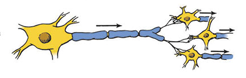
Converging circuit
several neurons impinge on one neuron
cerebellum converging info from 4 different brain regions
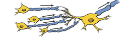
Diverging circuit
the axon branches of one neuron impinge on two or more neurons, then those impinge on two or more neurons amplification of signal
e.g. one neuron stimulating many muscles neurons
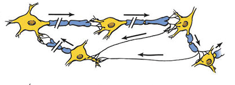
Reverberating circuit
each neuron in a series sends a branch back to the beginning neuron so that a volley of impulses is received at the final neuron
rhythmic activities
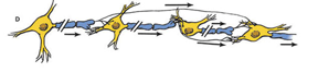
Parallel circuit
contains a number of neurons in a series, each neuron supplying a branch to the final neuron
e.g. reflex arcs
simple circuits
no more than 2 neurons
for their projection to the cerebral cortex
olfactory, optic
3 neuron circuit classic for conscious sensations
3 required to transmit a nerve impulse from periphery by spinal nerve to the cerebral cortex
reflexes def
an automatic or unconscious response of an effector organ to an appropriate stimulus
reflex arc
receptor
afferent limb
central connections
efferent limb
effector organ
myotatic (stretch) spinal reflex - knee jerk reflex
Striking the middle patellar ligament:
tendon of insertion for the quadriceps femoris and transmits its action to extend the tibia.
Stretches the quadriceps muscle
stimulates muscle spindles (receptors for muscle sense).
An impulse’s path
dorsal root of the spinal nerve
motor neuron in the ventral horn of the gray matter,
Muscle fibers of the quadriceps muscle, causing it to contract.
The purpose of the reflex is to oppose stretch of the muscle.
somatic reflexes
effector organs are composed of striated muscle
visceral reflexes
effector organs are either smooth or cardiac muscle, or glands
regulate visceral functions and are transmitted by the autonomic nervous system
postural reflexes
maintaining an upright position
muscle tonus is that state of muscle tension that enables and animal to assume and remain in the erect attitude
Standing reflex
Attitudinal reflexes
Righting reflex
Hopping reaction
Crossed extensor
Standing reflex
pushing down on the back of a dog causes muscle movements that compensate for and resist the displacement.
Attitudinal reflexes
displacement of one part of the body is followed by postural changes in other parts (e.g., lifting the head of a horse is followed by postural changes in the rear quarters so that a new attitude is assumed).
Righting reflex
dropping an inverted cat is followed by its landing in the upright position.
Hopping reaction
pushing a supported dog with three limbs elevated results in a placement correction of the intact leg to act as a rigid pillar.
Crossed extensor
stepping on something sharp causes leg to contract while other leg stays standing/ extends (postural)
meninges
covering of the brain and spinal cord
dura mater
most superficial
outer
touch dense regular fibrous CT
adheres to the skull
not present around the brain
epidural space
between dura mater and bone around spinal cord
arachnoid
delicate spider web like loose CT around brain
thin sheet around spinal cord
subarachnoid space filled with CSF
pia mater
delicate loose CT on surface of brain and spinal cord
ventricles of the brain - basics
Cavities or hollowed-out spaces within the substance of the brain
each of the four ventricles has a choroid plexus
tuft of capillaries that secretes CSF
ventricles of the brain - list
The lateral ventricles are paired cavities
right and left cerebral hemisphere.
Interventricular foramen
(foramen of Monro)
Third ventricle
The cerebral aqueduct
(mesencephalic aqueduct).
The fourth ventricle
is located beneath the cerebellum and above the medulla oblongata.
continuous caudally as the central canal of the spinal cord.
ependymal cells
glial cell
unite with the capillaries to form the choroid plexus
capillaries in pia mater
flow of CSF
Lateral ventricles
interventricular foramen
third ventricle
cerebral aqueduct
fourth ventricle
foramina of Luschka
Subarachnoid space and spinal cord
CNS metabolism
The CNS receives its energy principally from carbohydrates
glucose
CNS receives glucose by simple diffusion and insulin is not required.
Advantageous for the animal when insulin is lacking or in short supply
enables the CNS function to continue when other systems fail.
The relatively high rate of metabolism/oxygen consumption
the CNS constitutes only 2% of body mass
consumes approximately 20% of the total oxygen supplied to the body.
blood-brain barrier
Many substances in the blood do not readily enter the cells of the CNS
The capillaries of the CNS have tight junctions between their endothelial cells
limit the diffusion of substances from capillaries.
Lipid-soluble substances,
readily diffuse
oxygen and carbon dioxide
Transport for most substances is provided for by astrocytes (a glial cell)
selective
Some areas of the hypothalamus, as well as other portions of the brain that serve as chemoreceptor areas, lack a blood–brain barrier.
blood requirement
The CNS must have a continuous supply of blood for normal functioning.
Hypoxia (deficient oxygen)
Other tissues can be deprived of a blood supply for extended periods and recover to normal function when the blood supply resumes.
Five to 10 minutes of little or no blood to the brain injures higher brain cells (in the cerebrum) so that no recovery occurs.