Bone Disorders
1/131
Earn XP
Description and Tags
24 June 2025
Name | Mastery | Learn | Test | Matching | Spaced |
|---|
No study sessions yet.
132 Terms
Name the types of bone cells
Osteocyte
Osteoblast
Osteogenic cell
Osteoclast
What
What is an osteocyte responsible for
Maintaining bone tissue
What is an osteoblast responsible for
Forming bone matrix
What is an osteogenic cell responsible for?
Stem cell
What is an osteoclast responsible for?
Bone resorption
Name some fibro-osseous lesions
Fibrous dysplasia
Cemento-osseous dysplasia (COD)
What is fibrous dysplasia?
A tumor-like condition, characterized by replacement of normal bone by fibrous connective tissue intermixed with abnormal bone
What does dysplasia in bone look like?
Disorganized growth that is not malignant
What kind of a condition is fibrous dysplasia?
A sporadic condition resulting from a postzygotic mutation
Depending on when the mutation in fibrous dysplasia takes place, what processes can be involved?
One bone (monostotic)
Multiple bones (polyostotic)
Skin
Endocrine system
What does polyostotic fibrous dysplasia mean?
Isolated or with syndrome
What are syndromes involved with polyostotic fibrous dysplasia?
McCune-Albright Syndrome
Jaffe-Lichtenstein Syndrome
Mazabraud Syndrome
What is McCune-Albright Syndrome?
A syndrome of polyostotic fibrous dysplasia that is Polyostotic FD + café au lait + endocrinopathies*
What is Jaffe-Lichtenstein Syndrome?
A syndrome of polyostotic fibrous dysplasia that is Polyostotic FD + café au lait
What is Mazabraud Syndrome
A syndrome of polyostotic fibrous dysplasia that is Polyostotic FD + intramuscular
What gender does Monostotic Fibrous Dysplasia affect the most?
M = F

What age group does Monostotic Fibrous Dysplasia affect the most?
Teenage years (2nd or 3rd decade)

Where would you find Monostotic Fibrous Dysplasia?
Affects the maxilla more than mandible, monostotic accounts for 80% of all cases

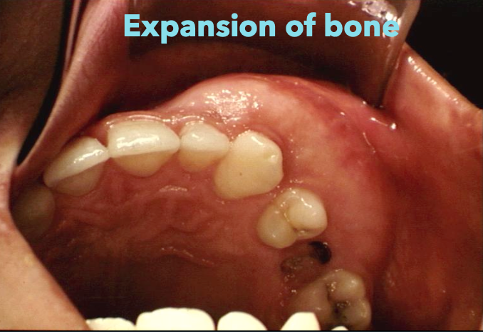
What is the most common feature of Monostotic Fibrous Dysplasia?
Painless, slowly-growing swelling of affected area
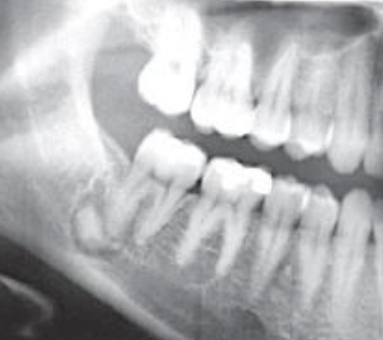
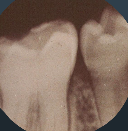
What does fibrous dysplasia look like on a radiograph?
In the early stages, the lesion may be radiolucent but with time it becomes radiopaque
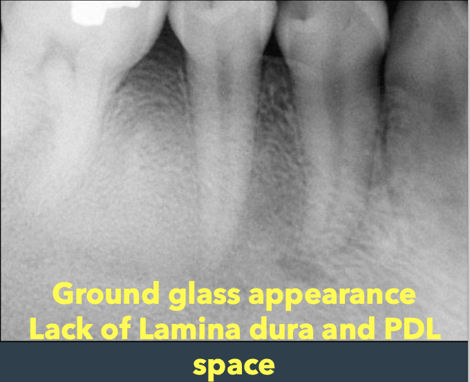
What are some characteristics of fibrous dysplasia on a radiograph?
“Ground-glass” opacification
Not well demarcated, blend
Narrowing of PDL with an ill-defined lamina dura
Expansion of both buccal and lingual plates
Obliteration of maxillary sinus
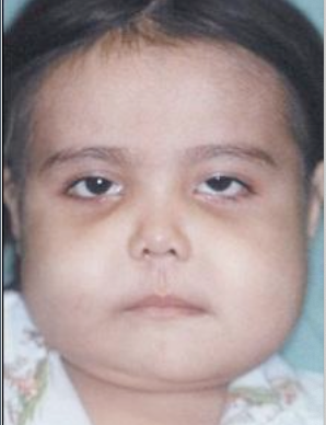
Fibrous Dysplasia Clinical and Radiographic Image
What is the involvement of polyostotic fibrous dysplasia?
Two or more bones; can involve up to 75% of skeleton
Who does polyostotic fibrous dysplasia mainly affect?
Children before 10 years old
What can happen if the jaw is involved in polyostotic fibrous dysplasia?
Facial asymmetry may result

What are some other features of polyostotic fibrous dysplasia?
Pain due to pathologic fracture of the long bones is common
Leg length discrepancy (hockey stick deformity)
What are some endocrinopathies seen in McCune Albright Syndrome in polyostotic fibrous dysplasia?
Sexual precocity (early puberty)
Pituitary adenoma
Hyperthyroidism
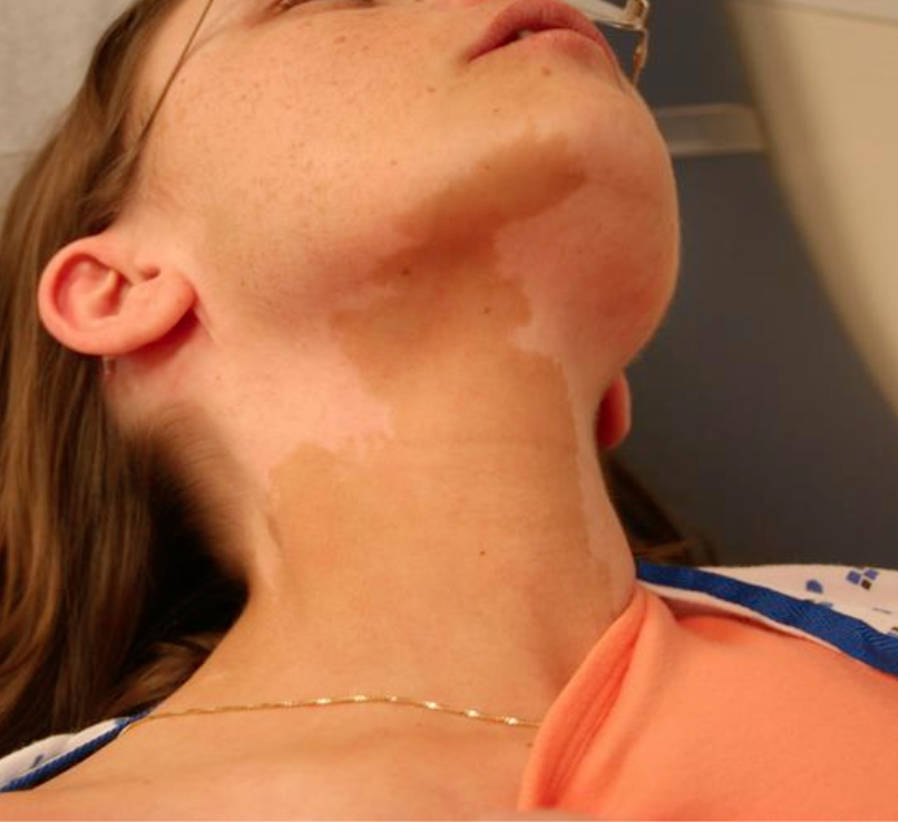
What is Neurofibromatosis 1 (NF1)?
Café au lait spots are smaller and higher in #
Borders are smooth and ovoid shape (coast of California)
Cross the midline
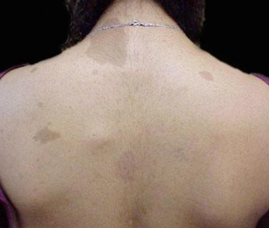
Cafe au lait spots in McCune Albright Syndrome
Café au lait spots are larger and fewer in #
Borders are jagged and irregular (coast of Maine)
Found in midline and does not cross the midline
What is the treatment for fibrous dysplasia?
Varies, medication, pain management, physical therapy and surgery as resection
Disease tends to stabilize and stop growing at skeletal maturity
Up to 50% recurrence
What is the most common fibro-osseous lesion encountered in clinical practice?
Cemento-osseous dysplasia (COD)
Where would you find cemento-osseous dysplasia?
In tooth-bearing areas of the jaws
In which demographic does cemento-osseous dysplasia show up?
Middle aged females, AA
What are the types of cemento-osseous dysplasia?
Focal
Periapical
Florid
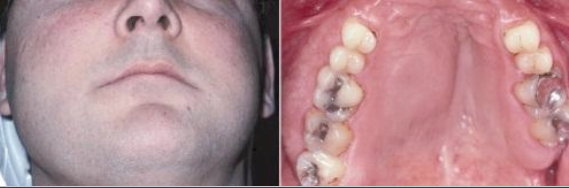
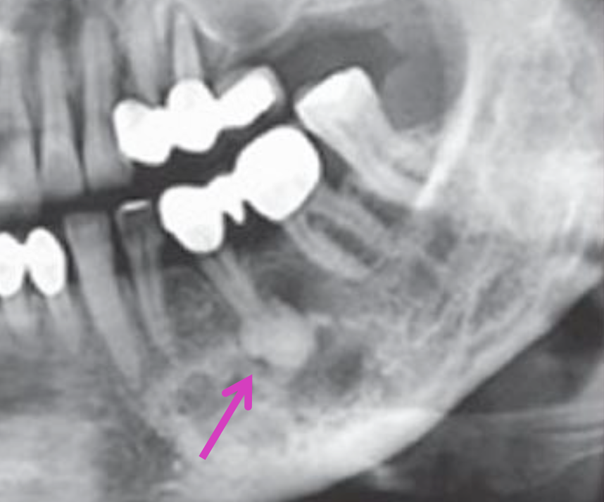
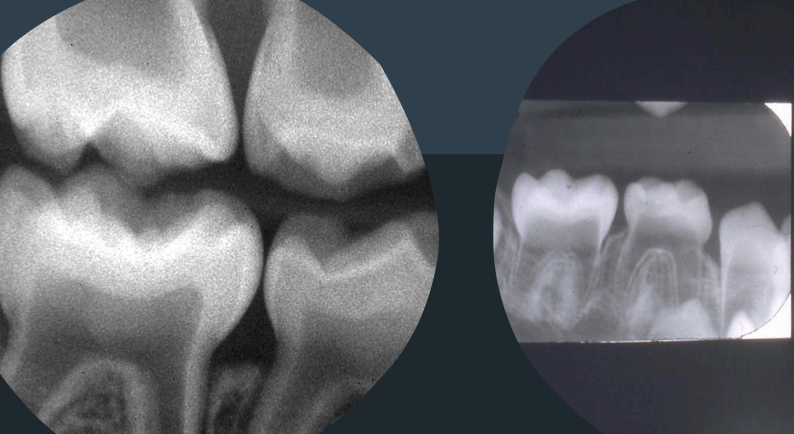
Focal cemento-osseous dysplasia
Periapical cemento-osseous dysplasia
Florid cemento-osseous dysplasia
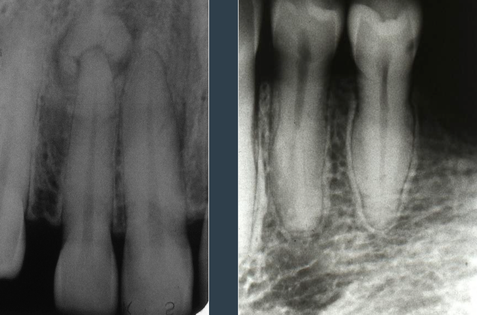
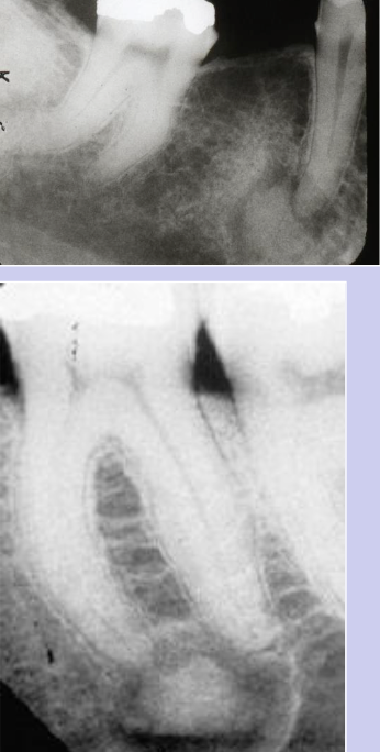
Features of periapical cemento-osseous dysplasia
Located near periapical region of the anterior mandible
Multiple foci are usually present
Teeth are invariably vital and asymptomatic
Early lesions are circumscribed areas of RL involving the apex of a tooth
looks identical to that of a periapical granuloma or cyst
The PDL will be intact; the lesion will NOT fuse to the tooth
Lesion is typically non-expansile self-limiting
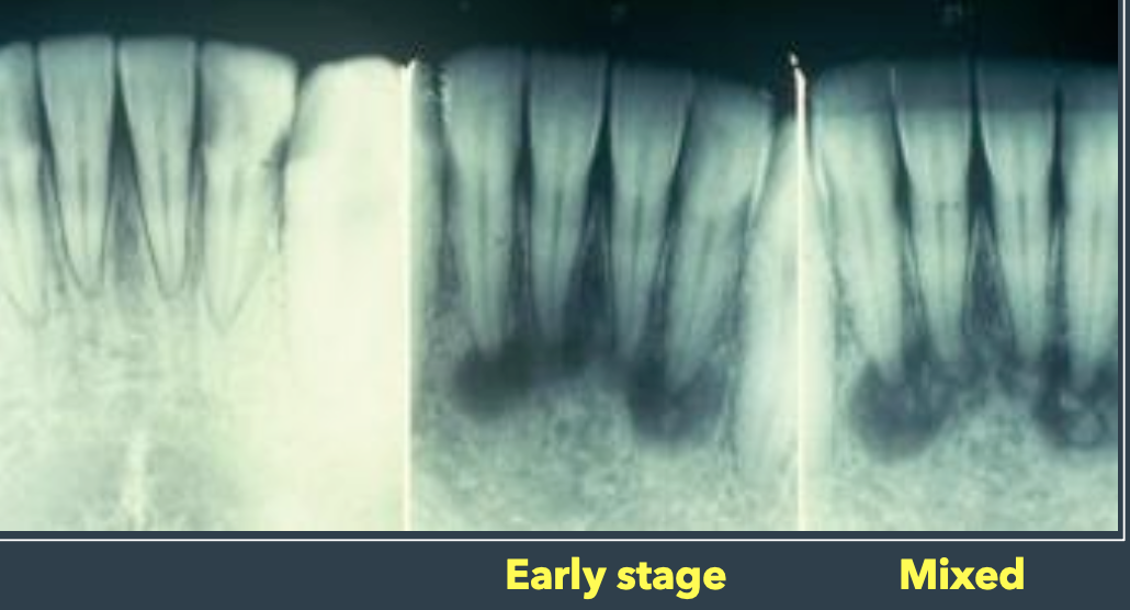
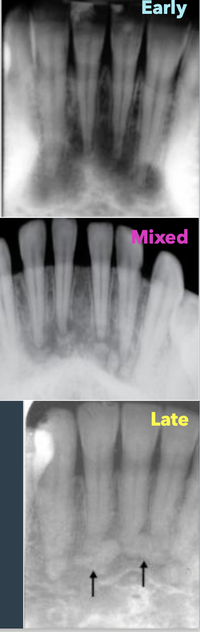
Stages of periapical cemento-osseous dysplasia
Early stage: Radiolucent (RL) lesions
Mixed stage: RL-RO appearance
Late-stage: Densely RO with a RL rim
In which demographic would you see periapical cemento-osseous dysplasia
90% are female
70% in African Americans
Middle age (40s)
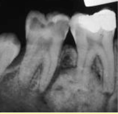
In which demographic do you see focal cemento-osseous dysplasia?
90% occur in females, AA
Middle age

What is the most common location of focal cemento-osseous dysplasia?
Posterior mandible
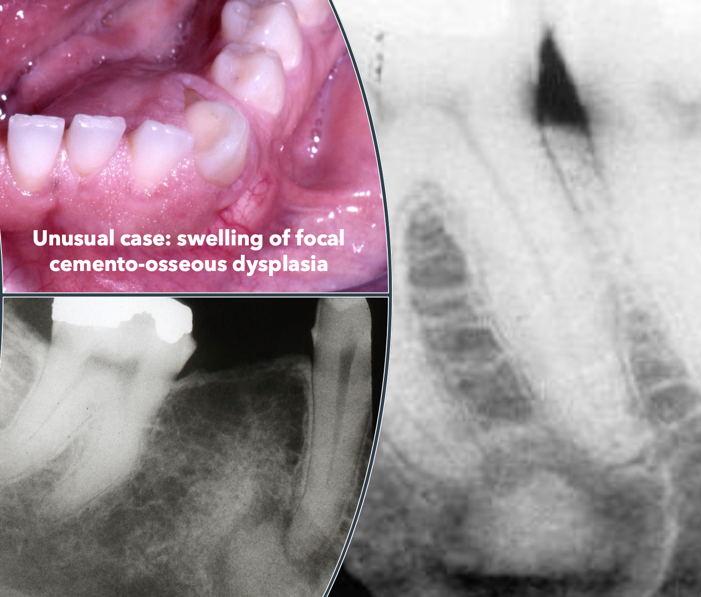
What are some clinical features of focal cemento-osseous dysplasia?
Single lesion
Asymptomatic
Lesions are smaller than 1.5cm
Radiolucent to radiopaque
Occurs in the tooth-bearing areas of the jaws
Well-defined rim during mixed stage

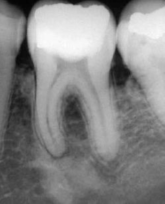
What can cemento-osseous dysplasia potentially be confused with?
Hypercementosis
What is the difference between cemento-osseous dysplasia and hypercementosis?
Cemento-osseous dysplasia is not within the PDL space whereas in hypercementosis, the radiodensity is all within the PDL
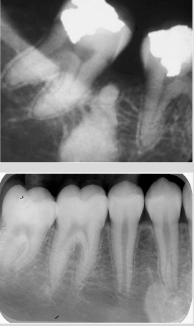
In which demographic do you see florid cemento-osseous dysplasia?
90% are female
90% are African American
Middle aged or older adults
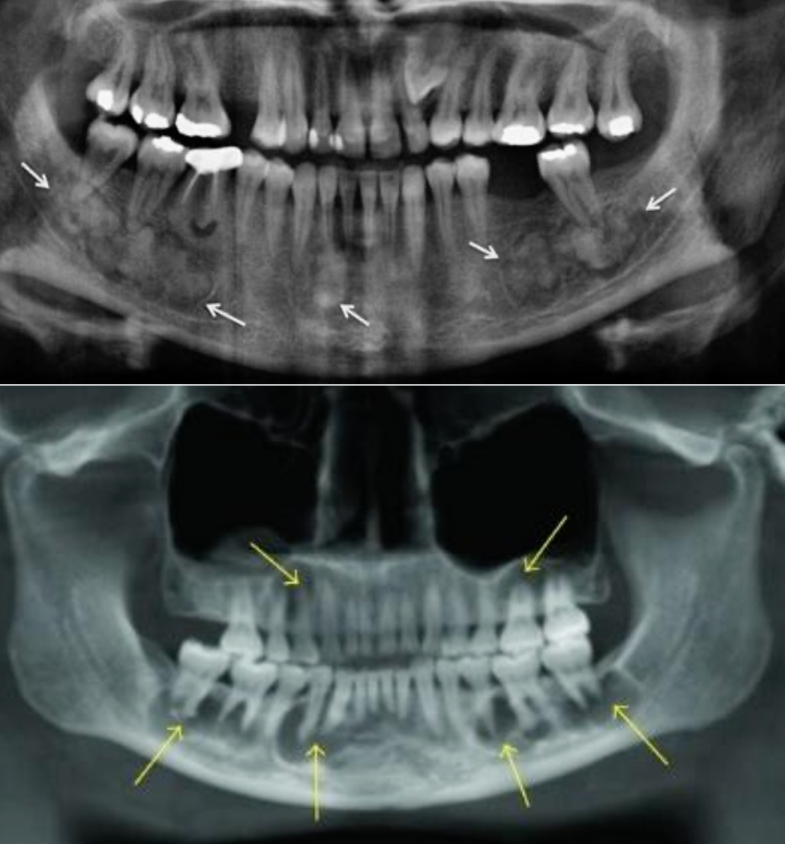
Where might you find florid cemento-osseous dysplasia?
Multiple focal involvement not limited to the anterior mandible
Patients may just have lesions in the posterior jaws but many patients have lesions throughout

What is a marked tendency of florid cemento-osseous dysplasia?
Bilateral and symmetrical, but teeth are vital and asymptomatic

What is the treatment for cemento-osseous dysplasia?
Periapical or florid COD, diagnosis can be made from the distinctive clinical & radiographic findings – do NOT need biopsy
Biopsy of florid COD may lead to necrosis due to the hypovascularity
Focal COD may require surgical investigation because the features are less specific
Follow up
Management: AB if osteomyelitis is present
Differential diagnoses of radiopaque lesion at apex
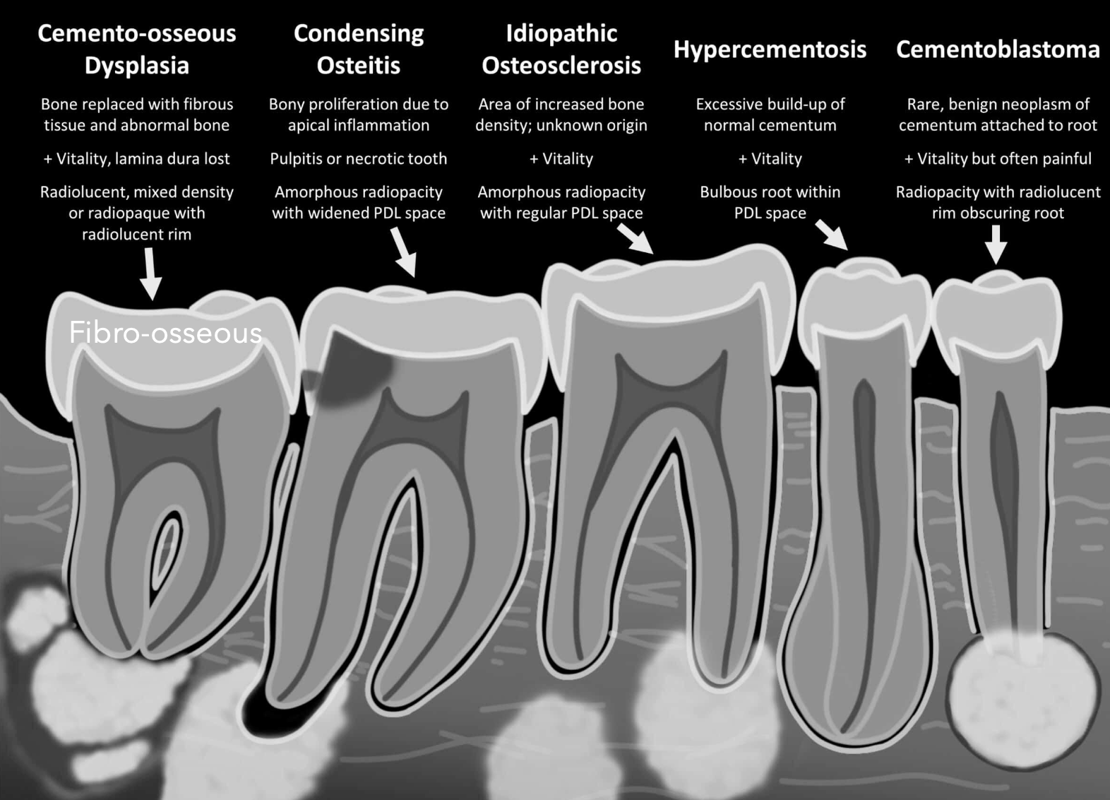
What are some radiographic features of focal COD
Rim is prominent during the mixed phase
What are some radiographic features of cementoblastoma (benign neoplasm of cementum)
Radiolucent rim is contiguous with PDL and PDL is not intact at the involved portion of the root, effacement of root
What are some radiographic features of condensing osteitis (focal sclerosing osteomyelitis)?
No radiolucent rim, borders blend with surrounding trabeculae due to pulpal involvement
What are some radiographic features of idiopathic osteosclerosis, dense bone, enostosis, bone scar?
No radiolucent rim, borders blend with surrounding trabeculae
Name some hereditary bone disorders
Osteogenesis Imperfecta (OI)
Osteopetrosis
Cleidocranial Dysplasia
Cherubism
What is another name for osteogenesis imperfecta?
Brittle bone disease
What is the cause of osteogenesis imperfecta?
Mutation in type 1 collagen
What is the mode of inheritance for osteogenesis imperfecta?
Autosomal dominant (AD = 90%)
Autosomal recessive (AR = 10%)
Some are sporadic
In which demographic does osteogenesis imperfecta show up?
Gender: M=F
Age: Infant, young children
Where would you find osteogenesis imperfecta?
Bone, teeth, ligament, skin, sclera
What is the prevalence of osteogenesis imperfecta?
Most common inherited bone disorder
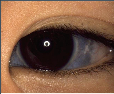
What condition is a blue sclera associated with?
Osteogenesis Imperfecta
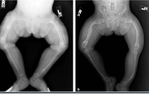
What are some clinical features of osteogenesis imperfecta?
Osteopenia (low bone density)
Short stature/bowing of legs/pathologic fractures
Irregular skill (Wormian skull)
Hearing deficit
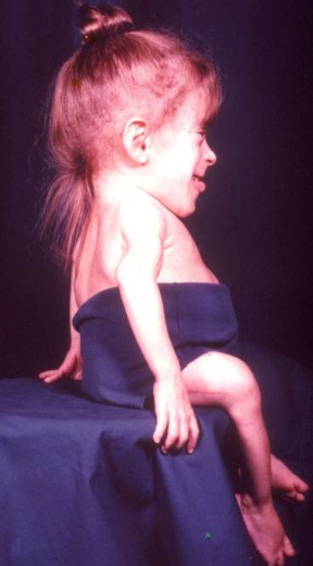
What are some craniofacial features of osteogenesis imperfecta?
Triangular face, maxillary hypoplasia, Class III malocclusion
What are some dental features of osteogenesis imperfecta?
Blue/grey translucent (opalescent teeth)
Different mutation to dentinogensis imperfecta
Both dentitions affected
Obliterated pulp chamber/ shell teeth
Fracture of enamel and dentin
Open bite, cross bite
Vertical dimension decrease
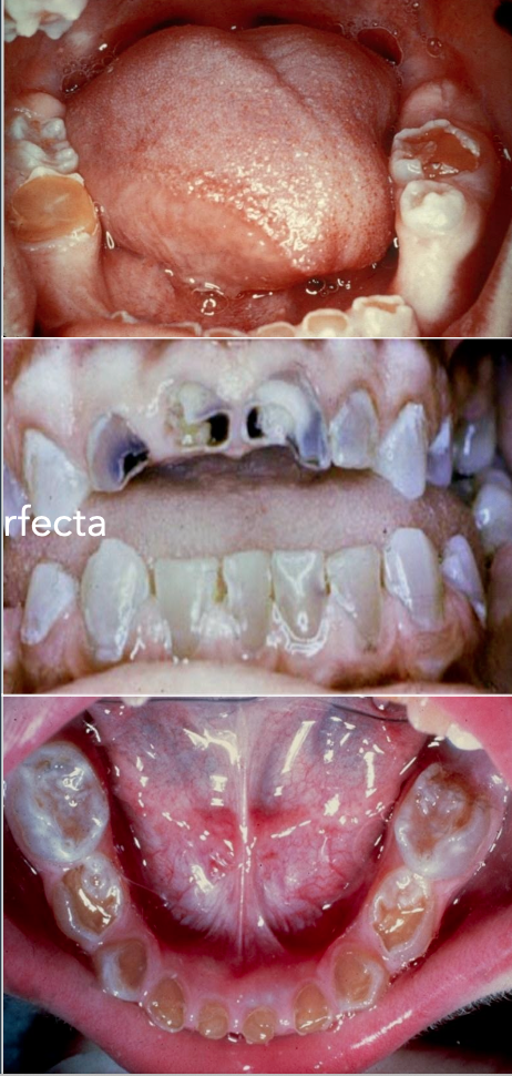
Obliterated pulp in osteogenesis imperfecta
Shell teeth in osteogenesis imperfecta
What is the dental management for osteogenesis imperfecta
Crown
Crown/bridge
Partial or complete denture
Implants
Orthognathic surgery
Orthodontics
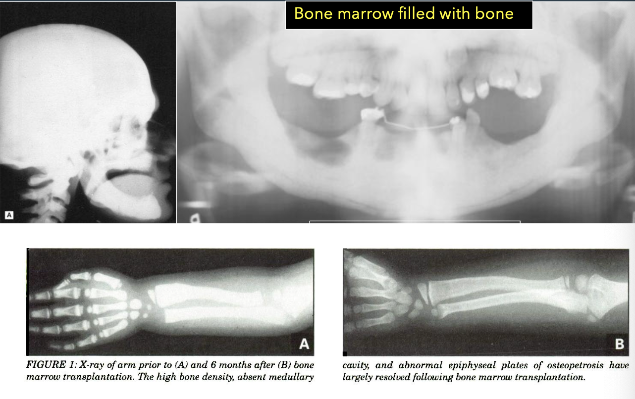
What is another name for osteopetrosis?
Albers-Schönberg Disease; Marble bone disease
What is osteopetrosis?
Inherited function of a decreased osteoclastic activity (greek: petros “rock”) can be AD or AR (fatal)
INCREASE in bone density

What are some clinical features of osteopetrosis?
Can show up anywhere and can be associated with anemia, pathologic fractures, infection, deafness, and blindness
In what demographic do you see osteopetrosis?
Juvenile vs adult
Gener: M=F
Age: infancy except adult form
Osteopetrosis can caused delayed tooth eruption
Osteopetrosis can cause osteomyelitis
What are some craniofacial features of osteopetrosis?
Frontal bossing
Hypertelorism
Broad face
Snub nose
What is the management of osteopetrosis?
Bone marrow transplant
Palliative
What is the prognosis of osteopetrosis?
Good for AD but fatal for AR

What is another name for cleidocranial dysplasia?
Cleidocranial dysotosis
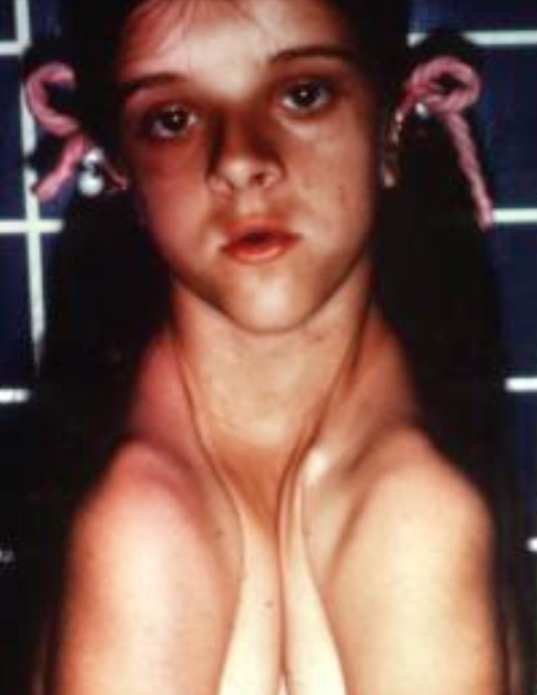
What is cleidocranial dysplasia?
Syndrome complex characterized by dental and clavicle abnormalities
What is the mode of inheritance for cleidocranial dysplasia?
AD inheritance

What do you see in cleidocranial dysplasia?
Bone defects chiefly affect skull and clavicles
Usually, clavicles are present with hypoplasia
Clavicles are absent in 10% of cases
Unusual mobility of shoulders
Name the clinical features of cleidocranial dysplasia
Big head
Pronounced frontal bossing
Ocular hypertelorism
Broad base of nose
Hypoplastic mid face
Long neck
Missing or hypoplastic clavicles
Short stature; Scoliosis
Delayed suture closure
Wormian bone
What are the dental features of cleidocranial dysplasia
Patients have a high-arched palate
Increased prevalence of cleft palate
Mandibular prognathism
Prolonged retention of deciduous teeth
Delay or failure of eruption of permanent teeth
Abnormally shaped teeth
**Numerous unerupted permanent and supernumerary teeth**
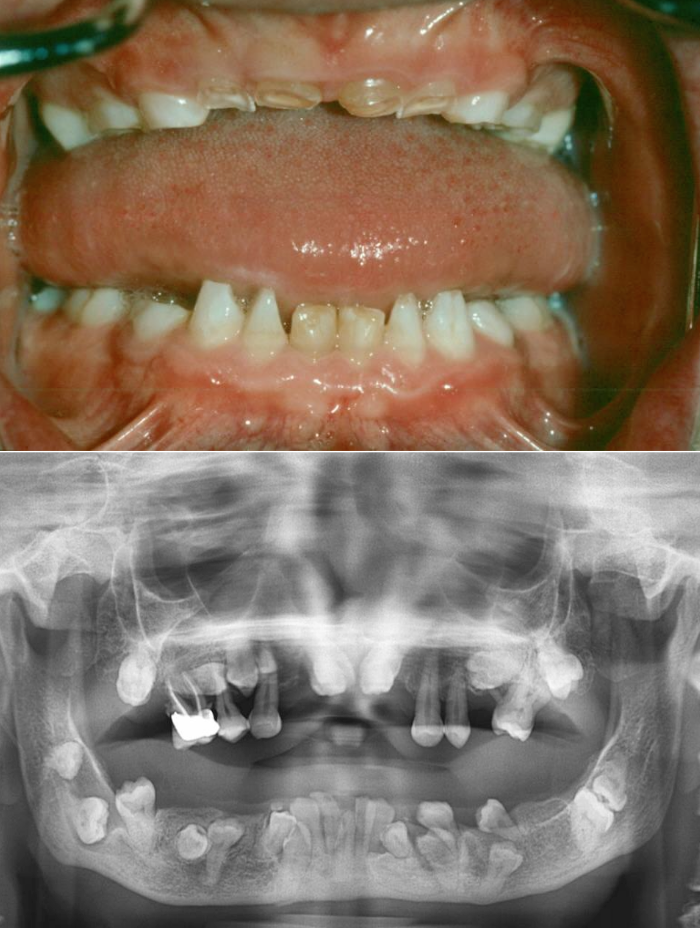
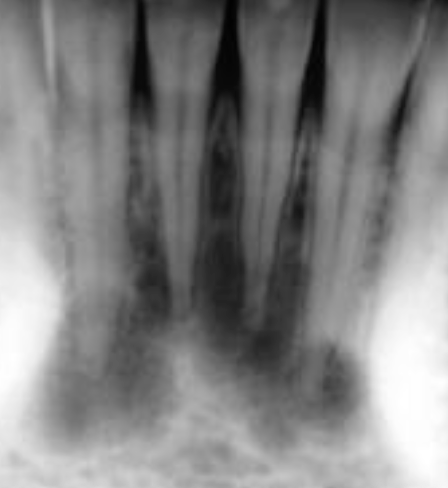
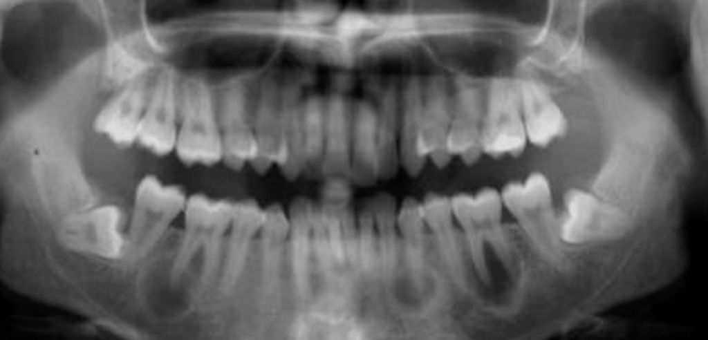
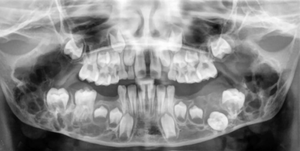
What is this a pano of?
Cleidocranial dysplasia

What is the mode of inheritance of cherubism?
Autosomal dominant inheritance
What mutation causes cherubism?
SH3BP2 chromosome #4p16
In which demographic do you see cherubism?
Gender: M=F
Age: 2-5 (children)

In which location does cherubism appear?
Bilateral posterior mandible (most common), maxilla

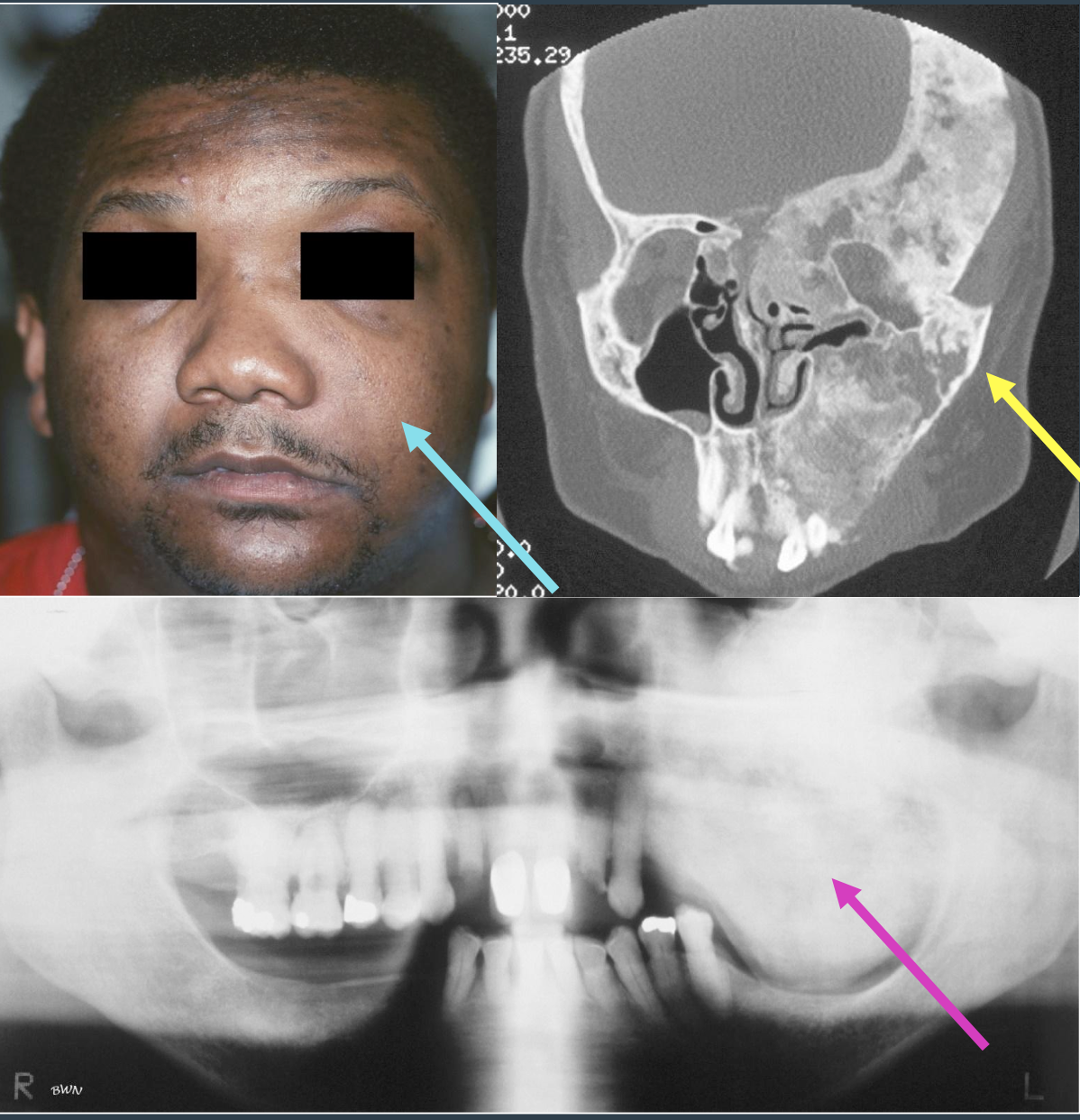
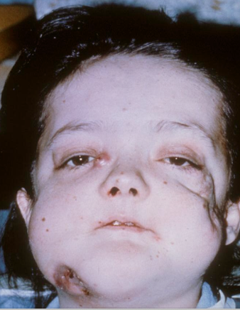
What are some characteristic features of cherubism?
Clinical alterations progress until puberty, then stabilize and slowly regress, asymptomatic
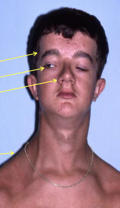
There is an “eyes upturned to heaven” appearance due to a wide rim of exposed sclera noted below the iris

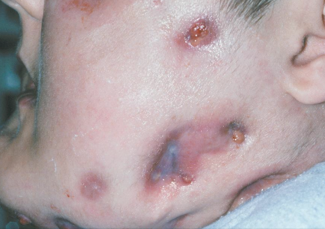
What are the clinical features of cherubism?
Mandibular lesions are painless, bilateral, posterior and expansile
Maxillary involvement occurs posteriorly as well
In severe cases, entire maxillary and mandible are involved
Distortion of the alveolar ridges
May lead to failure of tooth eruption
Microscopic findings are identical to those found in central giant cell granulomas (CGCG)
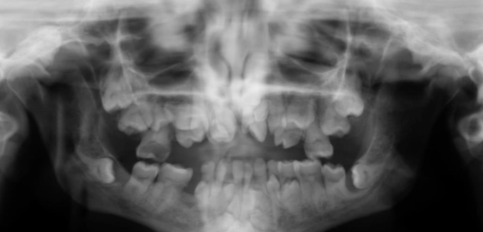
What are some radiographic features of cherubism?
Multilocular, radiolucent, expansile
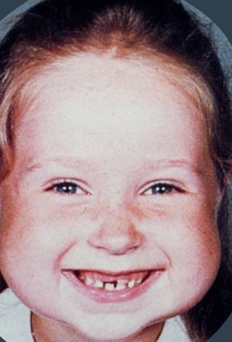
What is this a pano of?
Cherubism

What is this
Cherubism
What is the prognosis and management of cherubism?
Unpredictable
Usually, the lesions show varying degrees of remission & involution after puberty
By age 30, most patient’s facial features are normal
However, some patients are left with facial deformities
Early surgical intervention with curettage has lead to both good results or rapid regrowth with worsening deformity; therefore, optimal therapy hasn’t been determined
Radiation therapy is contraindicated due to risk of postirradiation sarcoma
What is another name for paget disease of bone?
“Osteitis Deformans”
What is paget disease of bone?
A metabolic bone disease characterized by abnormal resorption and deposition of bone of unknown cause (mainly osteoclast)
What is the cause of paget disease of bone?
30% hereditary, AD, sporadic, or paramyxovirus
In which demographic does paget disease of bone appear?
Gender: M (caucasian) > F
Age: Older adults (>45 years)
Where does paget disease of bone appear?
Affects more than one bone (polyostotic)