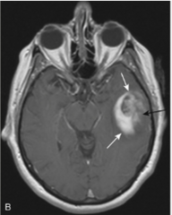
6.2 intracranial pathology
 Call Kai
 Learn
Learn Practice Test
 Spaced Repetition
 Match
Match Flashcards
 Knowt Play
1/119

There's no tags or description
Looks like no tags are added yet.
Name | Mastery | Learn | Test | Matching | Spaced | Call with Kai | Chat |
|---|
No analytics yet
Send a link to your students to track their progress
120 Terms
types of neuroimaging used for pathologies
- MRI is study of choice in most pts, can use CT
- can use US in infants
on non-contrasted CT of the head, how do the structures appear
- bone or blood = white (arrows = blood)
- air, water, CSF = dark
- metal causes streak artifact
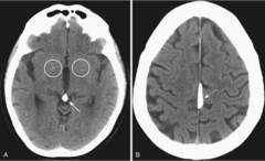
some _____ which show up white, can be normal in the brain as the incr w age
calcifications (circles)

CT vs MRI of the brain/skull
- MRI = better for detecting and staging intracranial + spainal cord abnormalities. Has superior contrast and soft-tissue resoltuon
- CT = better for calcifications or evaluating bone
the study of choice for acute head trauma
non contrasted CT
goal of noncontrasted CT for head trauma
look for mass effect and blood
mass effect- look for
displacement of normal structures
blood on head trauma CT will appear how
bright and settle in crevices or dependent structures
where are skull fx usually located
at point of impact
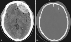
skull fx typically imply what?
additional intracranial injury
kinds of skull fx
- linear
- depressed
- basilar
to look for fx on CT you need to use
Bone windows
fractures of the cranial vault most likely occur where?
the temporal and parietal bones
most common type of skull fracture
linear skull fracture

clinical importance of linear skull fx
not much clinical importance, need to note any underlying injury
depressed skull fractures result from
high-energy blow to small area of skull (ex hammer)
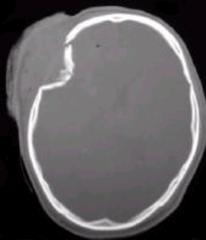
depressed skull fx have high risk for
underlying brain injury > may require surgical eval
depressed skull fx are usually where?
frontoparietal region and comminuted
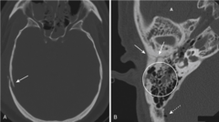
what does this picture show
- comminuted fx of right remporal bone (solid white arrows)
- fluid in the mastoid air cells (circle)
- air in the brain = pneumocephalus (dotted white arrow)
most serious skull fx
basilar skull fx
basilar skull fx consist of
linear fx at the base of the skull
basilar skull fx can be assoc w
tears in the dura mater w subsequent CSF leak > rhinorrhea and otorrhea

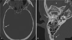
when to suspect basilar skull fx
1. air seen in the brain
2. fluid in the mastoid air cells
3. air-fluid level in sphenoid sinus
study of choice for facial fx
CT
when ordering a CT for facial fx, you must remember to
look at several contiguoius images as to ensure visualization of the entire fx
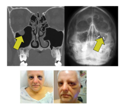
most common orbital fracture
blow-out fx
from direct impact to the orbit, ex: ball hitting eye

blow-out fx leads to
fx of the inferior orbital floor (into maxillary sinus) or the medial wall of the orbit (into ethmoid sinus)
tripod fracture is caused by
blunt force to the cheek
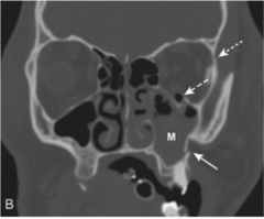
tripod fx
separation of the zygoma from the remainder of the facial bones
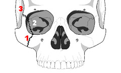
how is the zygoma separated from the remainder of the facial bones in a tripod fx?
- separation of the fronotzygomatic suture
- fx of the orbital floor
- fx of the lateral wall of the ipsilateral maxillary sinus
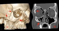
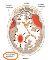
4 types of intracranial hemorrhage
- epidural hematoma
- subdural hematoma
- intracerebral hemorrhage
- subarachnoid hemorrhage
where is an epidural hematoma
b/w dura mater and the skull
epidural hematoma is usually from
blunt head trauma from an MVA
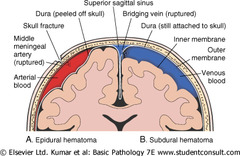
almost all epidural hematomas have an assoc _________ fx
temporal bone fx
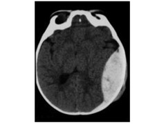
how do epidural hematomas appear
- high density, biconvex, lens-shaped "mass"
- most often found in the temporoparietal region
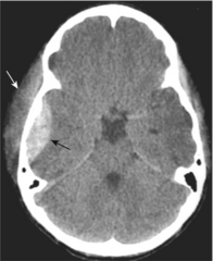
epidural hematomas DO NOT CROSS
suture lines

recall the meninges
- dura mater = outside layer
- arachnoid = avascular middle, separated by subdural space
- pia mater = closley applied to brain and spina cord, carries blood vessels for both. Separated by subarachnoid space

SDH is more common than epidural BUT is not assoc with
skull fracture typically
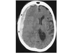
SDH usually occur with
deceleration injuries or falls

SDH causes hemorrhage into
space b/w dura mater and the arachnoid
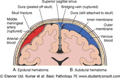
SDH is assoc with
higher mortality rate
SDH can cross ____ but cannot cross _____
can cross suture lines but cannot cross the midline
SDH freq signal severe _____
parenchymal brain injury and incr ICP
how do SDH appear on CT
- crescent-shaped
- extra cerebral bands of high attenuation that may cross suture lines and enter the interhemispheric fissure
- do not cross the midline (but can cause mass effect)
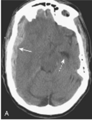
As time passes what happens to how SDH appear on imaging
- may appear isodense to the remainder of brain
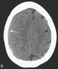
- look for compressed or absent sulci, or sulci dispalced away from the inner side of the skull (white arrow)
Subarachnoid hemorrhage is usually from
ruptured blood vessel
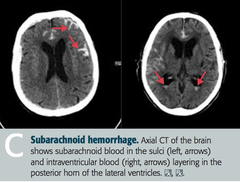
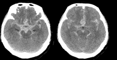
subarachnoid hemorrage occurs where?
b/w arachnoid and pia mater
subarachnoid hemorrhage can cause
intraventricular hemorrhage
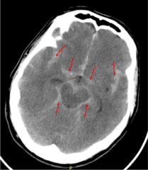
numerous causes for intracerebral hematoma
- trauam/shearing injury
- vascular disease or rupture
- amyloid deposits
CT findings of intracerebral hematomas over time
change over time and may not be immediately evident on the initial scan
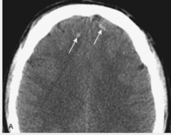
intracerebral hematoma can manifest by
multiple areas of high attenuation hemorrhage w/in brain parenchyma on CT
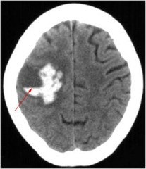
where is intracerebral hematoma usually found?
frontal or temporal lobes
what has the poorest prognosis of all head traumas
axonal injury
axonal injury
acceleration/deceleration forces diffusely injure axons deep to the cortex, producing unconsciousness from the moment of injury
axonal injury is responsible for
prolonged coma following head trauma
initial CT scan of axonal injury
may be normal or underestimate degree of injury
CT findings of axonal injury may be similar to
those described for intracerebral hemorrhage following head trauma
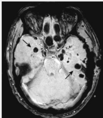
what is the study of choice for diffuse axonal injury
MRI: Gradient susceptibility MRI
Gradient susceptibility MRI is very sensitive to
bleeding at the gray/white matter junction and can detect v small lesions
how will axonal injury appear on imaging?
punctuate hemorrhages (dark spots)
incr ICP is caused by either ______ or ______
- cerebral edema = incr volume of the brain
- hydrocephalus = incr size of ventricles
most common cause of cerebral edema
HTN, masses
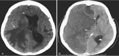
2 types of cerebral edema
- vasogenic = extracellular accumulation of fluid
- cytotoxic = cellular edema

vasogenic cerebral edema is assoc w
- malignancy and infx
- predominantly affects the white matter (inside)
cytotoxic cerebral edema is assoc with
- cerebral ischemia
- d/t cell death
- affects both gray and white matter
with cerebral edema, what can happen on imaging?
- compression or obliteration of the normal sulci
- ventricles may be compressed
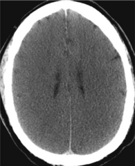
reasons for imaging strokes
- determine if another cause of neurologic impairment
- identify presence of blood > distinguish ischemic from hemorrhagic
- identify the infarct and characterize it
most strokes are ______ in origin
thromboembolic
acute strokes are initially imaged by
noncontrast CT
when would CT findings of a stroke appear?
- hemorrhagic = immediate
- ischmeic = w/in hours after onset of sx
MRI of strokes is more senstive and specific for detecting early infarction- how early can it detect?
w/in 20-30 mins of onset of the event

the most common finding of an acute less than 24h non-hemorrhagic stroke is
normal CT scan
CT stroke findings: 12-24 hrs
indistinct area of low attenuation in a vascular distribution
CT stroke findings: >24 hrs
better circumscribed lesion w mass effect that peaks at 3-5 days and usually disappears by 2-4 weeks
TLDR: abnormality peaks w/in 1st week, and resolves w/in a few weeks
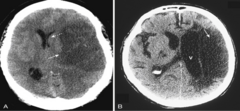
which type of stroke is assoc w higher morbidity and mortality?
hemorrhagic > ischemic
hemorrhage from stroke can occur into where?
brain parenchyma or subarachnoid space

ischemic stroke, if recognized early, can be tx w
intravenous-thrombolytics: tenectoplase (TNK) or mechanical thrombectomy
image-guided interventions for hemorrhagic strokes
CTA or MRA to pinpoint area and possibly coil or clip
most frequent CNS aneurysm is
berry aneurysm
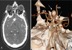
berry aneurysm forms from
congenital weakening in the arterial wall, usually at the sites of vessel branching in the circle of Willis
what incr growth of berry aneurysms
HTN and aging
larger berry aneurysms do what?
bleed more freq
someone w a ruptured aneurysm may say
"the worst headache of my life"
upon aneurysm rupture where does blood usually enter?
subarachnoid space
most aneurysms are detected by
CTA or MRA
hydrocephalus
expansion of ventricular system causing an incr in volume of CSF
hydrocephalus can be d/t
- overproduction of CSF
- under absorption of CSF
- restriction in outflow of CSF
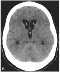
in hydrocephalus, compare the ventricles to the sulci
ventricles are usually disproportionally dilated compared w the sulci
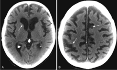
in cerebral atrophy, compare the ventricles and sulci
both ventricles and sulci are proportionately enlarged
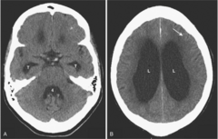
what is a form of communicating hydrocephalus
normal pressure hydrocephalus
normal pressure hydrocephalus has what classic triad of sx
1. gait
2. dementia
3. urinary incontinence
age of onset for normal pressure hydrocephalus
60-70 y/o
normal pressure hydrocephalus is easily treated w
ventriculoperitoneal shunt
image findings of normal pressure hydrocephalus
- enlarged ventricles with normal or flattened sulci
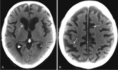
cerebral atrophy
loss of both gray and white matter
cerebral atrophy is assoc with
dementia ex: Alzheimer's disease
Alzheimer's disease
diffuse cortical atrophy, esp in the temporal lobes
ventricles dilate in both hydrocephalus and cerebral atrophy- what makes cerebral atrophy different?
ventricles dilate b/c loss of normal cerebral tissue. This vacant space is passively filled with CSF but CSF production and absorption are normal
image findings of cerebral atrophy
proportionate enlargement of both the ventricles and the sulci
gliomas
common intraaxial (w/in brain parenchyma) mass in an adult